Imaging cases
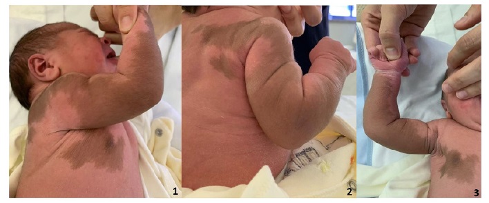
Large brown macula in a newborn
Mácula gigante num recém-nascido
1 Department of Pediatrics, Unidade Local de Saúde de Viseu Dão-Lafões. 3504-509 Viseu, Portugal. gisela_s_oliveira@hotmail.com; jnsmarques@campus.ul.pt; jdcfa82@gmail.com; pedromaneirasousa@gmail.com; isabelverdelho@hotmail.com
2 Department of Dermatology, Unidade Local de Saúde de Viseu Dão-Lafões. 3504-509 Viseu, Portugal. rita.cabral.5736@hstviseu.min-saude.pt
Abstract
The authors report the case of a male term newborn born after an uneventful monitored pregnancy. On physical examination, he presented with a light brown hyperpigmented homogeneous macule >15 cm in size compatible with a giant congenital melanocytic nevus (CMN). The boy was discharged with indication for Neonatology and Dermatology follow-up. Since then, he has shown adequate psychomotor growth and development and unremarkable neurologic examination and cranioencephalic magnetic resonance imaging.
CMN is a benign proliferation of melanocytes forming theca/nests in the dermal-epidermal junction and/or dermis. It may be present at birth or develop during the first two years of life in any body area. CMN can be classified as small, medium, large, giant, or multiple. Giant CMN is rare and is a risk factor for melanoma.
Management of CMN should be based on age, nevus size, and other factors. Because of the potential for malignancy, giant CMN requires clinical surveillance with neurodevelopmental monitoring and regular dermoscopy, as well as early surgical management of suspicious lesions.
Keywords: congenital melanocytic nevus; dermoscopy; newborn; risk for malignant transformation; skin lesion
Resumo
É descrito o caso de um recém-nascido de termo, do sexo masculino, fruto de gestação vigiada sem intercorrências. Ao exame objetivo, o rapaz apresentou uma mácula hiperpigmentada, de coloração castanho claro, homogénea, com mais de 15 cm, compatível com nevo melanocítico congénito (NMC) gigante, tendo sido referenciado para a consulta de Neonatologia e Dermatologia. Desde então, tem apresentado crescimento e desenvolvimento psicomotor adequados e exame neurológico e ressonância magnética cranioencefálica sem alterações.
O NMC é uma proliferação benigna de melanócitos, de coloração castanha, que forma tecas/ninhos na junção dermoepidérmica e/ou derme. Pode estar presente ao nascimento ou surgir nos primeiros dois anos de vida em qualquer localização. Os NMCs podem ser classificados em pequenos, médios, grandes, gigantes ou múltiplos. O NMC gigante é raro e representa um fator de risco para melanoma.
A abordagem do NMC depende da idade e do tamanho do nevo, entre outros fatores. Dado o potencial de malignidade, o NMC gigante requer vigilância clínica com acompanhamento do neurodesenvolvimento e dermatoscopia regular e abordagem cirúrgica precoce perante lesões suspeitas.
Palavras-chave: dermatoscopia; lesão cutânea; nevo melanocítico congénito; recém-nascido; risco de transformação maligna
The authors report the case of a term newborn boy born at 40 weeks postmenstrual age (maternal age 31 years) from an uneventful nonconsanguineous pregnancy monitored in primary health care. Blood group was A Rh-, both parents were healthy, and group B streptococcus screening test was negative. Prenatal ultrasound and serologies were unremarkable. The boy was born by vaginal delivery with a weight of 3530 g, height of 49 cm, head circumference of 33 cm, Apgar score of 9/10/10, good adaptation to extrauterine life, and no oxygen requirements.
Initial physical examination revealed a homogeneous, light brown, hyperpigmented macula more than fifteen cm in diameter on the right upper limb and thoracic region (Figures 1-3). Dermoscopy was performed, revealing a typical brown globular pattern with no irregular structures or visible atypia.
What is your diagnosis?
Diagnosis
Giant congenital melanocytic nevus (CMN) - Size at birth >15 cm, with predicted adult size >40 cm.
Discussion
After diagnosis, the newborn was discharged with indication for Neonatology and Dermatology follow-up. On reassessment, he showed adequate psychomotor growth and development, an unremarkable neurological examination, and no areas of concern on routine dermoscopy. Subsequent evaluation with cranioencephalic magnetic resonance imaging (MRI) at 14 months of age was normal.
CMN is a benign proliferation of neural crest-derived melanocytes that form theca or nests at the dermal-epidermal junction and/or in the dermis.1) Brown in color, it may be present from birth or appear during the first two years of life at any body surface area.1,2 Based on adult size projection, CMN are classified as small (<1.5 cm), medium (1.5-20 cm), large (20-40 cm), giant (>40 cm), or multiple.3 Compared to other CMN, giant CMN is a rare entity, with an incidence of 1:20,000 newborns, and has a higher and earlier risk for melanoma (6-20%).1,2,4,5 Melanomas arising from congenital nevi are rarer than spitzoid melanomas, but account for most melanoma-related deaths in childhood.5)
Management of CMN should be based on age, nevus size, and other factors such as psychosocial impact, association with vascular malformations, limb atrophy and asymmetry, neurocutaneous melanosis, and characteristic facial morphology.1,2,4,6) Given the potential for malignancy, giant CMN require clinical surveillance with neurodevelopmental follow-up, regular dermoscopy, and early surgical intervention (excision or biopsy) of suspicious lesions for timely diagnosis of melanoma.2,4-6 Surveillance with digital dermoscopy has optimized early diagnosis of melanoma and reduced the number of unnecessary excisions. Thus, in lesions considered atypical but with low suspicion of melanoma, dermoscopic surveillance may be preferred to surgery.2,4) A spectrum of central nervous system abnormalities has been described in association with congenital melanocytic naevi.6) In this context, cranioencephalic MRI should be performed for early detection of melanin deposition, especially in the temporal lobes, cerebellum, pons, and/or medulla, in neurologically asymptomatic patients with giant CMN.6,7 If cranioencephalic MRI shows no changes, it should be repeated only if suspicious symptoms arise.5,7
Authorship
Ana Gisela Oliveira - Bibliographical search; clinical case conception and design; Data curation; Formal analysis; Writing - original draft; Writing - review & editing; Validation
João Marques - Bibliographical search; Methodology; Data curation; Writing - original draft; Writing - review & editing; Validation
Joaquina Antunes - Bibliographical search; Methodology; Writing - original draft; Writing - review & editing; Validation
Pedro Maneira Sousa - Bibliographical search; Methodology; Writing - original draft; Writing - review & editing; Validation
Rita Cabral - Bibliographical search; Methodology; Writing - original draft; Writing - review & editing; Validation
Isabel Andrade - Bibliographical search; Methodology; Writing - original draft; Writing - review & editing; Validation
References
1. Pimenta R, Fernandes S, Filipe P, Laureano AO. Dermatoscopia na Idade Pediátrica - Parte I: Tumores Cutâneos. Journal of the Portuguese Society of Dermatology and Venereology. 2020;77(4):291-304. doi: https://doi.org/10.29021/spdv.77.4.1126.
[ Links ]
2. Chien JC, Niu DM, Wang MS, Liu MT, Lirng JF, Chen SJ, et al. Giant congenital melanocytic nevi in neonates: report of two cases. Pediatr Neonatol. 2010;51(1):61-4. doi: https://doi.org/10.1016/S1875-9572(10)60012-5.
[ Links ]
3. Krengel S, Scope A, Dusza SW, Vonthein R, Marghoob AA. New recommendations for the categorization of cutaneous features of congenital melanocytic nevi. J Am Acad Dermatol. 2013;68(3):441-51. doi: https://doi.org/10.1016/j.jaad.2012.05.043.
[ Links ]
4. Thomas L, Puig S. Dermoscopy, Digital Dermoscopy and Other Diagnostic Tools in the Early Detection of Melanoma and Follow-up of High-risk Skin Cancer Patients. Acta Derm Venereol. 2017;Suppl 218:14-21. doi: https://doi.org/10.2340/00015555-2719.
[ Links ]
5. JM Neves, B Duarte, MJ Paiva Lopes. Pediatric melanoma: Epidemiology, Pathogenesis, Diagnosis and Management. Revista SPDV 2020; 78 (2): 107-14. doi: https://doi.org/10.29021/spdv.78.2.1197.
[ Links ]
6. Kinsler VA, Chong WK, Aylett SE, Atherton DJ. Complications of congenital melanocytic naevi in children: analysis of 16 years' experience and clinical practice. Br J Dermatol. 2008;159(4):907-14. doi: https://doi.org/10.1111/j.1365-2133.2008.08775.
[ Links ]
7. Waelchli R, Aylett SE, Atherton D, Thompson DJ, Chong WK, Kinsler VA. Classification of neurological abnormalities in children with congenital melanocytic naevus syndrome identifies magnetic resonance imaging as the best predictor of clinical outcome. Br J Dermatol. 2015;173(3):739-50. doi: https://doi.org/10.1111/bjd.13898.
[ Links ]


