SciELO - Scientific Electronic Library Online

 
vol.30 número6Stent selado: uma complicação invulgar após a colocação de um novo stent metálico totalmente coberto com válvula anti-refluxo na hepaticogastrostomiaPolipose Linfomatosa Múltipla Silenciosa No Linfoma de Células Do Manto: Desde o Íleon Até Ao Estoma índice de autoresíndice de assuntosPesquisa de artigos
Home Pagelista alfabética de periódicos  

Serviços Personalizados

Journal

Artigo

Indicadores

Links relacionados

  • Não possue artigos similaresSimilares em SciELO

Compartilhar


GE-Portuguese Journal of Gastroenterology

versão impressa ISSN 2341-4545

GE Port J Gastroenterol vol.30 no.6 Lisboa dez. 2023  Epub 01-Fev-2024

https://doi.org/10.1159/000527586 

Endoscopic Snapshot

Endoscopic Submucosal Dissection of Subepithelial Lesion in the Cecum: Granular Cell Tumor

Disseção endoscópica da submucosa de lesão subepitelial do cego: tumor de células granulares

Diogo Bernardo Moura1 
http://orcid.org/0000-0002-8353-4619

Nuno Nunes1 

Carolina Chálim Rebelo1 

Francisca Côrte-Real1 

Ana Catarina Rego1 

Maria Antónia Duarte1 

1Gastroenterology Department, Hospital do Divino Espírito Santo de Ponta Delgada EPER, Ponta Delgada, Portugal


A 44-year-old female patient was subjected to total colonoscopy that revealed a 15 mm bulge in the cecum, covered by normal mucosa, compatible with subepithelial lesion, of hard consistency and no pillow sign (Fig. 1a). The lesion was evaluated by ultrasonography with mini-probe (Fig. 1b), which confirmed the presence of a sub-epithelial nodular hypoechoic lesion although it was not possible to safely distinguish between the second and third ultrasonographic wall layers.
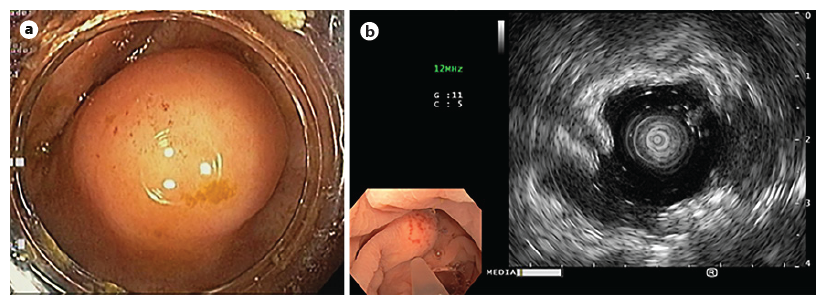
Fig. 1. Endoscopic findings. a In the cecum, a 15 mm bulge with normal mucosa was observed. b Ultrasonography - a subepithelial nodular lesion was confirmed, although it was not possible to safely distinguish between the second and third ultrasonographic wall layers. 

Endoscopic submucosal dissection (ESD) was pro-posed. The endoscopist had extensive experience in colorectal ESD. A glycerol solution with indigo carmine and adrenaline (1:50,000) was injected (Fig. 2a). Mucosal incision was performed (Fig. 2b), followed by submucosal endoscopic dissection using DualKnife JTM (Olympus, Tokyo, Japan) and IT Knife nanoTM (Olympus, Tokyo, Japan), with dry-cut current effect 2.5 and swift coagulation current effect 3 achieving total excision of the lesion at the end of the procedure (Fig. 2c-e). The scar was closed with Resolution 360TM ULTRA clips (Boston Scientific, Boston, USA) (Fig. 2f, g). The lesion was retrieved en bloc (Fig. 2h). No adverse events were observed.
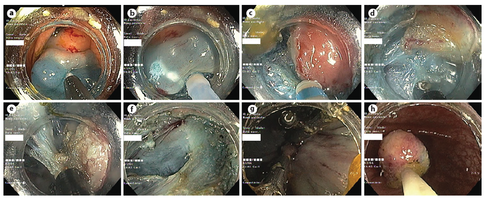
Fig. 2. Endoscopic submucosal dissection. a Submucosal injection. b Mucosal incision. c-e Submucosal dissection. f Dissection scar. g The scar was closed with clips. h En bloc lesion retrieval. 

Histological evaluation revealed a solid neoplasia of the submucosa consisting of epithelioid cells of vast granular citoplasma, centered by small, round, uniform nuclei (Fig. 3a, b). These cells were positive for S100 and inhibin - granular cell tumor (GCT) (Fig. 3c, d). The lesion was limited to the submucosa and was covered by normal colonic mucosa. The excision margins were free. A surveillance colonoscopy at 12 months is currently scheduled.

Fig. 3 Histological evaluation. a Submucosal expansion by epithelioid cells, covered by normal colonic mucosa (×20 magnification). b These cells have vast granular citoplasma, centered by small, round, uniform nuclei (×100 magnification). c, d Immunohistochemistry showing positivity for S100 (c) and inhibin (d) (×400 magnification). 

The authors present a case of a subepithelial lesion in the cecum evaluated by miniprobe ultrasonography and removed en bloc by ESD. GCT’s are a rare entity, whose pathological behavior is not fully understood, and are most frequently found incidentally. Granular cells have neuronal origin, and Schwann cells are precursors [1, 2].

The cases reported in the literature suggest a generally benign behavior but <2% have shown potential for malignancy, which is suggested by endoscopic features of ulceration or size >40 mm [3]. Histological proposed criteria for malignancy are the following: high number of mitosis, big nuclei, signs of lymphovascular invasion; evidence of metastization being the sole definitive criteria [2]. A case of local recurrence due to incomplete resection was reported [3]. ESD in the colon is a safe, technically demanding procedure that allows an en bloc resection and avoids surgery in the absence of features of malignancy. Endoscopic fullthickness resection using a full-thickness resection device is an alternative and developing method, with current evidence apparently showing a similar safety profile albeit with a lower complete resection rate [4]. To the best of our knowledge, this is the first case of GCT in the cecum treated by ESD in the West; all the other four cases were reported in China [5, 6].

References

1. Cha JM, Lee JI, Joo KR, Choe JW, Jung SW, Shin HP, et al. Granular cell tumor of the descending colon treated by endoscopic mucosal resection: a case report and review of the literature. J Korean Med Sci. 2009; 24(2): 337-41. [ Links ]

2. Mobarki M, Dumollard JM, Dal Col P, Camy F, Peoc'h M, Karpathiou G. Granular cell tumor a study of 42 cases and systemic review of the literature. Pathol Res Prac. 2020; 216(4): 152865. [ Links ]

3. Kawashima K, Hikichi T, Onizawa M, Gunji N, Takeda Y, Mochimaru T, et al. Colonic endoscopic submucosal dissection for a granular cell tumor with insufficient endoscopic manipulation in the hepatic flexure. Case Rep Gastroenterol. 2022; 16(1): 216-22. [ Links ]

4. Krutzenbichler I, Dollhopf M, Diepolder H, Eigler A, Fuchs M, Herrmann S, et al. Technical success, resection status, and procedural complication rate of colonoscopic full-wall resection: a pooled analysis from 7 hospitals of different care levels. Surg Endosc. 2021; 35(7): 3339-53. [ Links ]

5. Chen Y, Chen Y, Chen X, Chen L, Liang W. Colonic granular cell tumor: report of 11 cases and management with review of the literature. Oncol Lett. 2018; 16(2): 1419-24. [ Links ]

6. Take I, Shi Q, Qi ZP, Cai SL, Yao LQ, Zhou PH. Endoscopic resection of colorectal granular cell tumors. World J Gastroenterol. 2015; 21(48): 13542-7. [ Links ]

1Statement of Ethics Ethical Approval Statement: ethical approval was not required for this study in accordance with local/national guidelines. Written informed consent was obtained from the patient for publication of this case report and any accompanying images, according to Helsinki declaration.

3Funding Sources The authors have no funding sources to declare.

5Data Availability Statement All data generated or analyzed during this study are included in this case report. Further inquiries can be directed to the corresponding author.

Received: July 18, 2022; Accepted: October 03, 2022

Correspondence to: Diogo Bernardo Moura, diogobernardomoura@gmail.com

Conflict of Interest Statement The authors have no conflicts of interest to declare.

Author Contributions Diogo Bernardo Moura: article concept, literature review, and drafting of the manuscript. Nuno Nunes: main endoscopist of the described procedure, literature review, and critical review of the manuscript. Carolina Chálim Rebelo, Francisca Côrte-Real, Ana Catarina Rego, and Maria Antónia Duarte: critical review of the manuscript.

Creative Commons License This is an open-access article distributed under the terms of the Creative Commons Attribution License