SciELO - Scientific Electronic Library Online

 
vol.31 número3Hepatite autoimune induzida pelo vírus da hepatite delta: um quebra-cabeçasRemoção endoscópica de dois bezoares gigantes petrificados com recurso a litotrícia mecânica e por laser índice de autoresíndice de assuntosPesquisa de artigos
Home Pagelista alfabética de periódicos  

Serviços Personalizados

Journal

Artigo

Indicadores

Links relacionados

  • Não possue artigos similaresSimilares em SciELO

Compartilhar


GE-Portuguese Journal of Gastroenterology

versão impressa ISSN 2341-4545

GE Port J Gastroenterol vol.31 no.3 Lisboa jun. 2024  Epub 28-Jun-2024

https://doi.org/10.1159/000533498 

Endoscopic Snapshot

Cholangioscopy with Laser Lithotripsy in the Treatment of a Patient with Type II Mirizzi Syndrome

Colangioscopia com Litotrícia Laser no tratamento de doente com Síndrome de Mirizzi tipo II

Marta Moreira1 

Ana Catarina Carvalho2 

Isabel Tarrio1 

Alda João Andrade1 

Tarcísio Araújo1 

Luís Lopes1  3  4 

1Department of Gastroenterology, Hospital de Santa Luzia, Viana do Castelo, Portugal;

2Department of Gastroenterology, Centro Hospitalar Tondela-Viseu, Dão Lafões, Portugal;

3Life and Health Sciences Research Institute (ICVS), School of Medicine, University of Minho, Braga, Portugal;

4ICVS/3B’s - PT Government Associate Laboratory, Braga/Guimaraes, Portugal


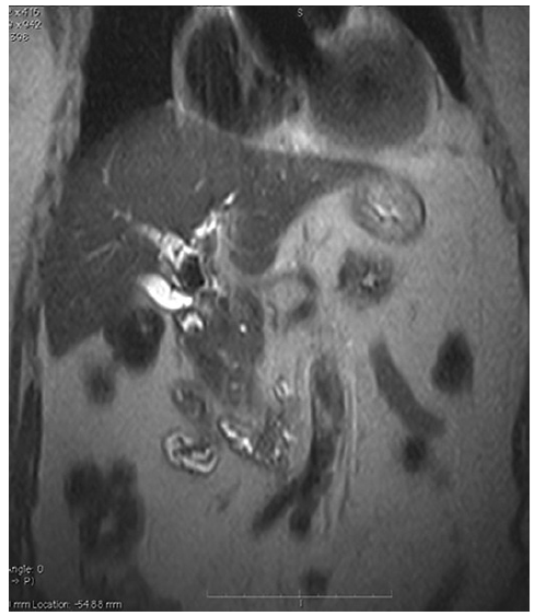
Eighty-Seven-year-old woman, without relevant medical past history, was referred to ERCP for treatment of acute cholangitis. Abdominal CT scan revealed dilatation of intra and extrahepatic bile ducts with hyperdense content in the cystic duct and in the common bile duct, suggestive of Mirizzi syndrome and choledocholithiasis. An abdominal MRI was performed revealing a large 4 cm stone in the infundibulum-cystic duct, a 2 cm stone in the hepatic duct (Fig. 1).

Fig. 1 MRI showing a large stone in the infundibulum-cystic duct, with about 4 cm. 

During the initial ERCP, cholangiography revealed a large stone in the cystic causing a stenosis of the hepatic duct with upstream dilation of the biliary tree, confirming the Mirizzi syndrome (Fig. 2). A plastic double pigtail stent (7 Fr × 4 cm) was inserted, after sphincterotomy. After multidisciplinary evaluation of the patient conditions, given the patient frailty it was decided to treat the condition using an endoscopic approach - laser lithotripsy assisted by cholangioscopy.

Fig. 2 Cholangiography showing a large stone causing compression of the common hepatic duct. 

SpyGlass cholangioscopy revealed two impacted large stones, one in the cystic duct and unexpectedly, another in the hepatic duct. Lithotripsy with laser Holmium was successfully performed in both stones; after both lithotripsies, a fistula between the cystic and the hepatic duct was noticed, involving less than one-third of the circumference of the hepatic duct (Mirizzi type II). The patient presented a favourable clinical evolution and was discharged 4 days after the procedure.

Discussion

Mirizzi syndrome is an uncommon cause of acute cholangitis [1]. ERCP allows the resolution of jaundice through the placement of stents and, in some cases, the removal of the cystic duct stone, although most patients are treated surgically [1-3]. The use of cholangioscopy-guided lithotripsy with the Spyglass platform was already described in the treatment of patients with Mirizzi syndrome in some case reports [3-5].

References

1. Chen H, Siwo EA, Khu M, Tian Y. Current trends in the management of Mirizzi Syndrome: a review of literature. Medicine. 2018; 97(4):e9691. [ Links ]

2. Ibrarullah M, Mishra T, Das AP. Mirizzi syndrome. Indian J Surg. 2008;70(6):281-7. [ Links ]

3. Salgado-Garza G, Hernandez-Arriaga P, Gonzalez-Urquijo M, Díaz-Elizondo JA, Flores-Villalba E, Rojas-Méndez J, et al. Singleoperator cholangioscopy and electrohydraulic lithotripsy for the treatment of Mirizzi syndrome. Ann Med Surg. 2021;62:274-7. [ Links ]

4. Tsuyuguchi T, Sakai Y, Sugiyama H, Ishihara T, Yokosuka O. Long-term follow-up after peroral cholangioscopy-directed lithotripsy in patients with difficult bile duct stones, including Mirizzi syndrome: an analysis of risk factors predicting stone recurrence. Surg Endosc. 2011 Jul;25(7):2179-85. Epub 2010 Dec 24. [ Links ]

5. Zimmer V, Bier B. Cholangioscopic characterization of type II Mirizzi syndrome associated with multifocal intraductal papillary neoplasm of the bile duct (with video). Gastrointest Endosc. 2021 Mar;93(3):771-2. Epub 2020 Sep 10. [ Links ]

1Statement of Ethics The subject has given her written informed consent to publish their case (including publication of images) that is stored in the archives of the department. This article is exempted from ethical approval due to local guidelines. The data regarding this case are not publicly available due to containing information that could compromise the privacy of the patient but are available from M.M. (corresponding author e-mail) upon reasonable request.

3Funding Sources This study did not require any funding.

Received: April 06, 2023; Accepted: July 21, 2023

Correspondence to: Marta Moreira, martaimamoreira@gmail.com

Conflict of Interest Statement Authors declare no conflict of interests for this article.

Author Contributions Marta Moreira, Ana Catarina Carvalho wrote the manuscript. Isabel Tarrio and Alda João Andrade helped in the video edition. Tarcísio Araújo and Luis Lopes were responsible for the revision of its contents.

Creative Commons License This is an open-access article distributed under the terms of the Creative Commons Attribution License