Introduction
Acute genital ulcers (AGU) are a frequent cause of emergency room visits and can be located in the anogenital area or vagina. Its diagnosis is mostly clinical and initially approached from a syndromic point of view. It is based on history, physical examination, and, in some cases, laboratory findings. AGU can be categorized into infectious and noninfectious groups1. Sexually transmitted infections are the main representative of the first group and include herpes simplex, syphilis, lymphogranuloma venereum, chancroid, and donovanosis. Among the most prevalent nonsexually transmitted infectious agents, the Epstein-Barr virus and Cytomegalovirus stand out1–7. In the group of noninfectious ulcers, the main etiologies are Behçet’s disease (BD), Chron’s disease, and Lipschütz’s ulcer1.
However, the etiological diagnosis of AGU is difficult since there is a wide variety of causative agents and in many cases, they are poorly recognized by the attending physicians3. BD is a multisystem inflammatory disease with vascular involvement, characterized by periods of outbreaks and remission, commonly addressed by rheumatologists8,9. However, AGU is one of its first manifestations and the dermatological evaluation is, most of the time, responsible for initially suspecting the diagnosis9. The disease can, on many occasions, develop into a serious clinical condition, compromising the patient’s health if the diagnosis is not made immediately.
The doctor in the emergency room is frequently faced with AGU and must be prepared to adequately identify its various etiologies. It is essential that they recognize the characteristics of Behçet’s ulcer in order to avoid misdiagnosis and delay in the treatment of the disease. Thus, the objective of the present study is to report a case of a patient with extensive and deep ulcers triggered by BD, as well as to discuss the clinical and pathological findings and to carry out a brief review of the literature on the syndromic approach to AGU.
Case description
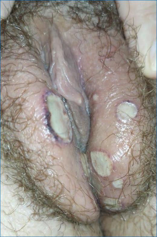
The present work was approved by the Research Ethics Committee of Irmandade de Misericórdia da Santa Casa de São Paulo Hospital and the patient signed a free and informed consent form for publication. A 31-year-old female patient sought the emergency department complaining of multiple painful vulvar ulcers that had started 1 day before. She was initially treated with oral acyclovir due to suspected genital herpes. After 3 days of treatment, she observed an important and progressive worsening of the condition and returned to the emergency room. On return, she presented herself with several ulcerative lesions on the vulva, with well-defined, deep, and very painful edges located in the labia majora, and introitus. The ulcers had a necrotic background, some with a violet halo around them (Figures 1 and 2). No changes were seen in the vagina and cervix.

Figure 1 Deep, extensive, confluent, and well-defined acute vulvar genital ulcers with well-defined edges characterize Behcet’s disease-related genital ulcers.

Figure 2 Deep, extensive, confluent, and well-defined acute vulvar genital ulcers with well-defined edges characterize Behcet’s disease-related genital ulcers.
The patient denied previous illnesses or surgeries, use of medication, or risky sexual behavior. She had a previous natural delivery and active sexual life with a steady partner, using oral hormonal contraception. The patient’s mother was diagnosed with systemic lupus erythematosus.
She was hospitalized and treated with intravenous corticosteroids due to suspicion of BD or Lipschütz ulcer, debridement of ulcers, and local hygiene were also performed. Serologies were performed for herpes simplex, human immunodeficiency virus, Cytomegalovirus, syphilis, and all results were negative. Complementary tests such as blood count, erythrocyte sedimentation, and antinuclear factor did not show relevant changes.
There was an improvement of the lesions, and she was discharged after 7 days; home use of prednisone 20 mg/day and referral to a rheumatologist were advised. Complete improvement of the vulvar lesions occurred in about 40 days. In the subsequent detailed anamnesis, she reported a history of recurrent oral ulcers since adolescence, with about seven episodes per year, in addition to nonspecific and intermittent polyarthralgia without a definitive diagnosis. Thus, her previous history and the presented evolution of the case confirmed the diagnosis as BD. Treatment with prednisone 20 mg daily was maintained and oral colchicine was introduced. The patient is doing well, with no further lesions.
Discussion
Among the noninfectious etiologies of AGU that can affect women, BD is important due to the high prevalence, as well as the severity of the disease. The most common clinical manifestations of BD are recurrent oral and genital ulcers, skin lesions (erythema nodosum, pustular lesions, pyoderma gangrenosum, and superficial thrombophlebitis), uveitis, in addition to multiple other less common systemic manifestations. It is not a disease with chronic and persistent inflammatory activity; recurrent attacks of acute inflammation are more common. Although most of its manifestations are considered benign and self-limiting, the involvement of the central nervous system and large vessels, a less usual manifestation, can be severe or progressive, with significant lethality8,10.
The AGU associated with BD is generally characterized by defined borders, with an erythematous or violet halo, associated with a yellowish or greyish pseudomembrane, and a necrotic background. Lesions tend to form scars and genital sequelae8. The pain component is also present but may be a confounding factor with other etiologies.
Behçet’s disease (BD) has its complete pathophysiology unknown. It is known that some viral and bacterial infectious agents can behave as instigators of the inflammatory response behind the disease. However, several theories are still being investigated and none completely justifies the development of the disease, which makes it difficult to standardize an effective diagnostic test. The genetic correlation with the HLA-B51 class I antigen is important in pathogenesis, but its presence alone is not sufficient to explain all of the observed symptoms8. Therefore, it is difficult to predict risk factors for the development of the condition, while an adequate anamnesis and physical examination are essential since the diagnosis is fundamentally clinical.
There are numerous diagnostic classifications proposed for the disease based on the patient’s symptoms, but there are no definitive satisfactory complementary tests to confirm the diagnosis11. The international criteria of the BD should always include the major criteria of recurrent oral ulcers (at least three times in 1 year) and at least two minor criteria, such as recurrent genital ulcers, eye lesions, skin lesions, or positive pathergy test, in the absence of any clinical explanation12. Other classifications described in the literature are widely used and present variations of the International criteria, such as those by C.G. Barnes and H. Yazici and the International Team for the Revision of the International Criteria for BD (2014)13,14.
In the case reported, the patient’s ulcer contemplated the characteristics of a lesion commonly described in BD. However, an erroneous initial etiological diagnosis of AGU was made since the pain component was valued and not associated with other characteristics of the lesion. Herpes simplex was first treated with acyclovir. The syndromic approach to AGU recommended by the Ministry of Health of our country initially includes the treatment of herpes simplex as it is one of the most common etiologic agents in cases of AGU1,2,15.
A complete anamnesis was performed later and personal and family history, such as oral ulcers in adolescence, intermittent polyarthralgia, and maternal autoimmune disease, were only considered after years of symptoms. This could have elucidated the condition and contributed to the delay in diagnosis and treatment.
The morbidity and mortality of BD are significant and early treatment can improve the quality of life, avoiding irreversible damage and exacerbation of the disease since there is no cure. The treatment regimen is varied and involves the use of immunomodulatory and anti-inflammatory drugs to induce and maintain the remission of the condition9. In the case under study, the proper use of medications promoted complete improvement of symptoms without recurrence of lesions.
Conclusion
AGU can be the first clinical manifestation of BD, and the dermatological evaluation can be responsible for initially suspecting the diagnosis when faced with this complaint in the emergency room. It is essential that everyone should be able to recognize the characteristics of Behçet’s ulcer and invest a few minutes of care in improving the anamnesis to avoid delay in the diagnosis. In that way, the patient’s quality of life will be assured. Thus, understanding the syndromic approach to AGU is extremely important in the medical field.


