COMENTÁRIO
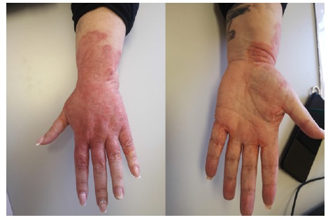
Sexo feminino, 48 anos, com lesões eritemato-descamativas pruriginosas e vesículas, com bordo bem definido, no dorso da mão e punho dominantes, com atingimento das regiões interdigitais poupando a face ventral (Figura 1), com um ano de evolução e crescimento centrípeto.
Tratava-se de uma enfermeira com dermatite de contacto alérgica ao látex confirmada, cujas lesões prévias eram bilaterais e resolveram com evicção, sem recorrência há mais de 5 anos1. A doente foi inicialmente tratada com corticoterapia oral e tópica, sem melhoria clínica. Pela suspeita de dermatite de contacto, foi referenciada ao Serviço de Imunoalergologia, tendo sido realizados testes epicutâneos com as séries Europeias standard, cosméticos, cabeleireiros, colas, acrilatos e desinfetantes, negativos às 48 e 72 horas2. Dadas as características das lesões (unilaterais, poupando áreas da mão, limites bem definidos), foi pedida avaliação de Dermatologia, que realizou raspagem cutânea e análise microscópica.
Foram identificados elementos fúngicos, tendo sido realizada uma cultura fúngica, positiva para Trichophyton interdigitale, levando à prescrição de antifúngicos tópicos e orais, com melhoria após 2 meses de tratamento3. Este caso destaca o desafio diagnóstico de infeções fúngicas atípicas em doentes com dermatites conhecidas, enfatizando a necessidade de colaboração entre especialidades em casos complexos.

Figure 1 Diffuse, uniform erythematous lesions on the dorsum of the hand, interdigital regions, and distal third of the forearm, with well-defined limits and peripheral desquamation, sparing the distal phalanges of the dorsum and ventral side of the hand and fingers. (No vesicular lesions at the time of photograph).
COMMENT
Female, 48 years old, with pruritic erythematous-scaly lesions and vesicles, with well-defined borders on the dorsum of the dominant hand and wrist, affecting the interdigital regions, sparing the ventral face (Figure 1), with one year of evolution and centripetal growth. She was a nurse with a confirmed latex allergic contact dermatitis, whose previous lesions were bilateral and resolved with avoidance, without recurrence for over 5 years 1.
Initial treatment consisted of oral and topical corticosteroids, without clinical improvement. Due to the suspicion of contact dermatitis, she was referred to our Allergy and Clinical Immunology
Department, and epicutaneous tests were performed with the standard European, cosmetics, hairdressing, glues, acrylates, and disinfectants series, all negative at 48 and 72 hours 2. Given the characteristics of the lesions (unilateral, sparing areas of the hand, well-defined limits), Dermatology was consulted, leading to skin scraping and microscopic analysis. Fungal elements were identified
and a fungal culture was conducted, which was positive for Trichophyton interdigitale, prompting the prescription of topical and oral antifungals, with improvement after 2 months of treatment3. This case highlights the diagnostic challenge of atypical fungal infections in patients with known dermatitis, emphasizing the need for collaboration between specialties in complex cases.


