A 17-year-old girl presented to the Emergency Department (ED) with painful inguinal lymphadenopathies associated with dyspareunia, leukorrhea, dysuria, pollakiuria, and hypogastric and lumbar pain with one week of evolution. She also mentioned asthenia and involuntary weight loss of 10 kg in the last six months and denied a history of ulcers, rashes, fever, night sweats, or other complaints.
On psychosocial assessment, the girl reported unprotected sexual intercourse with her boyfriend until the previous month, when he was observed in the ED for a genital ulcer. She denied other risk behavior such as alcohol or drug use.
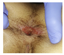
On physical examination, the girl presented with painful non-adherent bilateral inguinal lymphadenopathies of elastic consistency and small hypertrophic and macerated perianal erythematous moist plaques (Figure 1).
Soft tissue ultrasound confirmed inguinal lymphadenopathies, the largest on the right side with 24 mm. Abdominal ultrasound and chest radiography were normal. Laboratory evaluation showed C-reactive protein of 3.41 mg/dL and normal complete blood count, peripheral blood smear, erythrocyte sedimentation rate, lactate dehydrogenase, uric acid, ionogram, transaminases, urea, and creatinine. Rapid urinalysis showed no relevant changes. Serologic tests revealed the presence of total antibodies and positive immunoglobulin M for Treponema pallidum and reactive rapid plasma egain (RPR; titer 1:64). Other serologies (hepatitis C virus, hepatitis B virus, human immunodeficiency virus) were negative. Fecal calprotectin was elevated.
Discussion
The girl was treated with a single intramuscular (IM) dose (2.4 million units) of benzathine penicillin G. Her boyfriend had been treated for syphilis and urethritis, and although nucleic acid amplification test forChlamydia trachomatis(CT) andNeisseria gonorrhoeae(NG) was not available, the girl was empirically treated with 250 mg IM ceftriaxone and 1 g oral azithromycin.
At follow-up, the patient reported symptom improvement, weight gain, resolution of the anal lesion, and lymphadenopathies. In the fourth month of follow-up, laboratory evaluation showed a lower RPR titer (1:2) and normalization of calprotectin. The disease was reported.
Syphilis is a systemic disease caused by Treponema pallidum that is transmitted by direct contact with infected lesions or through the placenta, resulting in fetal infection.1 Its incidence has been increasing in Europe, with adolescents being particularly vulnerable. A report from the European Center for Disease Prevention and Control showed an incidence of 7 cases per 100,000 population in Europe in 2018, and 2.2 cases per 100,000 population in Portugal.2 Young people aged 15-24 years accounted for 13% of all reported cases. 2 The diagnosis of syphilis can be challenging because it can mimic other systemic diseases.3 Clinical presentation is variable and depends on the stage of the disease.4 The primary stage is defined by a typically painless chancre at the site of inoculation, which may heal spontaneously within weeks even without treatment. The secondary stage occurs approximately six to eight weeks after the onset of the primary stage and is characterized by a wide variety of signs and symptoms. It classically presents with a symmetric polymorphic maculopapular rash on the trunk, palms, and soles, lymphadenopathy, and constitutional symptoms. Mucosal lesions in the mouth and perineum, such as condylomata lata, have also been described.4 Clinical manifestations of the tertiary stage are highly variable, the most common being cardiovascular syphilis, gummatous lesions, and generalized paresis and tabes dorsalis.4
This report described a case of secondary syphilis in a sexually active adolescent with unprotected sexual intercourse with her boyfriend, who had been diagnosed with a genital ulcer. She reported lymphadenopathies and urinary and constitutional symptoms, but it was not until physical examination that an anal lesion was noted and the hypothesis of perianal condylomata lata was raised. Condylomata lata is one of the cutaneous manifestations of secondary syphilis, reported in 9-44% of cases.5 It is highly infectious and occurs predominantly in the perianal area and vulva, although atypical sites have been described.4,5 The diagnosis of syphilis is based on the patient´s history, physical examination, and serologic testing (nontreponemal and treponemal tests).6,8 Early disease detection, accurate treatment, and prevention of transmission are essential to reduce the burden of the disease.3,7 If left untreated, the disease can evolve to tertiary stage and symptoms can appear any time from 1-30 years after primary infection, with potentially serious multiorgan manifestations.7
Sexually transmitted infections (STIs) are one of the most important public health problems in adolescence.3 The emergence of new cases of STIs, mainly chlamydia infection, gonorrhea, and syphilis, warns of the potential failure of sexual education aimed at promoting healthy habits and modifying risky practices. It is important to maintain a high index of suspicion for syphilis in the presence of a sexually active adolescent with lymphadenopathies, anal lesions, and constitutional symptoms. International guidelines recommend universal screening for CT, NG, and syphilis in all sexually active females aged between 14 and 25 years, as these infections are often asymptomatic and have specific treatment.10 Cases of syphilis, chlamydia infection, and gonorrhea should be reported, and sexual contacts should be traced, reported, and treated.


