Introduction
Ovarian fibromatosis is a rare benign clinicopathological entity first described by Young and Scully in 1984, which causes ovarian enlargement, thus mimicking an ovarian neoplasm1,2,3. It is not considered as part of the more general group of abdominal fibromatosis5. This nonneoplastic disease can cause an increase in ovarian size up to 6-12 cm, presenting as solid or very rarely cystic-appearing masses, with smooth or lobulated surfaces1), (6. It is usually seen unilaterally, but a few bilateral cases have been reported1,6.
Due to its rarity, the pathogenesis of this disorder is not well understood1.
Patients are mostly young premenopausal women and common clinical manifestations include abdominal pain, menstrual irregularities, and rarely they may present some androgenic effects such as hirsutism or virilization1,2,3,4), (5,6. Histologically, it is characterized by a proliferation of collagen-producing spindle cells surrounding normal ovarian structures, not destroying the normal follicular structures, but causing thickening of the superficial cortex and enlargement of the ovary2,3,5,6.
Ovarian fibromatosis may mimic malignant ovarian tumors due to similar presentation and radiological findings1. Because of its rare occurrence, preoperative diagnosis is still a problematic issue as in other benign solid ovarian tumors such as fibromas, fibrotechomas, and massive ovarian edemas1.
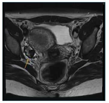
On MRI, the “black garland sign” refers to a marked T2 hypointense thick rim of fibrous tissue surrounding the ovary in the setting of ovarian fibromatosis2. The black garland appearance along with relative sparing of the central ovarian parenchyma helps to distinguish ovarian fibromatosis from other fibrous ovarian lesions including fibromas, fibrothecomas, and Brenner tumors2,5. On post-contrast imaging, fibromatosis demonstrates no significant enhancement in the parenchymal phase, owing to its fibrotic nature2.
Only a few case reports have been documented concerning computed tomography (CT) and magnetic MRI findings3. We report a case of characteristic MRI findings reflecting pathological features suggestive of ovarian fibromatosis3.
Case report
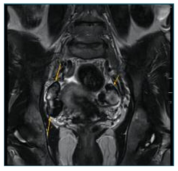
A 44-year-old woman, nulliparous, with regular menses and a personal history of primary Sjogren Syndrome, arterial hypertension, sinus bradycardia, hyperparathyroidism and androgenetic alopecia consulted her gynecologist to clarify a clinical condition of pelvic pain associated with an approximately 1cm heterogenous solid adnexal mass, Color Score 1 was found on ultrasound. Further characterization of the mass through MRI demonstrated moderate enlargement of the ovaries with lobulated margins. The ovarian cortex was thickened and demonstrated a homogeneous low-signal intensity on both T1 and T2-weighted images, suggestive of fibrous tissue, with a “black garland” appearance and consistent with ovarian fibromatosis.
In the left ovary, a cystic formation with 20mm, thick walls (4.5mm) and internal parietal enhancement was identified, likely in relation to the corpus luteum.
On the surface of the right ovary, a hypersignal focus (5.5mm) was identified in T1-weighted fat suppressed sequences, which may have corresponded to a small cyst with protein content. However, based on the signal pattern, a focus of superficial endometriosis cannot be excluded. In conclusion, suggestive aspects of ovarian fibromatosis were found.
In this particular case, clinical and ultrasound surveillance every six months was chosen. The patient preferred a watch and wait attitude once complete resolution of the clinical condition was not guaranteed with the excision of the ovary. Since the diagnosis, the adnexal mass is stable and pelvic pain is sporadic controlled by analgesics.

Figure 1 Black garland Sign. Axial T2-weighted image demonstrating a markedly T2 hypointense rim around the ovaries (right adnexa showed) without a central ovarian parenchymal mass. This hypointense rim is referred to as the ‘‘black garland’’ sign.
Discussion
Differential diagnosis of ovarian fibromatosis includes other fibromatous diseases, such as ovarian fibromas, fibrothecomas, Brenner tumors and Krukenberg tumors1,4. Ovarian fibromas and fibrothecomas can easily be distinguished because of a well-circumscribed mass compressing the underlying parenchyma, unlike the diffuse infiltrative pattern seen in fibromatosis1. Heterogeneously echogenic adnexal masses that show posterior wall attenuation are usually ovarian fibromas, dermoids, or degenerative leiomyomas4. The presence of ovarian infiltration, with partial sparing of normal ovarian structures, which is particularly visible on MRI, can help to distinguish fibromatosis from ovarian fibroma4. Low flow on Doppler sonography and poor enhancement on dynamic and delayed CT are suggestive of ovarian fibromas4. Ovarian fibromatosis appears relatively well limited, with no involvement of adjacent pelvic structures4.
Massive ovarian edema (MOE), as described by Young and Scully, is another nonneoplastic disease where enlargement of the ovaries is due to cortical and medullary edema. Microscopically, MOE may be associated with variable amounts of ovarian fibromatosis1. Ovarian fibromatosis is closely related to massive ovarian edema (residual normal ovarian structures are present) but differs from it by the presence of abnormal fibrous tissue in the former and edema in the latter4.
There are some reports of ovarian fibromatosis in the literature showing an association with omental fibrosis, idiopathic sclerosing peritonitis, and intraabdominal fibromatosis1. Ascites was also a common finding1. There are also reports of associated sex-cord type cellular proliferations, which can explain the androgenic manifestations in ovarian fibromatosis1. The sex cord structure rarely seen in ovarian fibromatosis contains granulosa cell-like or Sertoli cell-like elements. Androgenic manifestations correlated with the presence of lutein cells reported ovarian fibromatosis with minor sex cord elements who had amenorrhea and lower abdominal pain.
Retroperitoneal fibrosis (RF) is commonly associated with immunoglobulin G4 (IgG4)-related disease. This fibro-inflammatory condition is characterized by a lymphoplasmacytic infiltrate with IgG4-positive plasma cells, fibrosis, and oftentimes, elevated serum IgG4 levels, which could be associated with Sjögren’s syndrome7.
Although ovarian fibromatosis is a benign entity, it is often treated with salpingo-oophorectomy. Conservative surgery has been shown to result in normalization of menstrual cycles and increased fertility, and so the preoperative differentiation of ovarian neoplasms is extremely important2,3,4,8.
In this particular case, although we did not opt for surgical excision of the affected ovary, we considered it might have been relevant not only for diagnostic confirmation, but also for improving clinical symptoms, as well as the study of a possible correlation between hypertension and hyperandrogenism that ovarian fibromatosis is sometimes associated with. Histological confirmation would have been extremely important, because it’s the only way to confirm the diagnosis, although imaging findings are highly suggestive of ovarian fibromatosis.
In conclusion, ovarian fibromatosis should be considered in women with solid ovarian tumors with a predominant fibrous component4. Imaging studies with MRI may be helpful in the preoperative diagnosis of ovarian fibromatosis and entrapped follicles between the fibrous tissue is one of the most important discriminating features in the diagnosis of this disorder1,4. Black Garland-like appearance on T2-weighted images may not always be observed in ovarian fibromatosis but it may be a diagnostic clue to this rare entity3.


