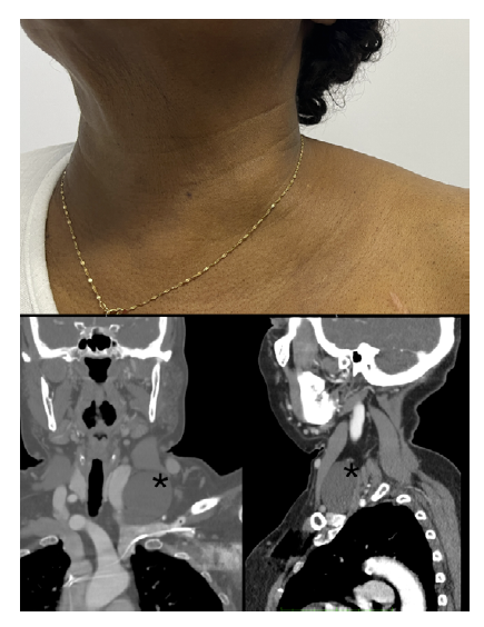
A female, 69 years old, with a prior history of rheumatoid arthritis, arterial hypertension, and dysrhythmia with a pacemaker, presented with a painless left supraclavicular mass, gradually growing for two years. The physical examination revealed a voluminous, regular, and mobile lesion, resulting in significant deformity (Figure, top). A computed tomography angiography was requested to clarify the etiology of the mass. This exam confirmed the presence of a non-contrasting cystic dilatation in the supraclavicular region (Figure, marked with “*”) adjacent to the confluence of the left subclavian vein, which corresponded to an aneurysmatic thoracic duct. No intervention was proposed in the absence of symptoms, and re-evaluation at six months is scheduled. A lymphoscintigraphy may be performed in case of new symptoms or volumetric increase. This image illustrates the importance of considering rare etiologies for supraclavicular masses.
fistula is large, it may lead to leg ischemia or high-output heart failure. Close post-procedural surveillance and early diagnosis are essential to avoid serious complications.


