Description
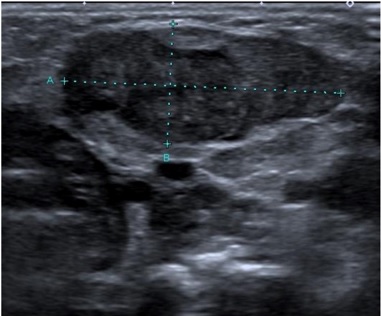
A 6 weeks old male infant, with a closely monitored pregnancy and eutocic birth without complications was observed in the Emergency Department (ED) presenting a left cervical mass first noticed three days before, without any accompanying symptoms. Physical examination revealed facial asymmetry with impaired left cervical rotation and a 3 cm wide mass in the left lateral-cervical region (zone III), firm, immobile and painless. A soft tissue ultrasound showed multiple adenopathy, the largest measuring 31x14 mm (Fig. 1). A full blood count, C-reactive protein and Epstein-Barr Virus serology were requested, which did not reveal any abnormalities. Reactive adenopathy was considered a probable cause, and so clinical reassessment was scheduled within 72 hours. The patient´s reassessment was identical and a second ultrasoundexamination of lymph nodes showed findings indicative of a reactive process. He was then referred for a pediatric consultation and, 1 week later, an ultrasound reassessment by a pediatric radiologist showed a fusiform thickening of the left sternocleidomastoid muscle that was suggestive of fibromatosis colli (Fig. 2). The patient then began physiotherapy with a significant improvement and the condition completely resolved within 4 months.

Figure 2: Fusiform thickening of the left sternocleidomastoid muscle. Contralateral sternocleidomastoid muscle with normal thickness and ecostructure
Fibromatosis colli is the most severe presentation of congenital muscular torticollis and a relatively rare cause of neck swelling in neonates and infants. This pathology occurs in 0.4% of newborns.1 The pathogenesis is still unclear, but it is believed that, after a complicated delivery, there may be neck compression leading to ischemia with consequent damage to muscle fibers.1,2Typically, it appears 2-4 weeks after birth, most commonly following a difficult delivery.3 But even in cases of infants with a normal delivery this diagnosis should be kept in mind. It is usually a self-limiting condition resolving within 4-8 months which requires only physical therapy and conservative management.3,4Fibromatosis colli is classified as a benign fibroblastic and myofibroblastic tumor according to the new WHO classification of Soft Tissue Tumors.5 The diagnosis is possible through the history and physical examination and, normally, it is possible to see the head of the neonate tilted toward the affected side, and there may be an associated jaw rotation to the contralateral shoulder due to sternocleidomastoid muscle contracture. The abnormal position of the neck may lead to facial asymmetry and positional plagiocephaly.6
The differential diagnosis of pediatric neck masses is extensive and should always be considered,7 the most frequent being the differential diagnosis with adenopathy as in our clinical case.
Ultrasound is the diagnosis imaging modality of choice if there is any doubt in the diagnosis. It shows focal or diffuse enlargement of the sternocleidomastoid muscle in a fusiform configuration.8 The mass moves synchronously with the rest of the sternocleidomastoid muscle on real-time sonography. Unlike fibromatosis colli, adenopathy is characterized by ultrasound as a conglomerate tangled mass with normal homogeneously low echogenicity parenchyma and preservation of fatty hyperechoic hilum alongside ovoid morphology.9Maddalozzo and Goldenberg reported that ultrasonography was 100% sensitive for diagnosis when there is a strong suspicion from the clinical history and physical examination of fibromatosis colli.10
The importance of an accurate clinical diagnosis is thus emphasized, and a confident diagnosis implies the need for an ultrasound examination by a radiologist experienced in pediatric imaging with knowledge of the sonographic characteristics of this clinical entity.8


