SciELO - Scientific Electronic Library Online

 
vol.30 número6O Novo Fibroscan-AST (FAST) Score: Reforçando o Impacto da Diabetes Mellitus no Fígado Gordo Associado a Disfunção MetabólicaPancreatite da goteira: Casos clínicos e revisão da literatura índice de autoresíndice de assuntosPesquisa de artigos
Home Pagelista alfabética de periódicos  

Serviços Personalizados

Journal

Artigo

Indicadores

Links relacionados

  • Não possue artigos similaresSimilares em SciELO

Compartilhar


GE-Portuguese Journal of Gastroenterology

versão impressa ISSN 2341-4545

GE Port J Gastroenterol vol.30 no.6 Lisboa dez. 2023  Epub 01-Fev-2024

https://doi.org/10.1159/000527209 

Research Article

Endoscopic Approach to Duodenal Adenomas in Familial Adenomatous Polyposis: A Retrospective Cohort

Abordagem endoscópica de adenomas duodenais na Polipose Adenomatosa Familiar: um coorte retrospetivo

Joana Lemos Garcia1 

Isadora Rosa1  2 
http://orcid.org/0000-0002-2953-5987

João Pereira da Silva1 

Pedro Lage1  2 

Isabel Claro1  2 

1Gastroenterology Department, Instituto Português de Oncologia de Lisboa Francisco Gentil E.P.E., Lisboa, Portugal;

2Familial Risk Clinic, Instituto Português de Oncologia de Lisboa Francisco Gentil E.P.E., Lisboa, Portugal


Abstract

Introduction:

Over 90% of the patients with familial adenomatous polyposis (FAP) will develop duodenal adenomas.

Aim:

The aim of this study was to evaluate the effectiveness and safety of endoscopic excision of large duodenal adenomas in FAP patients.

Methods:

All FAP patients from a familial risk clinic submitted to endoscopic therapy for duodenal adenomas ≥10 mm between January 2010 and February 2021 were included.

Results:

From 151 FAP families, 22 patients (50 lesions) were included: 54.5% female; median follow-up 8.5 (IQR: 5.8-12.3) years after the first endoscopy. First therapeutic endoscopy occurred at a median age of 41.0 years (IQR: 33.0-58.2). Repeat therapeutic endoscopy was required in 54.5% of patients. Median size of the largest adenoma was 15 mm (IQR: 10-18 mm); resection was piece-meal in 63.1% and en bloc in the remaining. In 2 cases, the resection was incomplete (fibrosis due to previous resection and difficult positioning). Complications occurred in 6.3% of the resected lesions (4 patients): 2 immediate (bleeding, perforation); 4 in the first week (1 bleeding, 2 mild pancreatitis, 1 perforation requiring surgery; the latter two after ampullectomy). Histology revealed low-grade dysplasia adenomas in 90.1%; no adenocarcinomas were found. One patient with Spigelman stage IV disease not amenable to endoscopic control underwent elective duodenopancreatectomy (without duodenal cancer).

Conclusion:

Endoscopic surveillance and treatment of duodenal adenomas in FAP patients was safe and effective in the prevention of duodenal cancer.

Keywords: Familial adenomatous polyposis; Duodenum; Adenomas; Endoscopy; Endoscopic mucosal resection

Resumo

Introdução:

Mais de 90% dos doentes com Polipose Adenomatosa Familiar (PAF) desenvolvem adenomas duodenais.

Objetivo:

Avaliar a eficácia e segurança da excisão endoscópica de adenomas duodenais em doentes com PAF.

Métodos:

Incluídos todos os doentes com PAF submetidos a terapêutica endoscópica de adenomas duodenais ≥10 mm entre janeiro/2010-fevereiro/2021.

Resultados:

Em 151 famílias com PAF, incluídos 22 doentes (50 lesões): 54.5% mulheres; mediana do follow-up 12.3 (IQR: 6.0-19.0) anos. Primeira endoscopia terapêutica (resseção de pólipos duodenais ≥10 mm) ocorreu numa mediana de idades 41.0 (IQR: 33.0-58.2) anos.Em 54.5% dos casos, foi necessária uma nova endoscopia terapêutica. Dimensão mediana do maior adenoma: 15 mm (IQR: 10-18 mm); resseção realizada em piecemeal em 63.1% e em bloco nos restantes. Em dois casos, a resseção endoscópica foi incompleta (fibrose em local de resseção prévia:1; posicionamento:1). Complicações em 6.3% das lesões ressecadas (4 doentes): 2 imediatas (hemorragia e perfuração, manejadas endoscopicamente); 4 na primeira semana (1 hemorragia controlada endoscopicamente, 2 pancreatites ligeiras tratadas conservadoramente, 1 perfuração com necessidade de cirurgia; as duas últimas após ampulectomia). A avaliação histológica revelou adenomas com displasia de baixo grau em 90.1%; nenhum adenocarcinoma. Um doente com doença Spigelman IV não controlável endoscopicamente realizou duodenopancreatectomia (sem cancro).

Conclusão:

A vigilância e tratamento endoscópicos de adenomas duodenais em doentes com PAF revelaram-se seguros e eficazes na prevenção de cancro duodenal.

Palavras Chave: Polipose adenomatosa familiar; Duodeno; Adenomas; Endoscopia; Mucosectomia

Introduction

Familial adenomatous polyposis (FAP) is an inherited autosomal-dominant condition caused by a mutation of the adenomatous polyposis coli (APC) gene, on the long arm of chromosome 5 [1]. It is marked by a high incidence of colorectal adenomas and cancer. Nevertheless, with adequate screening and prophylactic measures regarding colorectal cancer, duodenal disease has emerged as one of the most important causes of morbidity and mortality in affected patients [2]. Duodenal adenomas (DAs) occur in more than 90% and duodenal cancer in 3-5% of FAP cases [3-6]. The adenoma-carcinoma progression in this location may take up to 15-20 years [7]. Specific regions of the APC gene may be associated with severe disease, due to clustering of somatic mutations, and loss of the wild-type allele [2, 8].

International guidelines advocate regular endoscopic surveillance of the duodenum. Risk stratification, follow-up intervals, and therapeutic approaches are determined according to the Spigelman classification [9], which considers polyp number, size, and histology [9-14]. Excision is recommended for non-ampullary (and some ampullary) adenomas ≥10 mm [14], considering the balance between the risk of endoscopic/surgical resections and the risk of developing duodenal carcinoma. The chosen method is often endoscopic mucosal resection (EMR), which has proven to be safe and effective [15-18]. Significant recurrence rates have been reported, although it is not always straightforward whether they are true recurrences or simply disease progression [16-21]. Furthermore, despite these recommendations, it remains unknown whether DA resection truly changes the natural history of cancer risk since there is an underlying field defect in the duodenum [22].

When invasive disease is suspected, surgical approaches should be considered [14]. These include pancreas-sparing duodenectomy and pancreatoduodenectomy, which offer definitive therapy in preventing duodenal carcinoma, or segmental duodenal resection, for patients with dominant or limited disease, where removing a short segment allows safe endoscopic surveillance/treatment of the remaining bowel. Nevertheless, adenomatous disease may recur in the remaining small bowel, and these patients must be kept under regular surveillance [23, 24].

Additionally, medical treatment using the cyclooxygenase inhibitors sulindac and celecoxib has been studied, yielding conflicting results - due also to significant side effects, their use is not recommended in Europe [25-30]. The aim of this study was to evaluate the effectiveness and safety of endoscopic excision of large DAs in patients with FAP and to assess results in light of the most recent guidelines.

Methods

A retrospective study was developed in the familial cancer clinic of an oncological centre, where 151 families with FAP are currently accompanied. FAP families’ files from the familial cancer clinic were reviewed and all FAP patients submitted to endoscopic resection of DAs with at least 10 mm greatest axis, from January 2010 to February 2021, were included. These procedures were described as therapeutic endoscopies.

FAP patients undergo regular endoscopic surveillance according to international guidelines, having their first upper gastrointestinal endoscopy at the age of 20-25 or earlier in case of colectomy before 20 years old. Follow-up intervals are determined according to the Spigelman classification [9], which considers the number, size, and histological characteristics (architecture and dysplasia grade) of duodenal polyps. Spiegelman stage is then calculated by summing the points attributed to these criteria and patients with Spigelman stage 0/I, II, III undergo endoscopy every 5, 3, and 1 year, respectively; those with Spigelman stage IV must be considered individually, undergoing surgery or surveillance every 6 months. Surveillance intervals may be shortened after removal of polyps with higher risk of recurrence, such as those harbouring high-grade dysplasia or with a villous histology, especially if removed piecemeal. This is considered case by case.

Study Procedures

The exams were performed under propofol sedation by an An-aesthesiologist, in the Endoscopy Unit of the Gastroenterology Department of our institution in case of non-ampullary adenomas or in a tertiary hospital with expertise in endoscopic retrograde cholangiopancreatography in case of ampullary adenomas. Endoscopes and duodenoscopes belonged to Olympus series 190 and 180, respectively. EMR was usually performed after submucosal injection of a solution containing patent blue (25 mg/mL), adrenalin (1:100,000), and Gelafundin, but decision was made case by case, namely, in ampullary tumours, where submucosal injection was not always necessary. The choice of the snare varied according to endoscopist’s preference (10-25 mm snares were available). Current settings were cutting and coagulation of 120 W - Pulse Cut Slow (ESG-100, Olympus Inc., Tokyo, Japan) for non-ampullary lesions or Endocut 2 60 W (ICC 200, Erbe, Tübingen, Ger-many) for ampullary tumours.

Results

Study Population Characteristics

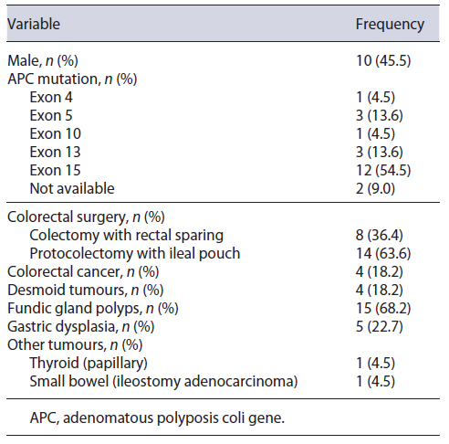
In a total of 151 FAP families, 22 patients from 21 families met the inclusion criteria (DAs with at least 10 mm greatest axis resected in the study period): 54.5% of the patients were female (Table 1), with a median follow-up time of 8.5 (IQR: 5.8-12.3) years after the first endoscopy and 3.7 (IQR: 1.0-5.3) years after the first therapeutic endoscopy. Most germline APC mutations occurred in exon 15 (54.5%). Eight (36.4%) patients had known family history of DAs. The highest Spigelman stage found in these relatives was I, II, III, and IV in 1, 2, 1, and 4 cases, respectively. Patient characteristics are summarized in Table 1. The first screening upper endoscopy happened at 38.0 years of age (median) (IQR: 28.8-52.3) in the study population and DAs were detected in the first exam in 18 (81.8%) of them - staged as Spiegelman I, II, and III in 3, 13, and 2 cases, respectively.

Table 1. Patient characteristics 

Endoscopic Therapeutic Procedures

First therapeutic endoscopy (resection of ≥10 mm duodenal polyps) occurred at a median age of 41.0 (IQR: 33.0-58.2) years, and 9.1% (n = 2), 40.9% (n = 9), 45.5% (n = 10), and 4.5% (n = 1) of the patients were staged as Spiegelman I, II, III, and IV, respectively. The median time interval between the first screening endoscopy and the first therapeutic endoscopy was 60.3 (±39.1) months, corresponding to a median number of three endoscopies (IQR: 1-5) during that period, in which smaller adenomas were resected in 15 patients (68.2%).

After the first therapeutic endoscopy, a new procedure was required in 12 (54.5%) patients, once in 5 cases, twice in 4, three times in 2, and five times in 1 (median number of therapeutic endoscopies = 2, IQR 1-3), corresponding to a total of 46 therapeutic endoscopies and 50 lesions removed. The median time interval between therapeutic procedures was 20 (IQR: 14-23) months.

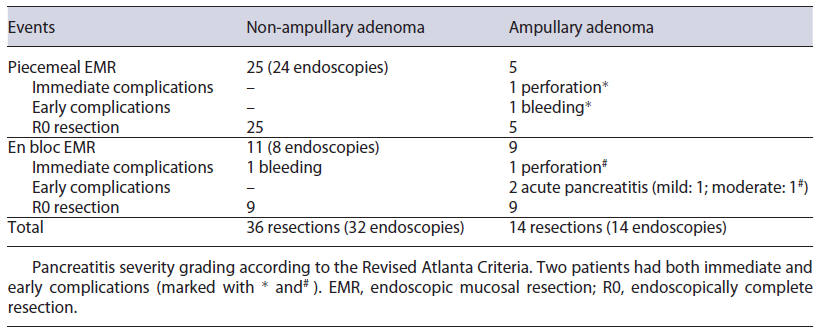
Most therapeutic procedures (69.6% of the procedures) included resection of only one large (≥10 mm) adenoma. The largest adenomas had a median size of 15 mm (IQR: 10-18 mm). The most frequently used technique was piecemeal and en bloc mucosectomy for non-ampullary and ampullary adenomas, respectively (Table 2). Prophylactic defect closure with clips was performed after resection of a 15 mm ampullary tumour and two non-ampullary lesions of 10 and 18 mm; visible vessels were coagulated with snaretip soft coagulation after resection of a 30 mm non-ampullary adenoma. Illustrating pictures can be seen in Figure 1. In 2 cases, resection was considered endoscopically incomplete - one due to scarring in previous resection site and the other due to difficult positioning. These patients were re-evaluated 3 and 5 months later and what was thought to be the residual lesion was successfully removed with cold snare in one and with biopsy forceps in the other case. Further endoscopic follow-up was per-formed annually and none developed adenocarcinoma. Complications occurred in 8.0% (n = 4) of the resected lesions - 3 after ampullectomy and one after a flat lesion mucosectomy (Table 2).
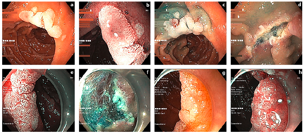
Fig. 1. Examples of resected DAs. a A 12-mm lesion (Paris 0-IIa) in white light examination (WLE). b Same lesion under narrow band imaging (NBI). c After submucosal injection. d During the resection procedure. e 15-mm DA (Paris 0-IIa) in WLE. f Post-polyp resection. g 12-mm lesion (Paris 0-IIa) in WLE. h Under NBI. 

Table 2. Endoscopy-related outcomes 

Two patients had both immediate (first 24 h) and early (first 7 days) complications; the others had early complications. Immediate complications consisted in intraprocedural bleeding after non-ampullary tumour resection, and perforation after ampullectomy, successfully managed endoscopically. Early (during the first week after the intervention) complications included 1 case of bleeding in a patient who had prophylactic defect closure with through-the-scope clips after ampullectomy, controlled in a repeat endoscopy; 2 cases of acute pancreatitis; one perforation after ampullary tumour resection that was undetected during the procedure. Both pancreatitis occurred after ampulloma resection, despite prophylactic pancreatic stent placement. According to the Revised Atlanta Criteria, one was mild and the other was moderate due to local complications. The latter happened in the same patient in whom a duodenal perforation was diagnosed more than 24 h after the procedure. This patient underwent surgery, with construction of a feeding jejunostomy and pancreatic necrosectomy. He did not require organ support and had a favourable outcome.

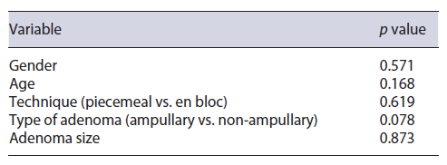
Occurrence of complications was not significantly associated with the technique (piecemeal vs. en bloc mucosectomy) (p = 0.619), type of adenoma (ampullary vs. non-ampullary) (p = 0.078), or adenoma size (p = 0.873) (Table 3). Histology revealed adenomas harbouring low-grade dysplasia in 89.1% (tubular adenomas 76.1%, tubulovillous 13.0%); high-grade dysplasia in 4.6% (n = 2) of cases; no adenocarcinomas were found.

Table 3. Procedure-related complications - statistical analysis 

One patient underwent elective duodenopancreatectomy, which did not harbour duodenal cancer. This patient had Spiegelman stage IV disease with three large (>30 mm) lesions that were considered to have a high risk of recurrence/treatment failure after endoscopic resection - one involving the bulbus with a bulky sessile component, one in the transition to the second portion of the duodenum, and the other adjacent to the papilla, close to a fibrotic area of previous resections. All other patients remain under active surveillance.

Discussion

Adenomatous duodenal disease is a known morbidity factor in FAP patients. Endoscopic resection of DAs has a high success rate, with reported complete resections in 86-100% of the cases [16, 21, 31-33]. Even though reported recurrence rates are 10-37% [18, 31-33], the natural history of DAs in FAP patients makes it difficult to distinguish disease progression from local recurrence. In our series, most patients required 2 therapeutic endoscopies during follow-up, reflecting this characteristic.

Endoscopic resection was a safe technique in our series, with an 8.0% complication rate, but with most cases amenable to conservative or endoscopic approaches. This rate is similar or even lower than that reported in other series, and it is also similar in terms of severity of the adverse events. As stated in the literature, most complications occurred after resection of ampullary adenomas, even though it did not reach statistical significance, probably due to our series’ small numbers. Particularly, acute pancreatitis occurred in 2 of 14 ampullary tumour resections despite pancreatic stent placement, in line with previous reports [18, 34-36]. Notably, intraprocedural bleeding rates were lower than expected from literature review [31, 32, 37].

The duodenum remains a challenging location for endoscopic therapy and mucosal resection is the first-line endoscopic resection technique for non-malignant large DAs [38, 39]. However, when EMR is not feasible and considering the risks associated with the surgical alternatives, endoscopic submucosal dissection can be considered by experienced endoscopists [39, 40]. In our series, duodenal surveillance started later than recommended in international guidelines since a significant number of patients were referred to our clinic only in adult age after a CRC diagnosis in the patient or in a family member.

There were no cases of duodenal cancer during follow-up, reflecting the effectiveness of endoscopic surveillance according to the Spigelman stage. Therefore, this work further strengthens current recommendations of DAs surveillance in FAP patients and legitimates the choice of endoscopic resection as the first-line treatment.

Acknowledgments

We thank all the members of the Gastroenterology Department and Familial Cancer Clinic for the support throughout the elaboration of this paper. We thank Dr. Tiago Bana e Costa and the staff at Hospital Egas Moniz’ Gastroenterology Department for their collaboration with the ampullectomies.

References

1. Mathus-Vliegen EMH, Boparai KS, Dekker E, Van Geloven N. Progression of duodenal adenomatosis in familial adenomatous polyposis: due to ageing of subjects and advances in technology. Fam Cancer. 2011; 10(3): 491-9. [ Links ]

2. Thomas LE, Hurley JJ, Meuser E, Jose S, Ashelford KE, Mort M. Burden and profile of somatic mutation in duodenal adenomas from patients with familial adenomatous- and MUTYH-associated polyposis. Clin Cancer Res. 2017; 23(21): 6721-32. [ Links ]

3. Bülow S, Christensen IJ, Højen H, Bjork J, Elmberg M, Jarvinen H, et al. Duodenal surveillance improves the prognosis after duodenal cancer in familial adenomatous polyposis. Colorectal Dis. 2012; 14(8): 947-52. [ Links ]

4. Bülow S, Björk J, Christensen IJ. Duodenal adenomatosis in familial adenomatous polyposis. Gut. 2004; 53(3): 381-6. [ Links ]

5. Heiskanen I, Kellokumpu I, Järvinen H. Management of duodenal adenomas in 98 patients with familial adenomatous polyposis. Endoscopy. 1999; 31(6): 412-6. [ Links ]

6. Vasen HF, Bülow S, Myrhøj T, Mathus-Vliegen L, Griffioen G, Buskens E. Decision analysis in the management of duodenal adenomatosis in familial adenomatous polyposis. Gut. 1997; 40(6): 716-9. [ Links ]

7. Okada K, Fujisaki J, Kasuga A, Omae M, Kubota M, Hirasawa T. Sporadic nonampullary duodenal adenoma in the natural history of duodenal cancer: a study of follow-up surveillance. Am J Gastroenterol. 2011; 106(2): 357-64. [ Links ]

8. Groves C, Lamlum H, Crabtree M, Williamson J, Taylor C, Bass S. Mutation cluster region, association between germline and somatic mutations and genotype-phenotype correlation in upper gastrointestinal familial adenomatous polyposis. Am J Pathol. 2002; 160(6): 2055-61. [ Links ]

9. Spigelman AD, Talbot IC, Williams CB, Domizio P, Phillips R. Upper gastrointestinal cancer in patients with familial adenomatous polyposis. Lancet. 1989; 334(8666): 783-5. [ Links ]

10. Vasen HFA, Möslein G, Alonso A, Aretz S, Bernstein I, Bertario L. Guidelines for the clinical management of familial adenomatous polyposis (FAP). Gut. 2008; 57(5): 704-13. [ Links ]

11. Herzig D, Hardimann K, Weiser M, Yu N, Paquette I, Feingold DL. The American Society of Colon and Rectal Surgeons clinical practice guidelines for the management of inherited polyposis syndromes. Dis Colon Rectum. 2017; 60(9): 881-94. [ Links ]

12. Stjepanovic N, Moreira L, Carneiro F, Balaguer F, Cervantes A, Balmana J. Hereditary gastrointestinal cancers: ESMO clinical practice guidelines for diagnosis, treatment and follow-up. Ann Oncol. 2019; 30(10): [ Links ]

13. Syngal S, Brand RE, Church JM, Giardiello FM, Hampel HL, Burt RW. ACG Clinical guideline: genetic testing and management of hereditary gastrointestinal cancer syndromes. Am J Gastroenterol. 2015; 110(2): 223-62. [ Links ]

14. van Leerdam ME, Roos VH, van Hooft JE, Dekker E, Jover R, Kaminski MF. Endoscopic management of polyposis syndromes: European Society of Gastrointestinal Endoscopy (ESGE) Guideline. Endoscopy. 2019; 51(9): 877-95. [ Links ]

15. Klein A, Tutticci N, Singh R, Bourke MJ. Expanding the boundaries of endoscopic resection: circumferential laterally spreading lesions of the duodenum. Gastroenterology. 2016; 150(3): 560-3. [ Links ]

16. Singh A, Siddiqui UD, Konda VJ, Whitcomb E, Hart J, Xiao SY, et al. Safety and efficacy of EMR for sporadic, nonampullary duodenal adenomas: a single U.S. center experience (with video). Gastrointest Endosc. 2016; 84(4): 700-8. [ Links ]

17. Mendonça EQ, Bernardo WM, de Moura EGH, Chaves DM, Kondo A, Pu LZCT. Endoscopic versus surgical treatment of ampullary adenomas: a systematic review and metaanalysis. Clinics. 2016; 71(1): 28-35. [ Links ]

18. Laleman W, Verreth A, Topal B, Aerts R, Komuta M, Roskams T. Endoscopic resection of ampullary lesions: a single-center 8-year retrospective cohort study of 91 patients with long-term follow-up. Surg Endosc. 2013; 27(10): 3865-76. [ Links ]

19. Ma T, Jang EJ, Zukerberg LR, Odze R, Gala MK, Kelsey PB. Recurrences are common after endoscopic ampullectomy for adenoma in the familial adenomatous polyposis (FAP) syndrome. Surg Endosc. 2014; 28(8): 2349-56. [ Links ]

20. Serrano PE, Grant RC, Berk TC, Kim D, Al-Ali H, Cohen Z. Progression and management of duodenal neoplasia in familial adenomatous polyposis: a cohort study. Ann Surg. 2015; 261(6): 1138-44. [ Links ]

21. Alexander S, Bourke MJ, Williams SJ, Bailey A, Co J. EMR of large, sessile, sporadic nonampullary duodenal adenomas: technical aspects and long-term outcome (with videos). Gastrointest Endosc. 2009; 69(1)66-73. [ Links ]

22. Yang J, Gurudu SR, Koptiuch C, Agrawal D, Buxbaum JL, Abbas Fehmi SM. American Society for Gastrointestinal Endoscopy guideline on the role of endoscopy in familial adenomatous polyposis syndromes. Gastrointest Endosc. 2020; 91(5): 963-82.e2. [ Links ]

23. Alderlieste YA, Bastiaansen BA, Mathus-Vliegen EMH, Gouma DJ, Dekker E. High rate of recurrent adenomatosis during endoscopic surveillance after duodenectomy in patients with familial adenomatous polyposis. Fam Cancer. 2013; 12(4): 699-706. [ Links ]

24. Augustin T, Moslim MA, Tang A, Walsh RM. Tailored surgical treatment of duodenal polyposis in familial adenomatous polyposis syndrome. Surgery. 2018; 163(3): 594-9. [ Links ]

25. Steinbach G, Lynch PM, Phillips RK, Wallace MH, Hawk E, Gordon GB. The effect of celecoxib, a cyclooxygenase-2 inhibitor, in familial adenomatous polyposis. N Engl J Med. 2000; 342(26): 1946-52. [ Links ]

26. Phillips RKS, Wallace MH, Lynch PM. A randomised, double blind, placebo controlled study of celecoxib, a selective cyclooxygenase 2 inhibitor, on duodenal polyposis in familial adenomatous polyposis. Gut. 2002; 50(6): 857-60. [ Links ]

27. Giardiello FM, Yang VW, Hylind LM, Krush AJ, Petersen GM, Trimbath JD. Primary chemoprevention of familial adenomatous polyposis with sulindac. N Engl J Med. 2002; 346(14): 1054-9. [ Links ]

28. Delker DA, Wood AC, Snow AK, Samadder NJ, Samowitz WS, Affolter KE. Chemoprevention with cyclooxygenase and epidermal growth factor receptor inhibitors in familial adenomatous polyposis patients: mRNA signatures of duodenal neoplasia. Cancer Prev Res. 2018; 11(1): 4-15. [ Links ]

29. Arber N, Eagle CJ, Spicak J, Racz I, Dite P, Hajer J. Celecoxib for the prevention of colorectal adenomatous polyps. N Engl J Med. 2006; 355(9): 885-95. [ Links ]

30. Samadder NJ, Neklason DW, Boucher KM, Byrne KR, Kanth P, Samowitz W. Effect of sulindac and erlotinib vs. placebo on duodenal neoplasia in familial adenomatous polyposis. JAMA. 2016; 315(12): 1266-75. [ Links ]

31. Lépilliez V, Chemaly M, Ponchon T, Napoleon B, Saurin J. Endoscopic resection of sporadic duodenal adenomas: an efficient technique with a substantial risk of delayed bleeding. Endoscopy. 2008; 40(10): 806-10. [ Links ]

32. Eswaran SL, Sanders M, Bernadino KP, Ansari A, Lawrence C, Stefan A, et al. Success and complications of endoscopic removal of giant duodenal and ampullary polyps: a comparative series. Gastrointest Endosc. 2006; 64(6): 925-32. [ Links ]

33. Fanning SB, Bourke MJ, Williams SJ, Chung A, Kariyawasam VC. Giant laterally spreading tumors of the duodenum: endoscopic resection outcomes, limitations, and caveats. Gastrointest Endosc. 2012; 75(4): 805-12. [ Links ]

34. Desilets DJ, Dy RM, Ku PM, Hanson BL, Elton E, Mattia A. Endoscopic management of tumors of the major duodenal papilla: refined techniques to improve outcome and avoid complications. Gastrointest Endosc. 2001; 54(2): 202-8. [ Links ]

35. Catalano MF, Linder JD, Chak A, Sivak MV, Raijman I, Geenen JE. Endoscopic management of adenoma of the major duodenal papilla. Gastrointest Endosc. 2004; 59(2): 225-32. [ Links ]

36. Bohnacker S, Seitz U, Nguyen D, Thonke F, Seewald S, deWeerth A, et al. Endoscopic resection of benign tumors of the duodenal papilla without and with intraductal growth. Gastrointest Endosc. 2005; 62(4): 551-60. [ Links ]

37. Ahmad NA, Kochman ML, Long WB, Furth EE, Ginsberg GG. Efficacy, safety, and clinical outcomes of endoscopic mucosal resection: a study of 101 cases. Gastrointest Endosc. 2002; 55(3): 390-6. [ Links ]

38. Vanbiervliet G, Strijker M, Arvanitakis M, Aelvoet A, Arnelo U, Beyna T. Endoscopic management of ampullary tumors: European Society of Gastrointestinal Endoscopy (ESGE) Guideline. Endoscopy. 2021; 53(4): [ Links ]

39. Vanbiervliet G, Moss A, Arvanitakis M, Arnelo U, Beyna T, Busch O. Endoscopic management of superficial nonampullary duodenal tumors: European Society of Gastrointestinal Endoscopy (ESGE) Guideline. Endoscopy. 2021; 53(5): 522-34. [ Links ]

40. Santos-Antunes J, Morais R, Marques M, Macedo G. Underwater duodenal ESD of a large adenoma using the Pocket-Creation method. GE Port J Gastroenterol. 2021; 28(5): 367-9. [ Links ]

1Statement of Ethics All procedures were done in accordance with the Helsinki Dec-laration. Ethical approval was not required for this study in accor-dance with local/national guidelines, and retrospective observational studies do not require specific authorization by our institution’s policy. All patients gave oral and written informed consent for every endoscopic procedure, and the research was carried out in accordance with the Helsinki Declaration.

3Funding Sources No funding was used.

5Data Availability Statement Research data are not shared due to confidentiality.

Received: May 15, 2022; Accepted: August 17, 2022

Correspondence to: Joana Lemos Garcia, joanalemosgarcia@outlook.com

Conflict of Interest Statement The authors have no conflict of interest to declare.

Author Contributions Joana Lemos Garcia and Isadora Rosa wrote the paper; João Pereira da Silva was responsible for the endoscopic treatment and for the referral of ampullectomy patients; Pedro Lage and Isabel Claro reviewed the paper; and Isadora Rosa, Pedro Lage, and Isabel Claro were responsible for the follow-up of these patients in the Familial Risk Clinic.

Creative Commons License This is an open-access article distributed under the terms of the Creative Commons Attribution License