


 Inglês (pdf)
 Artigo em XML
 Referências do artigo
 Enviar este artigo por email
 Citado por SciELO
  Similares em
SciELO
Similares em
SciELO 
 Permalink
Introduction
Alopecia areata (AA) is a non-scarring alopecia marked by immune-mediated damage to the hair follicles. It usually starts in childhood, with a higher prevalence in children than in adults1,2. Patients often present a patchy scalp alopecia that may also affect the body1,2. Trichotillomania (TT) is a psychodermatologic disorder characterized by repetitive hair removal. The urge to hair pulling is uncontrollable and leads to hair loss, with an increased distress and functional impairment. The age of onset is usually between 10 and 13 years and is more frequent in female patients3,4.
AA and TT are common causes of hair loss in children and may be triggered by anxiety and distressful events4,5. The rapid onset of these diseases can also exacerbate the psychological impairment in susceptible children, as well as psychiatric complications5. The simultaneous onset of AA and TT in children is rarely reported in the literature6. We describe the case of a 9-year-old male child with a 3-month history of hair loss. The clinical history associated with a detailed trichoscopy led to the diagnosis of concomitant AA and TT.
Clinical case
A healthy 9-year-old boy presented to dermatological consultation with a 3-month history of alopecic area and crusts in the frontal scalp. The mother referred patient’s anxiety after his grandfather’s death a year ago. He had no relevant medical history. When questioned, he denied hair pulling. The patient identified worsening of the alopecic area over the weeks, with occasional pruritus. His mother noticed progression of the hair loss. Recently, he started therapy sessions with a psychologist to manage his feelings.
Dermatological examination revealed an irregular patch of alopecia in the frontotemporal region with scarce hemorrhagic crusts (Figs. 1A and B). Trichoscopy indicated broken hairs, irregular black dots, exclamation mark hairs, coudability hairs, hairs of different lengths, follicular and interfollicular erythema, short vellus hair, trichoptilosis, hair powder, and hemorrhagic crusts (Figs. 2A-D). The pull test was negative. Eyebrows, nails, and other body areas did not present any alterations. These clinical and trichoscopic findings suggested the association of AA and TT, with signs of scratching. Histopathological study was not performed due to the patient’s young age and the risk of anesthetic scar. We prescribed clobetasol propionate emulsion 0.05% daily and topical minoxidil 5%. We also recommended regular psychological appointments. After this therapeutical approach, the lesions showed satisfactory improvement after 3 months.
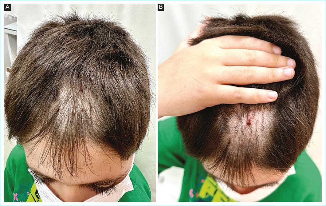
Figure 1 A: static assessment of the scalp hair loss showing an irregular patch of alopecia partially covered by hair shafts. B: evaluation after uncovering the alopecic area revealing an irregular patchy alopecia in the frontotemporal region with hemorrhagic crusts.

Figure 2 Trichoscopic features of the alopecic area. A: broken hairs, perifollicular, and interfollicular erythema (orange arrow), black dots, hair powder, and short vellus hair. B: hairs of different lengths, irregular black dots (green circle), perifollicular and interfollicular erythema (orange arrow), trichoptilosis, and short vellus hair. C: broken hairs, irregular black dots, exclamation mark hairs (yellow arrow), coudability hairs (blue arrow), hairs of different lengths, hair powder, and short vellus hair. D: broken hairs, irregular black dots, coudability hairs (blue arrow), hair powder, and hairs of different lengths.
Discussion
The diagnosis of AA involves medical and family history and a full examination of the scalp, face, and body. Nails should also be analyzed. The hair pull test is recommended, and a positive test indicates an active disease1. In the case described, the hair pull test was negative and can be interpreted as a stable alopecia. The most common trichoscopic findings in AA are yellow dots (60-94% of cases), exclamation mark hairs (12-71%), black dots (50%), broken hairs (0-71%), and short vellus hairs (34-100%)1,7. These features were identified in the reported patient, as shown in figure 2. Scalp biopsy is rarely necessary and is only needed in inconclusive cases, especially when scarring alopecia cannot be excluded1.
The diagnosis of TT includes clinical findings, medical and psychiatric history. The physical examination should include trichoscopic analysis8. Typical signs are broken hairs of different lengths (80-100% of cases), hair powder (10-88.9%), tulip hairs (10-47.7%), trichoptilosis (34.1-100%), irregular black dots (27.3-100%), flame hairs (25-100%), v-sign (20-70%), hook hairs (11.1-56.8%), and coiled hairs (4.3-100%)7,9,10. Scratching and signs of hemorrhage can be often identified, as in the case reported. Biopsy can be used to confirm the diagnosis in undefined cases8. In the patient presented, the clinical history and the trichoscopic findings were sufficient to conclude the diagnosis of AA e TT. There is an important overlap between the trichoscopic features of the two conditions. For instance, black dots and broken hairs are identified in both diseases. We believe we faced both AA and TT because the patchy alopecia presented exclusive findings of AA (exclamation mark hairs and coudability hairs) simultaneously with signs of scratching, hairs of different lengths, and trichoptilosis — typical of TT. This set of alterations could not be explained by an isolated AA or TT, but only by both together.
The first-line treatment for pediatric AA includes topical corticosteroids, as prescribed in this case1. Topical clobetasol propionate 0.05% is safe and well-tolerated, with good cosmetic acceptance1. Intralesional corticosteroids are also recommended for patchy AA, but the procedure was not the first choice in this case due to the patient’s young age and fear of needle. Topical minoxidil is usually indicated as an adjuvant intervention, as used in this case1. Other therapy options are topical irritation treatment with diphenylcyclopropenone or anthralin, systemic corticosteroids, phototherapy, methotrexate, hydroxychloroquine, and JAK inhibitors1,11.
Considering TT, psychotherapy is considered the first-line therapy for pediatric patients3,12. Topical anesthetics, corticosteroids, or capsaicin can be helpful to control the urge of scratch12. Some studies indicate benefits in the use of n-acetylcysteine, clomipramine, olanzapine, and dronabinol in adults, but there are no specific doses for children3,12. The association of AA and TT is rare, but possible and can occur in children. For both diseases, dermatologists and psychologists should work together to manage the anxiety (triggering factor) and reduce the emotional impairment of the hair loss.

