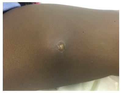
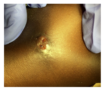
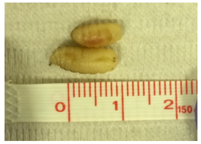
A 17-month-old boy presented to the pediatric Emergency Department with a six-day history of painful swelling of the left thigh and scrotum. His grandmother stated that she had seen a “worm” coming out of one of the lesions that morning. There was no history of trauma or previous skin lesions. The boy had travelled from Guinea-Bissau to Portugal seven days earlier to visit his family. On examination, the patient was afebrile and appeared healthy. Two red, hot, painful, furuncle-like swellings were noted: one measuring 3 x 5 cm with a 0.2 cm central ulcer on the medial aspect of the left thigh (Figure 1A) and another measuring 3 x 3 cm on the base of the scrotum (Figure 1B). There was no drainage when the lesions were squeezed. While the child was under observation, one larva spontaneously emerged from the thigh ulcer, and a second was pulled using tweezers (Figure 2).
What is your diagnosis?

Figure 1A Furuncle-like lesion on the thigh, with the posterior spiracles of the maggot visible in the central pore.
Discussion
Human myiasis is the infestation of humans with dipterous larvae. It can affect the skin, dead tissue, or natural cavities and is classified according to the site of infestation as cutaneous, ophthalmic, nasopharyngeal, enteric, oral, or urogenital. Cutaneous myiasis is the most common form. The infection also occurs in animals and is a major cause of economic loss in animal production and agriculture.1,2
In the present case, removal of larvae from the thigh ulcer suggested the diagnosis. Plastic bandages were applied to the lesions to suffocate any other larvae that might have been inside the lesions. The patient was kept on observation for 12 hours, but no other larvae were found. He was also treated with oral amoxicillin and clavulanate for seven days to prevent secondary bacterial infection. The removed maggots were preserved in a plastic container at 4ºC and sent to an entomologist at a specialized laboratory. The patient was reassessed three days later and was asymptomatic with good healing of the skin lesions. Entomological analysis of the maggots revealed Cordylobia anthropophaga larvae and confirmed the diagnosis.
Most fly species that cause myiasis are endemic to tropical and subtropical countries, especially in poor socioeconomic regions with inadequate sanitary conditions.1-3 Although uncommon in other parts of the world, the expansion of international air travel has made myiasis one of the most common travel-associated skin diseases, making it a knowledge priority for emergency medicine physicians.1
The diagnosis of cutaneous myiasis should be suspected when furuncle or boil-like skin lesions are observed in patients travelling from endemic regions.1-3 When the patient presents with visible maggots, as in the present case, the diagnosis reveals itself. However, other forms of cutaneous myasis, such as migratory and cavitary disease without visible maggots, may be more difficult to recognize. The importance of accurate diagnosis is to prevent the spread of myasis-causing flies to regions where they are not endemic and may pose a public health problem, in addition to relieving the patient’s symptoms.
Cordylobia anthropophaga is endemic to sub-Saharan Africa and is one of the most common etiological agents of furuncular myiasis. It infects children more frequently than adults.1,2 Although most types of cutaneous myiasis are uncommon in intact skin, the larvae of C. anthropophaga can penetrate the healthy skin of the host when lying on the ground or wearing contaminated clothing.1,2 Preventive measures include ironing clothes and preventing children from playing or sleeping in direct contact with the soil, especially during the rainy season. Skin wounds and lesions should be regularly cleaned and covered. Symptoms of infestation usually begin within two days of larvae penetration and range from pruritus to severe pain. Over the following days, the inflammatory response progresses, mimicking bacterial soft tissue infections such as cellulitis, and may cause fever. After eight to twelve days, a mature larva emerges from a furuncle-like lesion with the intention of reaching the ground and pupating to complete its life cycle.1,2
Treatment may include application of toxic substances to the larvae, covering the lesion to suffocate the larvae and force them to emerge, or surgical removal to achieve complete extraction of the larvae. Careful manual compression to remove the larvae may be useful when C. anthropophaga is suspected, but should be avoided with other species, so a detailed history and knowledge of the most common flies in the area where the patient has been is paramount.1-3 Antibiotic prophylaxis is not routinely recommended. In the present case, the decision was to start oral antibiotics because the extension of the lesions suggested cellulitis and, given the lack of experience with myiasis, there was no certainty that larval extraction was complete, which is a risk factor for secondary bacterial infection.
Correct identification of the larvae is important for treatment and public health. In this case, the entomologist’s instructions were followed, and the maggots were preserved in a dry container in the refrigerator until they could be sent to the laboratory. Some authors suggest killing the maggots by immersing them in hot water and then preserving them in a 70% ethanol solution.1 It is crucial to preserve the maggots in a way that preserves their appearance and color, since identification depends on macroscopic analysis.
Authorship
Ana Castelbranco Silva - Conceptualization; Data curation; Visualization; Writing - original draft; Writing - review & editing
Ana Lança - Data curation; Writing - original draft; Writing - review & editing
Tomás Nunes - Data curation; Writing - original draft; Writing - review & editing Rita Martins - Supervision; Writing - review & editing
Maria Gomes Ferreira - Supervision; Writing - review & editing


