Imaging cases
Tumefação no dedo do pé de uma criança
1 Department of Pediatrics, Unidade Local de Saúde de Trás-os-Montes e Alto Douro, 5000-508 Vila Real, Portugal. susctx@gmail.com
2 Department of Pediatrics, Unidade Local de Saúde do Alto Minho. 4904-858 Viana do Castelo, Portugal. a.pedro.marinho@gmail.com
3 Department of Dermatology, Unidade Local de Saúde de Santo António.4099-001 Porto, Portugal. miguel.o.oliveira@gmail.com, susanamlmachado@gmail.com
Abstract
SE is a slow and benign growth of normal bone under the nail. The most common site is the hallux. This growth can cause nail disorders, masking the true problem and often leading to misdiagnosis. The pain caused by SE, especially with shoe contact, is significant and can affect the patient’s quality of life, so a prompt diagnosis is important.
Keywords: nail; Pediatrics; subungual exostosis
Resumo
A exostose subungueal (ES) é um crescimento de osso normal, lento e benigno por baixo da unha. A localização mais frequente é o halux. Esta proliferação pode originar distúrbios ungueais, mascarando o verdadeiro problema e levando frequentemente a erros de diagnóstico. A dor provocada pela ES, sobretudo quando ocorre fricção promovida pelo calçado, é relevante e pode interferir com a qualidade de vida dos doentes, pelo que o rápido diagnóstico é importante.
Palavras-chave: exostose subungueal; Pediatria; unha
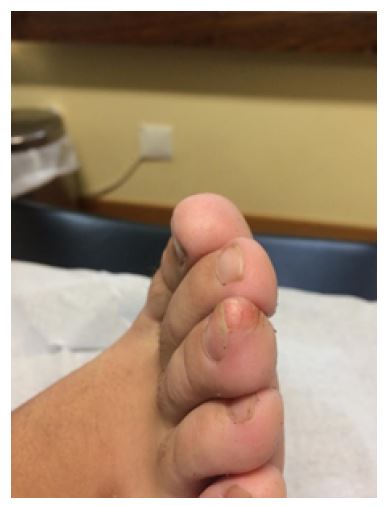
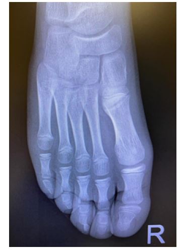
An otherwise healthy 10-year-old boy presented with a 2-year history of a painful nodule on the third finger of the right foot (Figure 1). There was no history of trauma. He had undergone multiple wart and antifungal treatments without improvement. Physical examination revealed a firm nodular lesion under the nail plate measuring 1 cm in diameter with regular borders and nail deformity. No similar nodules were found elsewhere. Right foot radiograph showed a bony prominence arising from the terminal end of the distal phalanx (Figure 2).
What is your diagnosis?
Diagnosis
Subungual exostosis (SE)
Discussion
The patient was diagnosed with SE and referred to orthopedic consultation. Excision of the nodule was performed without intercurrences. Anatomopathology confirmed the diagnosis of SE.
SE is a painful benign osteochondral proliferation of the distal phalanx of the fingers or toes, most commonly affecting the hallux.1-5 Progressive growth of the lesion may cause nail lifting and deformity.4 The etiology of SE is unknown, although trauma and chronic infection seem to play a role.3,5 Half of the cases occur in patients under the age of 18 years, and females are predominantly affected.2,5 The differential diagnosis is broad and should include other subungual masses such as osteochondroma, pyogenic granuloma, keratoacanthoma, wart, or fibroma.4 Radiograph of the affected digit is essential for diagnosis and shows a dorsal or dorsomedial surface proliferation of the distal phalanx.1,5 Histopathology typically shows endochondral ossification with lamellar trabeculae covered by a fibrocartilaginous cap.1,4 As this is usually a progressive condition, treatment consists of complete marginal surgical excision.1-5 Postoperative nail deformity may persist, and recurrence has been reported.1,2
Although SE is not a rare entity, a diagnostic delay of 12 months is common,2 highlighting the importance of a high index of suspicion for this entity by all physicians working with children, as it can affect their quality of life.3
Authorship
Susana C Teixeira - Conceptualization; Writing - original draft; Writing - review & editing
Pedro Marinho - Writing - review & editing
Miguel Oliveira - Writing - review & editing
Susana Machado - Patient´s assistant physician; Validation; Writing - review & editing
References
1. Ardebol J, Mubarak A. Subungual exostosis recurrence in a 16-year-old athletic male, Oxford Medical Case Reports. 2020; 4: 1-2.
[ Links ]
2. DaCambra MP, Gupta SK, Ferri-de-Barros F. Subungual exostosis of the toes: a systematic review. Clin Orthop Relat Res. 2014; 472: 1251-9.
[ Links ]
3. Piccolo V, Russo T, Rezende LL, Argenziano G. Subungual exostosis in an 8-year-old child: clinical and dermoscopic description. An Bras Dermatol. 2019; 94: 233-5.
[ Links ]
4. Russell JD, Nance K, Nunley JR, Maher IA. Subungual exostosis, Cutis. 2016; 98: 128-9.
[ Links ]
5. Carmona FJG, Huerta JP, Morato DF. A proposed subungual exostosis clinical classification and treatment plan. J Am Podiatr Med Assoc. 2009; 99: 519-24.
[ Links ]


