SciELO - Scientific Electronic Library Online

 
vol.36 número1Massa Intramuscular numa Lesão Proximal do Músculo Reto Femoral - Hematoma ou Neoplasia?Espondilodiscite Brucelar - Relato de Caso índice de autoresíndice de assuntosPesquisa de artigos
Home Pagelista alfabética de periódicos  

Serviços Personalizados

Journal

Artigo

Indicadores

Links relacionados

  • Não possue artigos similaresSimilares em SciELO

Compartilhar


Acta Radiológica Portuguesa

versão impressa ISSN 2183-1351

Acta Radiol Port vol.36 no.1 Lisboa abr. 2024  Epub 30-Abr-2024

https://doi.org/10.25748/arp.30305 

Imagens de Interesse

Colonic Obstruction due to Biliary Calculus: A Rare Case

Obstrução Cólica por Cálculo Biliar: Um Caso Raro

1Radiology Department, Centro Hospitalar Tondela-Viseu, Viseu, Portugal


Abstract

Colic obstruction due to a gallstone impacted at the sigmoid colon is a rare entity.

Despite its rarity, the radiologist should be aware of its existence, allowing a prompt diagnosis and treatment.

This article describes an 83-year-old male patient with a six-day history of lower quadrant abdominal pain and constipation.

In this context an abdominal ultrasound was performed where colonic distension and free interloop fluid was seen. Therefore, a CT was obtained and an impacted gallstone was seen at the sigmoid colon. Removal of the calculus through colonoscopy was attempted but without success, then an exploratory laparotomy was performed, and the calculus was successfully removed.

Keywords: Colonic obstruction; Biliary colon; Bowel; CT.

Resumo

Obstrução intestinal causada pela impactação de um cálculo biliar ao nível do sigmóide é uma entidade rara, mas que deve estar presente na mente do radiologista, de modo a permitir um diagnóstico atempado e a instituição precoce do tratamento adequado.

Este artigo descreve um paciente de 83 anos, do sexo masculino com história de dor abdominal ao nível dos quadrantes inferiores há cerca de 6 dias. Associadamente o paciente refere obstipação.

No presente contexto foi realizada uma ecografia abdominal onde se observou marcada distensão do cólon, bem como líquido livre interansas, posteriormente foi realizado TC abdominal onde se identificou um cálculo impactado ao nível do sigmóide. Foi realizada colonoscopia para tentar remover o cálculo, mas sem sucesso. O cálculo foi removido com sucesso através de laparotomia exploradora.

Palavras-chave: Obstrução cólica; Cólon biliar; Cólon; TC.

Case report

We present a case of an 83-year-old man with a six-day history of lower quadrant abdominal pain and constipation.

In this context an abdominal radiography and an abdominal ultrasound were performed. The first showed colonic loops distension until the sigmoid level and the latter demonstrated intestinal loops distension, with non-progressive bowel movements. Free interloop fluid was also observed, suggesting an occlusion.

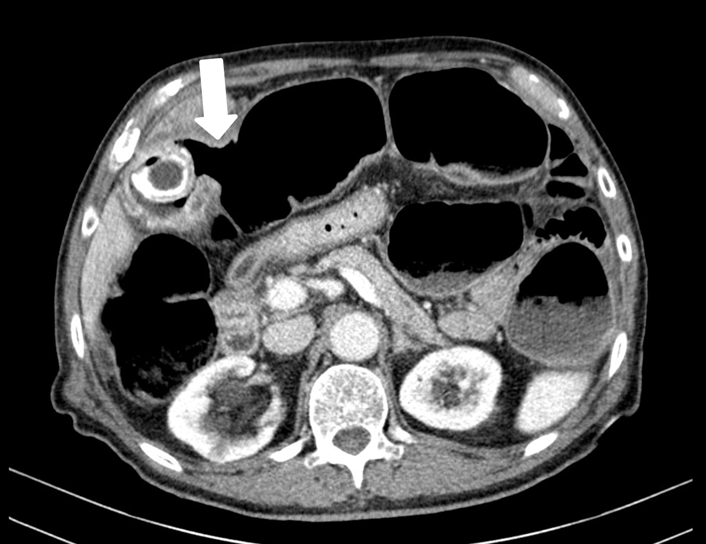
Therefore, a computed tomography (CT) was obtained to determine the obstruction level, in which a fistula between the gallbladder and the transverse colon was seen. The gallbladder had calculus and air. Practically all bowel loops were distended, and a biliary calculus measuring 36 mm was seen in the sigmoid bowel. At this point the bowel wall was thickened.

A colonoscopy for calculus extraction was performed but the inflammatory stenosis did not allow it. Therefore, an exploratory laparotomy was performed successfully removing the calculus.

Discussion

Gallstone ileus is a rare complication of biliary gallstones, representing only 1-4% of mechanical intestinal obstructions1 and is caused by the migration of gallbladder calculus through a biliodigestive fistula.2 This is more common in patients who have a history of acute cholecystitis, because the inflammatory process may approach the serosas of the biliary and intestinal tract, due to distention. With repeating inflammatory episodes, the serosas adhere and fistulisation may occur.1 Fistulisation to the gastrointestinal tract allows the passage of the calculi to the intestinal lumen until it becomes impacted at any level. Cholecystocolonic fistulisation is rare and only occurs in 10-25%, allowing direct passage of the stone to the colonic lumen.3

The stone size is also an important aspect, as stones smaller than 2 cm have a low probability of causing a colonic obstruction, unless there is some other process altering the colonic calibre.2 In this case the stone measured 36 mm and no underlying disease was found.

Most of these patients present with symptoms of mechanical obstruction, such as abdominal pain, distension, constipation and vomiting.2 Abdominal radiography and abdominal ultrasound are the first line exams in these scenarios.

A combination of intestinal distension, pneumobilia and ectopic gallstone constitute the pathognomonic Rigler’s triad of this disease in the radiography.1 However, these findings are only present in 40-50% of the patients.3 In this case only intestinal distension was seen in the radiography (Figure 1).
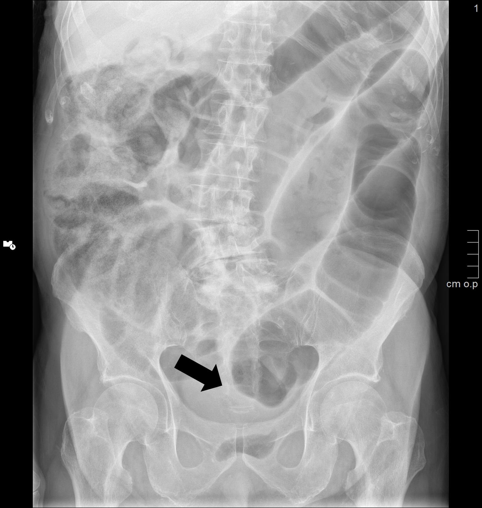
Figure 1: Abdominal radiograph showing diffuse intestinal loops distension (aster with stop signal at the sigmoid level (black arrow). 

Ultrasound sometimes can reveal the ruptured state of the gallbladder. Free interloop fluid and non-progressive intestinal movements can also be identified, suggesting an occlusive episode.

CT is the best method to assess the obstruction cause. In this case, the marked intestinal loops distension was identified and a 36 mm calculus was found in the sigmoid. At this level the colonic wall was markedly thickened (Figure 2). At the gallbladder level a continuity solution between the gallbladder wall and the colon was observed (Figure 3).
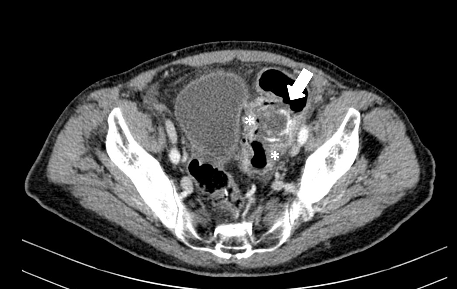
Figure 2: Calculus in the sigmoid’s lumen (white arrow) and inflammatory thickening of the colonic wall (asterisk) 

Figure 3: Cholecystocolonic fistula (white arrow) and diffuse intestinal loop distensions 

In these cases, enterolithotomy is the treatment of choice. Because this condition affects mostly elderly people with poor condition, gallstone ileus has a poor prognosis, with mortality ranging from 12% to 27%.2

Therefore, the radiologist should be aware of the clues to the diagnosis, allowing a prompt diagnosis and treatment and consequently minimizing the risk of complications.

References

1. Melo EL, de Paula FT, Siqueira RA, Ribeiro SC. Biliary colon: an unusual case of intestinal obstruction. Radiol Bras. 2015;48:127-8. doi: 10.1590/0100-3984.2014.0073. PMID: 25987755; PMCID: PMC4433305. [ Links ]

2. Rigler LG, Borman CN, Noble JF. Gallstone obstruction: pathogenesis and roentgen manifestation. J Am Med Assoc. 1941;117:1753-9. [ Links ]

3. Carlsson T, Gandhi S. Gallstone ileus of the sigmoid colon: an extremely rare cause of large bowel obstruction detected by multiplanar CT. BMJ Case Rep. 2015;2015:bcr2015209654. doi: 10.1136/bcr-2015-209654. PMID: 26682834; PMCID: PMC4691945. [ Links ]

Ethical disclosures

Financing Support: This work has not received any contribution, grant or scholarship.

Received: March 19, 2023; Accepted: November 09, 2023

Address Marta Vaz Dias, Serviço de Radiologia, Hospital de São Teotónio, Av. Rei Dom Duarte, 3504-509 Viseu, Portugal, e-mail: martaisavaz@gmail.com

Conflicts of interest: The authors have no conflicts of interest to declare.

Confidentiality of data: The authors declare that they have followed the protocols of their work center on the publication of data from patients.

Protection of human and animal subjects: The authors declare that the procedures followed were in accordance with the regulations of the relevant clinical research ethics committee and with those of the Code of Ethics of the World Medical Association (Declaration of Helsinki).

© Author(s) (or their employer(s)) and ARP 2024. Re-use permitted under CC BY-NC. No commercial re-use.

Creative Commons License This is an open-access article distributed under the terms of the Creative Commons Attribution License