SciELO - Scientific Electronic Library Online

 
vol.36 número1Obstrução Cólica por Cálculo Biliar: Um Caso RaroManifestações Esqueléticas Radiográficas da Mucopolissacaridose Tipo VI índice de autoresíndice de assuntosPesquisa de artigos
Home Pagelista alfabética de periódicos  

Serviços Personalizados

Journal

Artigo

Indicadores

Links relacionados

  • Não possue artigos similaresSimilares em SciELO

Compartilhar


Acta Radiológica Portuguesa

versão impressa ISSN 2183-1351

Acta Radiol Port vol.36 no.1 Lisboa abr. 2024  Epub 30-Abr-2024

https://doi.org/10.25748/arp.31290 

Imagens de Interesse

Brucellar Spondylodiscitis - A Case Report

Espondilodiscite Brucelar - Relato de Caso

Adriana Oliveira Guilarde1  2 
http://orcid.org/0000-0002-5032-8085

Ricardo Vieira Teles-Filho1 
http://orcid.org/0000-0003-4822-1526

Moara Alves Santa Barbara Borges1  2 
http://orcid.org/0000-0003-2461-2063

Giovanni Ferreira Viggiano1 
http://orcid.org/0000-0003-2623-4342

Paulo Sérgio Sucasas da Costa1 
http://orcid.org/0000-0001-9370-9139

Gustavo Abrahão Afiune1 
http://orcid.org/0000-0002-1510-262X

1Hospital das Clínicas, Universidade Federal de Goiás; Goiânia, Goiás,Brazil

2Instituto de Patologia Tropical e Saúde Pública, Universidade Federal de Goiás; Goiânia, Goiás,Brazil


Abstract

Brucellosis, a disease caused by bacteria of the genus Brucella, presents a wide range of symptoms in humans, which makes diagnosis challenging, especially when atypical manifestations occur. This article reports a rare case of spondylodiscitis and highlights the importance of investigating brucellosis in patients with prolonged spondylodiscitis who do not respond to conventional antibiotic treatments. Furthermore, it highlights the need for a complete epidemiological e radiologic analysis and the performance of specific serological tests for an accurate diagnosis of the underlying cause. Early suspicion, based on a detailed history and careful diagnosis, together with advances in imaging techniques, is crucial for planning effective treatment.

Keywords: Brucellosis; Spondylodiscitis; Infection; Radiology.

Resumo

A brucelose, uma doença causada por bactérias do gênero Brucella, apresenta uma ampla gama de sintomas em seres humanos, o que torna o diagnóstico desafiador, especialmente quando ocorrem manifestações atípicas. Este artigo relata um caso raro de espodilodiscite e ressalta a importância de investigar a brucelose em pacientes com espodilodiscite prolongada que não respondem aos tratamentos antibióticos convencionais. Além disso, destaca a necessidade de uma análise epidemiológica e radiológica completa e a realização de testes sorológicos específicos para um diagnóstico preciso da causa subjacente. A suspeita precoce, baseada em uma anamnese detalhada e em um diagnóstico cuidadoso, juntamente com os avanços nas técnicas de imagem, é crucial para o planeamento de um tratamento eficaz.

Palavras-chave: Brucelose; Espondilodiscite; Infecção; Radiologia.

A 74-year-old farmer presented to the emergency room with complaints of low back pain. Previously diagnosed with spondylodiscitis of unknown etiology, the patient experienced no improvement with empirical antibiotic therapy. The low back pain persisted for over a year, worsening in the last eight months. Within the last six months, a fistula with drainage to the right flank developed, accompanied by the onset of fever a week prior to hospital admission. Recent imaging revealed significant L5-S1 involvement by spondylodiscitis, along with paravertebral and muscular collections in the right psoas, iliac, and gluteus regions, featuring fistulous tracts to the skin. Incidentally, CT scans uncovered a fusiform aneurysm of the abdominal aorta extending to the right iliac artery, with associated mural thrombosis (Figure 1-2). Bacterial, fungal, and mycobacterial blood cultures remained negative. Positive seroagglutination tests for brucellosis prompted the initiation of gentamicin (14 days), doxycycline, and rifampicin, forming a 3-month therapeutic plan with the latter two antimicrobials. Despite the surgical staff's evaluation opting against surgery, the combined antimicrobial therapy resulted in a significant improvement in the infectious condition, prompting scheduled follow-up appointments. This case presents a rare case of spondylitis in a patient with brucellosis, underscoring the importance of investigating brucellosis in cases of prolonged spondylodiscitis resistant to conventional antibiotic therapy. It also emphasizes the need for heightened suspicion, detailed epidemiological investigation, and specific serological tests to establish the etiological diagnosis of brucellosis.

Figure 1: (A-B) axial and sagittal sections of contrast-enhanced CT showing a collection with capsular enhancement with heterogeneous content (pointed by the arrows), anterior to the vertebral bodies of the sacrum, with extension into the spinal canal, associated with erosion of the vertebral body of L5 and, notably, of S1. (C-D) gadolinium-enhanced T1 sequences showing extension of the collection to the right psoas muscle and erosions, sequestration and bone abscess in the vertebral body of S1 (pointed by the arrows). 
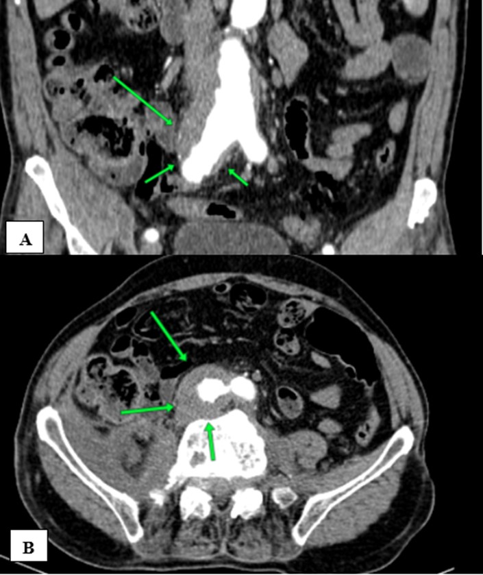
Figure 2: (A-B) axial and coronal contrast-enhanced CT, in the arterial phase, showing as additional finding a fusiform abdominal aorta aneurysm with extension to the right iliac artery. 

Radiology assumes a pivotal role in diagnosing brucellosis. While lacking pathognomonic radiographic features, imaging tests can reveal highly suggestive manifestations, including bone erosions, especially in the vertebrae, osteomyelitis, abscess collections, and joint changes. The confluence of clinical, epidemiological, and radiological findings is essential for a definitive diagnosis.1 Literature indicates that the formation of abdominal aneurysms can be a complication of brucellosis infection. In very rare cases, there is an unusual and potentially fatal combination of Brucella infection with spondylodiscitis and abdominal aneurysms. In this case, the absence of signs of aneurysm infection on imaging tests, such as densification of peri-aneurysmal fat, irregularity and thickening of the aortic wall, and peri-aortic soft tissue mass, coupled with no changes in aneurysm configuration post-antibiotic therapy, led to considering it an incidental finding with Brucella spondylodiscitis, rather than a complication of the infection.2 However, we underline the rare possibility of association between these findings.3,4

In the discussed case, radiological exams revealed severe spondylodiscitis at L5-S1, accompanied by paravertebral and muscular collections in the psoas, iliac, and gluteal regions with fistulous tracts extending to the skin. Additionally, computed tomography identified a fusiform aneurysm in the abdominal aorta, extending to the right iliac artery with mural thrombosis. These findings emphasize the pivotal role of radiology in accurate diagnosis.

References

1. Esmaeilnejad-Ganji SM, Esmaeilnejad-Ganji SMR. Osteoarticular manifestations of human brucellosis: A review. World J Orthop. 2019;10:54-62. doi:10.5312/wjo.v10.i2.54 [ Links ]

2. Li X, Cheng Z. Brucellosis involving the aorta and iliac arteries: a systematic review of 130 cases. Frontiers in Bioengineering and Biotechnology. 2023;11:1326246. doi: 10.3389/fbioe.2023.1326246 [ Links ]

3. Tali ET, Koc AM, Oner AY. Spinal brucellosis. Neuroimaging Clinics of North America. 2015;25:233-245. doi: 10.1016/j.nic.2015.01.004 [ Links ]

4. Rizkalla JM, Alhreish K, Syed IY. Spinal brucellosis: a case report and review of the literature. Journal of Orthopaedic Case Reports. 2021;11:1-5. doi: 10.13107/jocr.2021.v11.i03.2060 [ Links ]

Ethical disclosures

Financing Support: This work has not received any contribution, grant or scholarship.

Received: May 24, 2023; Accepted: December 19, 2023

Address Ricardo Vieira Teles Filho, Federal University of Goiás, Primeira Avenida, s/n, Setor Leste Universitário, Goiânia, GO, Brazil Zip Code - 74605-020, E-mail: ricardovteles@gmail.com

Conflicts of interest: The authors have no conflicts of interest to declare.

Confidentiality of data: The authors declare that they have followed the protocols of their work center on the publication of data from patients.

Protection of human and animal subjects: The authors declare that the procedures followed were in accordance with the regulations of the relevant clinical research ethics committee and with those of the Code of Ethics of the World Medical Association (Declaration of Helsinki).

© Author(s) (or their employer(s)) and ARP 2024. Re-use permitted under CC BY-NC. No commercial re-use.

Creative Commons License This is an open-access article distributed under the terms of the Creative Commons Attribution License