SciELO - Scientific Electronic Library Online

 
vol.31 número3Colangioscopia com Litotrícia Laser no tratamento de doente com Síndrome de Mirizzi tipo IISigmoidopexia endoscópica percutânea: um caminho ainda a percorrer índice de autoresíndice de assuntosPesquisa de artigos
Home Pagelista alfabética de periódicos  

Serviços Personalizados

Journal

Artigo

Indicadores

Links relacionados

  • Não possue artigos similaresSimilares em SciELO

Compartilhar


GE-Portuguese Journal of Gastroenterology

versão impressa ISSN 2341-4545

GE Port J Gastroenterol vol.31 no.3 Lisboa jun. 2024  Epub 28-Jun-2024

https://doi.org/10.1159/000533931 

Endoscopic Snapshot

Endoscopic Extraction of Two Giant Stone Bezoars Using Mechanical and Laser Lithotripsy

Remoção endoscópica de dois bezoares gigantes petrificados com recurso a litotrícia mecânica e por laser

Carla Oliveira1 

Gonçalo Nunes1  2 

Francisco Vara-Luiz1  2 

Gabriel Paiva de Oliveira3 

Ana Nunes1 

Jorge Fonseca1  2 

1Gastroenterology Department, Hospital Garcia de Orta, Almada, Portugal;

2PaMNEC - Grupo de Patologia Médica, Nutrição e Estudos Clínicos, CiiEM, Centro de Investigação Interdisciplinar Egas Moniz, Monte da Caparica, Portugal;

3Surgery Department, Hospital Garcia de Orta, Almada, Portugal


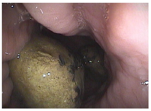
An 80-year-old woman with iron-deficiency anaemia underwent an outpatient upper gastrointestinal endoscopy, presenting two large petrified bodies with 60 mm and 90 mm length in the gastric lumen, associated with a 12 mm ulcer in the incisura (Fig. 1). Past medical history was relevant for type 2 diabetes mellitus and ruled out previous gastric surgeries.

Fig. 1 Two petrified bodies with 60 mm and 90 mm length in the gastric lumen. 

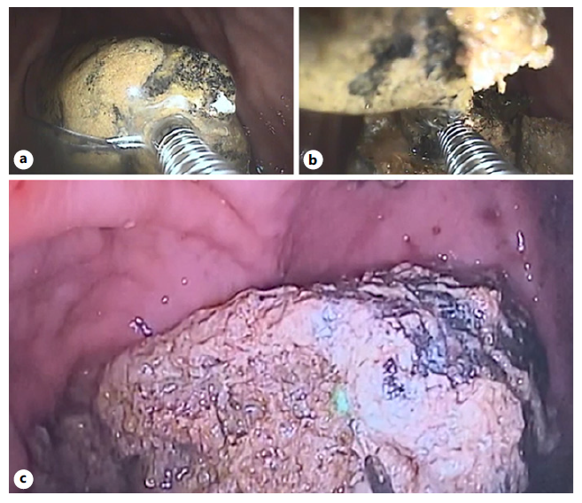
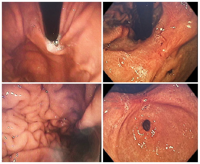
The patient was referred to our hospital and although liver tests and abdominal ultrasound were normal, she performed a magnetic resonance cholangiopancreatography that excluded a cholecystogastric fistula or any fistulous tract between the biliary tree and the stomach. In spite of being asymptomatic, considering the advanced age and high surgery risk, endoscopic extraction was scheduled. A double-channel gastroscope was used for the procedure, which was performed in two sessions (online suppl. video 1; for all online suppl. material, see https://doi.org/10.1159/000533931). Endoscopic re-assessment confirmed the presence of two bulky, hard, and rounded stones with smooth surface impossible to be removed directly using standard retrieval devices. The60mmstone wasfragmentedfromthe periphery using the LithoCrush V™ mechanical lithotriptor (Fig. 2a, b), and all fragments were extracted with a RothNet® during approximately 30 min. However, the 90 mm stone was too thick and round and could not be grasped with any of the available commercially baskets. Fulguration using bipolar and argon plasma coagulation probes at high potencies (120 W) was unsuccessful, with rapid loss of the endoscopic field of view due to smoke. Thus, laser lithotripsy was programed for a second intervention after 3 weeks. Using Auriga™ XL holmium laser and LightTrail™ fibre (600 μm; 120 W) from Boston Scientific, usually applied in urologic surgery, the stone was dissolved, producing smaller fragments that could be further fragmented with the lithotriptor basket and safely removed (Fig. 2c). The procedure was performed in na outpatient setting, under deep sedation and has taken almost 3 h. No associated complications were observed and the patient was further discharged. Pathological analysis was suggestive of phytobezoar. Low-fibre diet was recommended and proton-pump inhibitor prescribed for ulcer healing. One month later, the control endoscopy presented no residual stones or mucosa ulceration (Fig. 3).

Fig. 2 a-c  Mechanical lithotripsy using a Dormia basket and laser lithotripsy using holmium laser. 

Fig. 3. Gastric lumen with no residual lesions after the endoscopic procedure. 

Gastric bezoars are foreign bodies resulting from accumulation of undigested material, most commonly vegetable fibres, being rarely associated with severe gastrointestinal complications. Treatment includes chemical dissolution with agents like CocaCola® or acetylcysteine, prokinetics, endoscopic removal, and surgery. Endoscopic therapy involves fragmenting the bezoar using different mechanisms, most commonly forceps, snares, baskets, and argon plasma coagulation [1]. Although endoscopic treatment is not standardized, a minority of cases managed using laser lithotripsy were reported [2-5]. The authors describe a unique case of two giant gastric bezoars successfully treated through laser and mechanic lithotripsy.

References

1. Nunes G, Patita M, Barosa R, Canhoto M, Ramos LR, Lages P, et al. Small bowel obstruction after endoscopic fragmentation of a gastric bezoar: an unexpected complication. Turk J Gastroenterol. 2018;29(5): [ Links ]

2. Mao Y, Qiu H, Liu Q, Lu Z, Fan K, Huang Y, et al. Endoscopic lithotripsy for gastric bezoars by Nd:YAG laser-ignited mini-explosive technique. Lasers Med Sci. 2014;29(3): 1237-40. [ Links ]

3. Grande G, Manno M, Zulli C, Barbera C, Mangiafico S, Alberghina N, et al. An alternative endoscopic treatment for massive gastric bezoars: Ho:YAG laser fragmentation. Endoscopy. 2016;48(Suppl 1):E217. [ Links ]

4. Zheng YX, Prasoon P, Chen Y, Hu L, Chen L. Sandwich treatment for diospyrobezoar intestinal obstruction: a case report. World J Gastroenterol. 2014 Dec 28;20(48): 18503-6. [ Links ]

5. Hendriks, S, Verseveld, MM, Boevé, ER, Roomer, R. Successful endoscopic treatment of a large impacted gallstone in the duodenum using laser lithotripsy, Bouveret's syndrome: a case report. World J Gastroenterol. 2020; 26(19):2458-63. [ Links ]

1Statement of Ethics The authors have no ethical conflicts to disclose. Ethical ap-proval was not required for this study, in accordance with local/national guidelines. The patient has given the informed consent for publication of the present case.

3Funding Sources The authors have no funding sources to disclose.

5Data Availability Statement All data generated or analysed during this study are included in this article. Further enquiries can be directed to the corresponding author.

Received: May 21, 2023; Accepted: July 31, 2023

Correspondence to: Gonçalo Nunes, goncalo.n@hotmail.com

Conflict of Interest Statement The authors have no conflicts of interest to declare.

Author Contributions Carla Oliveira was responsible for material preparation, data collection, and the first draft of the manuscript. Gonçalo Nunes performed the endoscopic procedure, read, and ap-proved the final manuscript. Francisco Vara-Luiz, Gabriel Oliveira, Ana Nunes, and Jorge Fonseca read and approved the final manuscript.

Creative Commons License This is an open-access article distributed under the terms of the Creative Commons Attribution License