SciELO - Scientific Electronic Library Online

 
vol.31 número4Pancolite induzida por Obinutuzumab: a propósito de um caso clínicoSchwanoma da via biliar biliar comum: Um diagnóstico clinico-patologico volúvel índice de autoresíndice de assuntosPesquisa de artigos
Home Pagelista alfabética de periódicos  

Serviços Personalizados

Journal

Artigo

Indicadores

Links relacionados

  • Não possue artigos similaresSimilares em SciELO

Compartilhar


GE-Portuguese Journal of Gastroenterology

versão impressa ISSN 2341-4545

GE Port J Gastroenterol vol.31 no.4 Lisboa ago. 2024  Epub 30-Set-2024

https://doi.org/10.1159/000533959 

Clinical Case Study

Desmoid Tumor after Sleeve Gastrectomy: Case Report and Literature Review

Tumor desmóide após gastrectomia vertical: caso clínico e revisão da literatura

Renato Medas1  2 

Rosa Coelho1  2 

Renato Bessa-Melo3 

Pedro Pereira1  2 

Guilherme Macedo1  2 

1Gastroenterology Department, Centro Hospitalar Universitário São João, Porto, Portugal;

2Faculty of Medicine of the University of Porto, Porto, Portugal;

3General Surgery Department, Centro Hospitalar Universitário São João, Porto, Portugal


Abstract

Desmoid tumor is a rare mesenchymal neoplasm of un-known etiology. Despite rare, the diagnosis of desmoid tumors after bariatric surgery is increased over the last few years. We report a case of a 26-year-old male with complains of abdominal pain and postprandial fullness, diagnosed with a locally advanced large intra-abdominal mass (40 × 21 × 11.7 cm) centered in the mesentery, developed 3 years after sleeve gastrectomy. Percutaneous biopsy was suggestive of a mesenquimatous tumor and the patient underwent surgery. R0 surgical resection was achieved, despite intimal contact and common vascularization with a jejunal loop. Histopathology examination of the surgical specimen revealed fusiform to stellate cells with mild atypia, thin-walled vessels, and diffuse beta-catenin expression (negative for DOG-1, CD117, CD34, S100, desmin, and alpha-actin). The diagnosis of a desmoid tumor was made. The patient remained asymptomatic, and no recurrence occurred over a 4-year follow-up. With the increasing number of bariatric surgeries, owing to the alarming growing incidence of obesity and related conditions, it is expected that desmoid tumors reports will gradually increase over the next few years. Thus, both gastroenterologists and surgeons should be aware of the potential for desmoid tumor development shortly after surgery, to offer a prompt diagnosis and treatment.

Keywords: Desmoid tumor; Mesenteric tumor; Sleeve gastrectomy; Bariatric surgery

Resumo

O tumor desmóide é uma neoplasia mesenquimatosa rara de etiologia desconhecida. Apesar de raros, temos assistido a um aumento do número de diagnósticos, ao longo dos últimos anos, de tumores desmóides que se desenvolvem após cirurgia bariátrica. Descrevemos o caso de um homem de 26 anos com queixas de dor abdominal e enfartamento pós-prandial, diagnosticado com uma massa intra-abdominal centrada no mesentério (40 × 21 × 11.7 cm), localmente avançada, 3 anos após ter realizado gastrectomia vertical. Foi efetuada biópsia percutânea, cujo resultado foi sugestivo de tumor mesenquimatoso e o paciente foi referenciado para cirurgia. O doente foi submetido a cirurgia e o tumor foi passível de ressecção cirúrgica R0, apesar de contacto íntimo e vascularização comum com uma ansa jejunal. O exame anatomopatológico revelou células fusiformes a estreladas com atipia ligeira e vasos de parede fina, bem como expressão difusa de beta-catenina (na ausência de expressão de DOG-1, CD117, CD34, S100, desmina e alfa-actina), sendo compatível com o diagnóstico de um tumor desmóide. O doente permanece assintomático e sem evidência de recidiva ao longo de 4 anos de vigilância. Com o aumento do número de cirurgias bariátricas devido ao aumento alarmante da incidência de obesidade e condições relacionadas, espera-se que os diagnósticos de tumores desmóides aumentem nos próximos anos. Assim, tanto gastroenterologistas quanto cirurgiões devem estar alerta para o desenvolvimento desta entidade, de forma a oferecer um diagnóstico e tratamento adequado e atempado.

Palavras Chave: Tumor desmoide; Tumor mesentérico; Gastrectomia vertical; Cirurgia bariátrica

Introduction

Desmoid tumor (DT) is a rare mesenchymal neoplasm of unknown etiology. Most cases are sporadic (85-90%) and related to somatic mutations in CTNNB1 gene, while 5-10% arises from a germline mutation in APC gene, in the context of familial adenomatous polyposis (FAP) [1]. Previous trauma, abdominal surgery, pregnancy, and hormonal therapy are known risk factors for DT development in sporadic cases [2]. Typically, DT is characterized by slow growth and no potential of metastasis but is known to be locally aggressive, leading to significant morbidity and mortality. Despite margin-free surgical resection, DTs have a high recurrence rate, ranging between 5 and 67% [3]. A recent meta-analysis reported a recurrence rate of 17.7% [4]. Over the last years, an increased number of DT development after bariatric surgery have been reported [5-9]. The increasing number of bariatric surgery procedures performed worldwide, owing to the alarming growing incidence of obesity and related conditions [10], may explain the raise in DT diagnosis related to bariatric procedures. Herein, we report a case of an intra-abdominal DT developed 3 years after sleeve gastrectomy.

Case Report

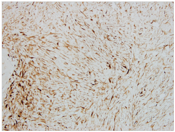
A 26-year-old male with a past medical history of vertical sleeve gastrectomy 3 years before, presented to the Gastroenterology consultation with abdominal pain and postprandial fullness of progressive worsening during the last 2 months. He denied associated symptoms such as nausea, vomits, fever, or weight loss. He also denied other medical conditions or a family history of FAP. Abdominal ultrasound revealed a bulky intra-abdominal mass that prompted further investigation. Abdominal computed tomography confirmed the presence of an intra-abdominal homogeneous mass (40 × 21 × 11.7 cm), centered in the mesentery, causing compression of the adjacent organs but without radiological signs of local invasion. No signs of bowel distension/obstruction or suspicious adenopathy were observed (Fig. 1). Percutaneous biopsy with an 18-gauge tru-cut needle was per-formed. Histopathological examination revealed fusiform cells arranged in bundles with beta-catenin expression (negative for desmin, DOG-1, CD117, and CD34), suggestive of a mesenquimatous tumor. Considering the tumor dimension and the symptomatic course of the disease, after discussion of the treatment options with the patient, elective surgery was decided. Laparotomy revealed a large mass arising from the mesentery root in intimal contact with a jejunal loop. After ligation of the vessels responsible for the tumor supply, segmental ischemia of the adjacent jejunal loop occurred. Thus, a 10 cm segmental enterectomy was also performed. The postoperative course was uneventful, and the patient was dis-charged 5 days later. The resected specimen measured 40 × 22.5 × 14.5 cm and weighed 5,550 g. Macroscopic evaluation revealed a smooth and brownish-white capsule with a homogeneous and whitish gelatinous core, with focal hemorrhagic areas. Histopathological examination showed fusiform to stellate cells with mild atypia and thin-walled vessels (Fig. 2). Immunohistochemical staining demonstrated diffuse beta-catenin expression, in the absence of expression for DOG-1, CD117, CD34, S100, desmin, and alpha-actin, consistent with desmoid-type fibromatosis (Fig. 3). Invasion of adipose tissue and small intestine wall (muscularis propria and submucosa) was present. Resection margins were negative. After 4-year follow-up, the patient remains asymptomatic without evidence of local recurrence.
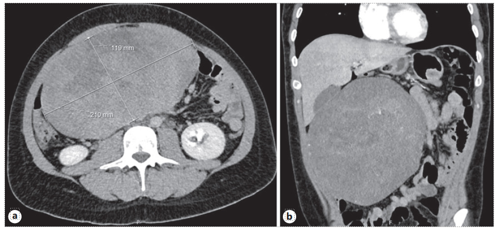
Fig. 1 a Bulky intra-abdominal mass, measuring 21.0 × 11.9 cm in axial axis, centered in mesentery. b Causing compression in the small bowel without imaging features suggestive of intestinal occlusion. 
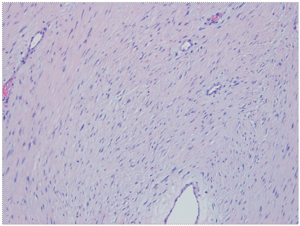
Fig. 2 Fusiform to stellate cells with mild atypia and thin-walled vessels. Presence of myxoid stroma, without mitosis or necrosis (×200). 

Fig. 3 Diffuse beta-catenin expression on immunohistochemical staining (×100). 

Discussion

DTs are a rare neoplasm derived from mesenchymal origin, with an estimated incidence of 2-4per million, accounting for 0.03% of all neoplasms [11]. The peak of incidence occurs between 30 and 40 years and is more common among women [12]. DTs etiology is not fully understood, but Wnt pathway plays a key role in DT pathogenesis [13]. Both sporadic DT and FAP-associated DT are linked to constitutive activation of the Wnt signaling pathway, resulting in cytoplasmic accumulation of β-catenin, followed by its translocation into the nucleus. Ultimately this results in the overexpression of genes involved in proliferation, fibrosis, and angiogenesis [14]. Since Wnt pathway has an active role in wounding repair [15], emerging data suggest that there is a dysregulated healing process after trauma (surgical or not) that favors DT development [14]. DTs can occur anywhere in the body but are more frequent in the abdominal wall, intra-abdominal cavity, and limbs [12]. Clinical presentation is variable, depending on its location, size, and growth rate. Intra-abdominal tumors are initially asymptomatic until they reach large dimensions, causing abdominal pain, palpable abdominal mass, bowel obstruction, ischemia, and rarely perforation or bleeding [12]. Focusing on DTs after bariatric surgery, Table 1 summarizes the described cases available in the literature. Abdominal pain is the most frequent symptom (100%), followed by palpable abdominal mass (40%)[5-9]. All previously reported cases revealed an abdominal mass of at least 10 cm, confirming the potential of local aggressiveness. These tumors mostly occurred in women (80%) and the median age at diagnosis was 44 years (interquartile range 10). Median time from surgery to diagnosis was 2 years (interquartile range 0.5) [5-9].

Table 1 Summary of described cases of desmoid tumors after bariatric surgery in the literature 

Imaging studies are usually the initial diagnostic method, followed by histopathological characterization of tissue sample to confirm the diagnosis [16]. When feasible, surgical resection used to be the first-line approach. More recently, The Desmoid Tumor Working Group suggest active surveillance for asymptomatic patients regardless of tumor size [1]. Surgery may be considered ad initium in symptomatic patients or critical locations such as mesentery or head and neck[1].Duringactive surveillance, an active treatment, either surgery or medical therapy, should only be considered in case of persistent progression (especially if become symptomatic or is located in critical sites) [1]. However, the choice of treatment should be individualized according to patient and lesion characteristics. In some cases, radiotherapy or chemotherapy alone or combined with surgery may also be an option [1].

All previously reported cases were approached surgically, without significant morbidity or mortality [5-9]. Negative margins on surgical specimens (R0 resection) were reported in 3 cases [5, 7, 8]. Absence of recurrence was reported in only 1 case, but follow-up time was not reported [8]. In our case, R0 resection was achieved, and the patient did not develop complications related to surgery despite a locally advanced tumor. No recurrence was seen 4 years after DT removal.

To our knowledge, this is the first literature review approaching the development of DTs after bariatric surgery. With the increasing number of bariatric procedures performed worldwide, it is expected that DTs reports will gradually increase over the next few years. Thus, both gastroenterologists and surgeons should be aware of the potential for DT development shortly after surgery, to offer a prompt diagnosis and treatment.

Acknowledgments

The authors have nothing to declare.

References

1. Desmoid Tumor Working Group. The management of desmoid tumours: a joint global consensus-based guideline approach for adult and paediatric patients. Eur J Cancer. 2020;127:96-107. [ Links ]

2. Shinagare AB, Ramaiya NH, Jagannathan JP, Krajewski KM, Giardino AA, Butrynski JE, et al. A to Z of desmoid tumors. AJR Am J Roentgenol. 2011;197(6):1008-14. [ Links ]

3. van Broekhoven DL, Grunhagen DJ, Verhoef C. Abdominal desmoid tumors: hands off? Ann Surg Oncol. 2016;23(7):2128-30. [ Links ]

4. Moore D, Burns L, Creavin B, Ryan E, Conlon K, Kelly ME, et al. Surgical management of abdominal desmoids: a systematic review and meta-analysis. Ir J Med Sci. 2023;192(2):549-60. [ Links ]

5. Mahnashi Y, Alotaibi AS, Aldakhail M, Alshahrani A. Desmoid tumor at the gastrointestinal anastomosis after a one-anastomosis gastric bypass (mini-gastric bypass): a case report. J Surg Case Rep. 2020;2020(1):rjz411. [ Links ]

6. NavariniD,Albuquerque A, Rovani A, SilvaV, Ranzolin B, Reffatti D, et al. Mesenteric desmoid tumor after Roux-en-Y gastric bypass: a case report. Clin Biomed Res. 2020;40:54-7. [ Links ]

7. Perez YL, Webber JD, Edelman DA. Mesenteric fibromatosis of the small bowel mesentery after gastric bypass surgery. CRSLS. 19(2), 2015. e2013.00264. [ Links ]

8. Sedeyn J, Nauts M, Faynberg T, Neff M. A hidden danger: case report ofmassive desmoid tumor in a post-bariatric surgery patient. Surg Obes Relat Dis. 2015;11(6):S148-9. [ Links ]

9. Sierra-Davidson K, Anderson G, Tanabe K, Meireles OR. Desmoid tumor presenting 2 years after elective Roux-en-Y gastric bypass: a case report and review of the literature. J Surg Case Rep. 2020;2020:rjz379. [ Links ]

10. Welbourn R, Hollyman M, Kinsman R, Dixon J, Liem R, Ottosson J, et al. Bariatric surgery worldwide: baseline demographic description and one-year outcomes from the fourth IFSO global registry report 2018. Obes Surg. 2019;29(3):782-95. [ Links ]

11. Nieuwenhuis MH, Casparie M, Mathus-Vliegen LM, Dekkers OM, Hogendoorn PC, Vasen HF. A nation-wide study comparing sporadic and familial adenomatous polyposis-related desmoid-type fibromatoses. Int J Cancer. 2011;129(1):256-61. [ Links ]

12. Fiore M, MacNeill A, Gronchi A, Colombo C. Desmoid-type fibromatosis: evolving treatment standards. Surg Oncol Clin N Am. 2016;25(4):803-26. [ Links ]

13. Ganeshan D, Amini B, Nikolaidis P, Assing M, Vikram R. Current update on desmoid fibromatosis. J Comput Assist Tomogr. 2019;43(1):29-38. [ Links ]

14. Federman N. Molecular pathogenesis of desmoid tumor and the role of ?-secretase inhibition. NPJ Precis Oncol. 2022;6(1):62. [ Links ]

15. Whyte JL, Smith AA, Helms JA. Wnt signaling and injury repair. Cold Spring Harb Perspect Biol. 2012;4(8):a008078. [ Links ]

16. Rosa F, Martinetti C, Piscopo F, Buccicardi D, SchettiniD,Neumaier CE, et al.Multimodality imaging features of desmoid tumors: a headto-toe spectrum. Insights Imaging. 2020;11(1):103. [ Links ]

Statement of Ethics Informed consent was obtained from the patient for publication of the medical case and any accompanying images. Ethical approval by Ethical Committee was not required due to local laws.

Conflict of Interest Statement None of the authors acted as Reviewer or Editor of this article. None of the authors disclosed personal conflicts of interest or financial relationships relevant to this publication.

Funding Sources The authors have no funding sources to declare.

Author Contributions Renato Medas collected the patient data, did the literature review, and created the first draft. Rosa Coelho planned and reviewed the manuscript. Renato Bessa-Melo and Pedro Pereira did a critical expert review of the manuscript. Guilherme Macedo did a critical expert review and approved the final version.

Data Availability Statement All data generated or analyzed during this study are included in this article. Further inquiries can be directed to the corresponding author.

Received: May 10, 2023; Accepted: August 19, 2023

Correspondence to: Renato Medas, renatogmedas@gmail.com

Creative Commons License This is an open-access article distributed under the terms of the Creative Commons Attribution License