Introduction
Hepatocellular adenoma (HCA) is a rare benign liver tumor[1]. It occurs predominantly in young women who use exogenous estrogens [2, 3]. The clinical presentation is heterogeneous, and the diagnosis is usually done incidentally on abdominal imaging. When they are symptomatic, the most common presentation is mild and non specific abdominal pain. However, the pain may be severe as a result of bleeding [4]. The main risk factor associated with a higher bleeding risk is size (larger than 5 cm) [5]. HCA also carries a risk of malignant transformation which also correlates strongly with size, as well as male gender and β-catenin activation [6-8]. Magnetic resonance imaging (MRI) with hepatocyte-specificcontrast agentsisthe bestimaging modality for its diagnosis [9].
Bleeding from a liver tumor is a rare but potentially life-threatening complication, with an estimated prevalence of 1% in Western countries [10]. It is a medical emergency that requires prompt management. The patient should be hemodynamically resuscitated, and an urgent contrast-enhanced abdominopelvic computer tomography (CT) should be performed. If active bleeding is detected, arterial embolization is preferable over surgery [11]. After the bleeding is controlled, an imaging study should be repeated to determinate etiology and define further management [12].
Case Report
A 40-year-old Caucasian woman presented with right hypochondrial pain with 24 h of evolution. The patient denied fever, nausea, vomiting, jaundice, gastrointestinal or constitutional symptoms. Her past medical and family histories were irrelevant, with no history of intravenous drugs or alcohol use. She was on oral contraceptive pill for 10 years.
On physical examination, she was normotensive, tachycardic, with a painful hepatomegaly and a tender abdomen, with an otherwise unremarkable examination. Her body mass index was 27 kg/m2.
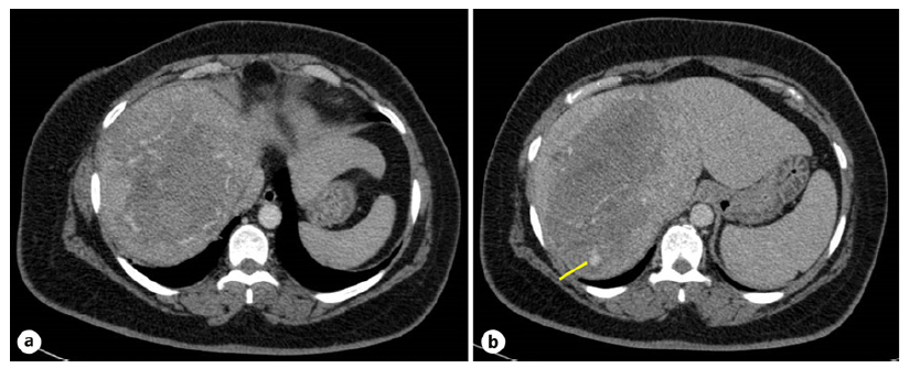
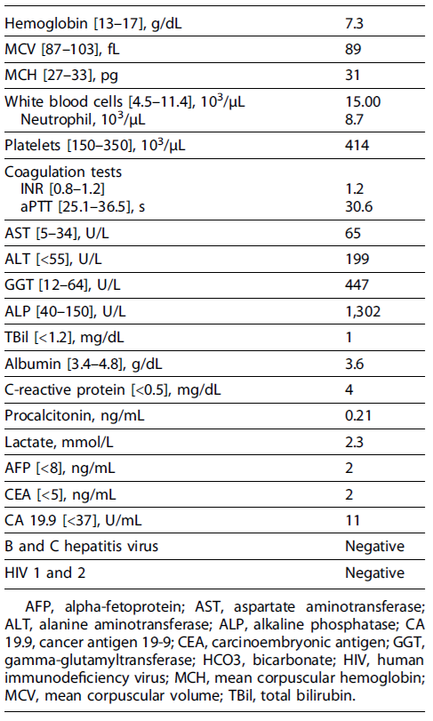
Prompt resuscitation with intravenous fluids and red blood cell units was initiated. Laboratory tests revealed a normocytic normochromic anemia with platelet count and coagulation profile within normal range and a cholestatic liver in jury (shown in Table1). Urgent abdominal CT angiography revealed 10 × 21 × 20 cm tumor of uncertain etiology in the right liver lobe with intratumoral active bleeding, with no signs of chronic liver disease (shown in Fig. 1).

Fig. 1 Abdominal contrast-enhanced CT. a A 10 × 21 × 20 cm tumor of uncertain etiology in the right liver lobe with intratumoral spontaneously hyperdense foci suggestive of blood products. b Active bleeding (arrow) in the early arterial phase in segment VII.
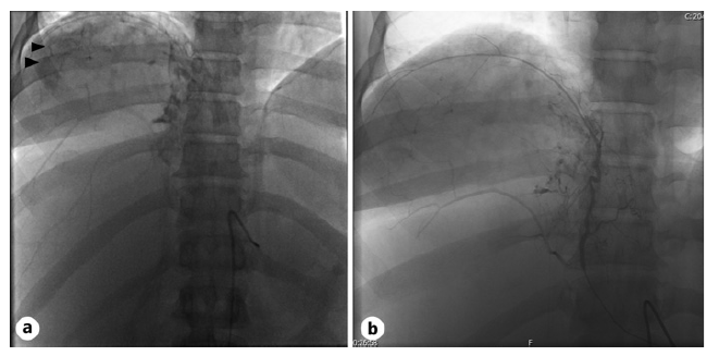
The case was discussed with Interventional Radiology and the Hepato-Biliary-Pancreatic Surgical team, and an urgent arterial embolization was performed, halting the bleeding (shown in Fig. 2). She was transfused with a total of 3 units of red blood cells, resulting in a final hemoglobin level of 8.3 g/dL.

Fig. 2 Angiography images before (a) and after (b) embolization. Angiography was used to identify the vascular supply to the tumor, with the phrenic artery showing significant vascular supply (arrows). Supra-selective arterial embolization of the right and medium hepatic and phrenic arteries with polyvinyl alcohol embolic particles was performed, with cessation of bleeding.
Further workup revealed negative serologies for hepatitis B and C virus. Regarding tumor markers, alpha-fetoprotein, carcinoembryonic antigen, and carbohydrate antigen 19-9 were within normal range (shown in Table 1). To determine etiology, an abdominal MRI with hepatobiliary-specific contrast was performed, with findings compatible with a 7.2 × 6.1 × 4.6 cm HCA, more likely inflammatory or hedgehog subtype (shown in Fig. 3). The patient was discussed in a multidisciplinary team meeting, and surgery was proposed.

Fig. 3 Abdominal MRI with hepatobiliary-specific contrast. Peripheral solid component showing enhancement on arterial phase (a), isointense (no washout) on portal phase (b), and hypointense on hepatobiliary phase (c) mass with 7.2 × 6.1 × 4.6 cm in segments VII, VIII, and I with a central hematic area secondary to the previous bleeding, suggestive of an HCA, more likely inflammatory or hedgehog subtype.
A robot-assisted laparoscopic extended right hepatectomy was performed with no complications. Histologic examination confirmed the definite diagnosis of an inflammatory HCA (shown in Fig. 4).

Fig. 4 Histologic examination. Low- (a) and high-power views (b) showing hepatocellular proliferation with trabecular growth pattern, sinusoidal dilatation, and inflammatory infiltrates. Immunohistochemistry staining of the tumor was liver fatty acid binding protein (LFABP) normal and heat shock protein 70 (HSP70) negative, excluding HCC. Furthermore, reticulin staining was intact, serum amyloid A (SAA) staining was positive (c), with normal nuclear β-catenin and glutamine synthetase staining, confirming the definite diagnosis of an HCA of inflammatory subtype.
Follow-up was uneventful, with normalization of hepatic biochemistry and an unremarkable abdominal ultrasound performed 6 months after surgery. The patient opted for a non-hormonal intrauterine device.
Discussion
HCA is a rare benign epithelial tumor of the liver with a reported prevalence of 0.001-0.004%. It occurs predominantly in women of reproductive age, with a re ported female:male ratio of 10:1 [12]. It is well established that exogenous estrogens are a risk factor for HCA, accounting for a 30-40-fold increase in the incidence of HCA, with long-term users bearing the highest risk [2, 3]. Furthermore, the incidence of HCA has increased in anabolic androgenic steroid users, noticeably in men for sport performance enhancement [2]. HCA is also associated with genetic syndromes, including glycogen storage disease type I and type III, with frequencies of 22-75%and 25%, respectively, and familial adenomatous polyposis [1, 5, 12]. Rarer associations include MODY3 diabetes and McCune-Albright syndrome [12], as well as conditions involving high levels of endogenous androgens or estrogens, like Klinefelter syndrome [13]. In recent years, metabolic syndrome and obesity are also emerging as risk factors [12].
HCA is usually solitary. However, up to half of patients may have multiple HCA, a condition termed liver adenomatosis [1, 5, 12]. This condition is associated with germline and somatic mutations in HNF-1α and MODY3 diabetes [12], although obesity, steatosis, and metabolic syndrome are also linked with hepatic adenomatosis [5, 6, 14].
Regarding genetic and pathological features, HCA is categorized into four subtypes with implications in management: HNF1-A mutations, inflammatory, β-catenin mutations, and unclassified. The clinical presentation is heterogeneous. Most HCA is found incidentally on abdominal imaging, though up to 14% of cases may exhibit abnormal serum liver tests. When they are symptomatic, the most common presentation is mild and nonspecific right hypochondrial pain, observed in 37% of patients. However, the pain may be severe as a result of bleeding [15].
While hemorrhage is an infrequent complication asso-ciatedwith hepatictumors [10], HCA ranksasthesecond most prevalent tumor connected with bleeding, following hepatocellular carcinoma (HCC), with up to 30% of HCA patients experiencing spontaneous bleeding [5, 16]. When bleeding occurs, it may be intratumoral or the tumor may rupture, leading to subcapsular or intraperitoneal hemorrhage presenting as an acute hemoperitoneum. Hemodynamic instability occurs in fewer than 10% of cases and is more commonly observed in patients with intraperitoneal bleeding rather than intratumoral bleeding, in contrast to the presented case [17]. Rupture is more likely in patients with large (>5cm), solitary, and superficial tumors. Additional risk factors include inflammatory subtype, hormone use, and pregnancy [5]. Our patient presented multiple risk factors associated with a higher bleeding risk, namely, the size, hormone use, and HCA subtype.
HCA also has a risk of malignant transformation. Surgical series report a 4-10% incidence of HCC within resected adenomas [16, 17]. Known risk factors include activating mutations in β-catenin, occurring in up to 5-10% of cases, male gender, and tumor diameter larger than 5 cm [6-8].
Concerning diagnosis, non-ruptured HCA is usually an incidental finding in abdominal imaging. Ultrasonography reveals variable echogenicity depending on the fat content, with a sensitivity of only 30%[18]. Contrast-enhanced ultrasound may help differentiate from other tumors [19]. Computed tomography can be diagnostic in cases of typical HCA, showing an isoattenuating or hypodense lesion with arterial phase enhancement, returning to near isodensity on portal venous and delayed phase images. However, only 75% of cases have typical features [20]. MRI with hepatocyte-specific contrast agents is the best imaging modality for its diagnosis with 80-90% specificity [21], allowing identification of the HCA subtype in up to 80% of patients and differentiation from other tumors [5,12].Despitethesignificant variation in MRI findings associated with HCA subtypes, the predominant observation is hypointensity on hepatobiliary sequences [5, 22]. However, β-catenin activated HCA and its distinction with unclassified HCA and HCC is not possible by any imaging technique. Furthermore, it also can be difficult to distinguish HCA from focal nodular hyperplasia in some cases [9]. Although imaging alone establishes a diagnosis in most cases of HCA, if it is inconclusive, referral to specialized centers with experts in this field is recommended. Performing a liver biopsy in this setting carries a low but non-negligible hemorrhagic risk given the vascular nature of HCA and other tumors which are part of the differential diagnosis. In a retrospective review involving 60 patients who underwent percutaneous biopsy, complications were documented in 12% of individuals, with a single episode of severe bleeding [23]. For these reasons, international guidelines suggest reserving liver biopsy for cases in which imaging is inconclusive and histology results will significantly impact treatment decisions [12, 24].
All patients should be advised to discontinue oral contraceptives, hormone-containing intrauterine devices, and anabolic steroids. Further management is dependent on gender, tumor size, the presence of symptoms, and whether bleeding or malignancy is suspected.
In non-ruptured HCA, the most relevant factors to consider are gender, size, and growth pattern. In asymptomatic nonpregnant women, discontinuing ex ogenous estrogens, control of body weight, and imaging reevaluation with contrast-enhanced MRI after 6 months are recommended. If the tumor size is or decreased to less than 5 cm, it can be managed conservatively. There is no consensus in the definition of stable disease and its follow-up. Most societies recommend interval imaging every 6 months for 12-24 months, followed by annual imaging for stable lesions [9, 12, 24]. However, if the HCA is persistently greater than 5 cm or increased in size, surgery is recommended given the risk of hemorrhage and malignancy. Furthermore, surgery is also recommended in patients who are symptomatic and male or have a proven β-catenin mutation, in cases where a biopsy was performed to establish the diagnosis. The last two are indications for surgery irrespective of tumor size given their higher risk of malignant transformation [9, 12, 24]. Surgery is the first line of therapy for curative treatment. Nonsurgical modalities are reserved for high surgical risk patients or tumors in challenging anatomical locations [9, 24].
If bleeding occurs, it is a medical emergency that requires prompt management. Intravascular volume replacement should be immediately started and an abdominopelvic CT with angiography should be performed. If active bleeding is detected, arterial embolization is preferred over surgery since it is minimally invasive and has lower morbidity. Surgery should be reserved for patients with persistent and severe hemodynamic instability or when embolization is ineffective or unavailable. When surgery is unavoidable, a damage control surgery with an abbreviated laparotomy with perihepatic packing is recommended [11]. Once the bleeding is controlled, etiology must be determined and an imaging study should be repeated since hemorrhage induces significant changes that may hinder a diagnosis in the acute setting. After establishing the diagnosis of a HCA, further management is dependent on its persistence. Surgical resection is advised if there is residual viable lesion on follow-up imaging [5, 12], as it happened in the case described. The management of HCA is determined by gender, tumor size, presentation, and progression pattern, and, importantly, requires the expertise of an experienced multidisciplinary team.


