SciELO - Scientific Electronic Library Online

 
vol.82 número3Nódulo no ombro de uma idosa índice de autoresíndice de assuntosPesquisa de artigos
Home Pagelista alfabética de periódicos  

Serviços Personalizados

Journal

Artigo

Indicadores

Links relacionados

  • Não possue artigos similaresSimilares em SciELO

Compartilhar


Portuguese Journal of Dermatology and Venereology

versão impressa ISSN 2795-501Xversão On-line ISSN 2795-5001

Port J Dermatol Venereol. vol.82 no.3 Lisboa set. 2024  Epub 21-Fev-2024

https://doi.org/10.24875/pjdv.23000105 

DERMATOLOGY IMAGES

Clues for eosinophilic fasciitis: groove sign and orange peel appearance

Pistas para fasceíte eosinofílica: sinal do sulco e aparência em casca de laranja

José Ramos1  * 

António Silva1 

Ângela Roda1 

João Alves1 

1Serviço de Dermatologia, Hospital Garcia de Orta, Almada, Portugal


A 45-year-old female was referred to dermatology with a 1-year history of progressive skin induration of the limbs. She denied joint mobility restrictions and systemic symptoms.

Clinical examination revealed symmetrical cutaneous induration and thickening of the legs, thighs, forearms, and arms with areas of orange peel-like appearance and skin depressions along the course of the superficial veins (“groove sign”) (Fig. 1A and B). Hands and feet were spared. Raynaud’s phenomenon was absent, and nail fold capillaroscopy was unremarkable. Laboratory findings demonstrated blood eosinophilia (1.9 × 10⁹/L) and increased erythrocyte sedimentation rate (120 mm/h).
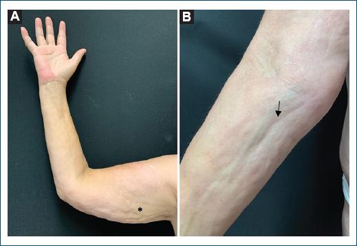
Figure 1 Cutaneous induration and thickening of the right arm and forearm, with a “pseudo-cellulite” appearance on the arm (A: asterisk) and “groove sign” on the forearm (B: arrow). 

A full-thickness incisional biopsy was consistent with eosinophilic fasciitis, establishing the diagnosis (Fig. 2). The patient started treatment with prednisolone 1 mg/kg/day. However, considering the poor response, recently, weekly methotrexate was added.
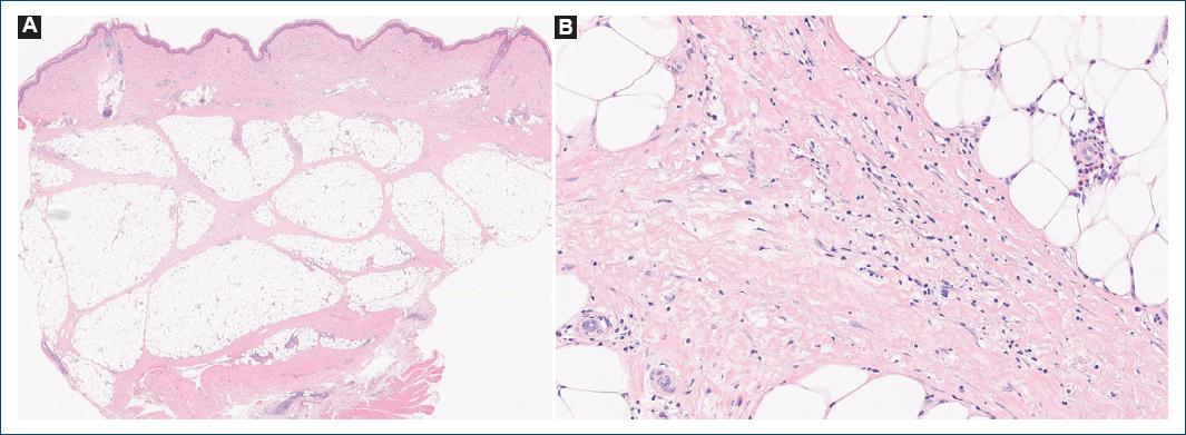
Figure 2 A full-thickness incisional biopsy showed hypodermis septa thickness with a lymphohistiocytic infiltrate, plasma cells, and occasional eosinophils extending into the lower dermis (H & E, magnification: A: ×1; B: ×40). 

Eosinophilic fasciitis is a rare fibrosing disorder of muscle fascia of unknown etiology. Clinically, areas of orange peel-like (“pseudo-cellulite”) and linear depressions along the course of the superficial veins (“groove sign”) are characteristic1. This last physical finding is probably due to the relative sparing of the epidermis and superficial dermis around the vessels by the fibrotic process compared to the deep tissue2. A full-thickness biopsy including the fascia and/or magnetic resonance showing increased signal intensity within the fascia is crucial for diagnosis. The first line of treatment is systemic corticosteroids, which may be associated with corticosteroid-sparing agents, like methotrexate3.

Recognizing the clinical clues of eosinophilic fasciitis is important, as prompt diagnosis and treatment are essential to prevent the development of joint contractures, which are responsible for the high morbidity of this condition.

References

1. Asaoka K, Watanabe Y, Itoh K, Hosono N, Hirota T, Ikawa M, et al. A case of eosinophilic fasciitis without skin manifestations:a case report in a patient with lupus and literature review. Clin Rheumatol. 2021;40:2477-83. [ Links ]

2. Camard M, Maisonobe T, Flamarion E. The groove sign in eosinophilic fasciitis. Clin Rheumatol. 2022;41:3919-20. [ Links ]

3. Jinnin M, Yamamoto T, Asano Y, Ishikawa O, Sato S, Takehara K, et al. Diagnostic criteria, severity classification and guidelines of eosinophilic fasciitis. J Dermatol. 2018;45:881-90. [ Links ]

FundingNone.

Ethical disclosures

Protection of human and animal subjects. The authors declare that no experiments were performed on humans or animals for this study.

Confidentiality of data. The authors declare that no patient data appear in this article. Furthermore, they have acknowledged and followed the recommendations as per the SAGER guidelines depending on the type and nature of the study.

Right to privacy and informed consent. The authors have obtained the written informed consent of the patients or subjects mentioned in the article. The corresponding author is in possession of this document.

Use of artificial intelligence for generating text. The authors declare that they have not used any type of generative artificial intelligence for the writing of this manuscript nor for the creation of images, graphics, tables, or their corresponding captions.

Received: December 17, 2023; Accepted: January 27, 2024

*Correspondence: José Ramos E-mail: jalramos@campus.ul.pt

Conflicts of interest

None.

Creative Commons License Portuguese Society of Dermatology and Venereology. Published by Permanyer. This is an open access article under the CC BY-NC-ND license