


 English (pdf)
 Article in xml format
 Article references
 Send this article by e-mail
 Cited by SciELO
  Similars in
SciELO
Similars in
SciELO 
 Permalink
Case
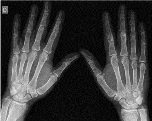
A 54-year-old male with a past medical history of cutaneous sarcoidosis (Lupus Pernio) was admitted to the rheumatology consultation complaining of pain, tumefaction, and functional limitation of the second finger of the left hand.
Radiographic imaging of the hands (Fig. 1) revealed a lace-like pattern with multiple well-defined cyst-like lesions in the phalanges, with greater expression in the distal and mild ones. A soft tissue thickness was observed in the left index finger (known as “sausage dactylitis”). The combination of clinical and radiologic features in the hands is virtually diagnostic of osseous sarcoidosis (Perthes disease).
Discussion
Osseous involvement in sarcoidosis occurs in 1-13% of patients, with an estimated average occurrence of 5%.1 It is more frequent in patients of African descent and it is usually accompanied by infiltrative skin lesions (i.e. Lupus Pernio).1,2
Bone lesions are frequently asymptomatic, although some patients present with symptomatic dactylitis (swelling, pain and stiffness).3
Osseous sarcoidosis can involve any bone from the axial to the appendicular skeleton. Nevertheless, the phalanges in the hands and feet are most frequently affected.2
Radiologic features usually include multiple cystic lesions, sometimes resulting in a typical lacy pattern (these classic lesions, which affect the hands/feet, are known as Perthes disease/Jungling’s disease).1
There are three radiological types of sarcoid bone disease: type I, big cystic lesions (rare); type II, multiple small cysts; type III, tunneling of the cortex of the phalanges, which leads to remodeling of the cortical and trabecular architecture.1
This case showed that the combination of radiologic features in the hands/feet is virtually diagnostic for osseous sarcoidosis, in a patient with a history of Lupus Pernio.

