


 English (pdf)
 Article in xml format
 Article references
 Send this article by e-mail
 Cited by SciELO
  Similars in
SciELO
Similars in
SciELO 
 Permalink
Case
We present a case of a 57-year-old male, previous athlete, who was referred to our Radiology Department to perform a left hip MRI to further study a femoroacetabular impingement previously diagnosed on conventional radiography.
As an incidental finding, a well-defined mass measuring 22x18x21 mm was detected, arising from the symphysis pubis joint space and projecting into its posterosuperior side. It demonstrated high signal intensity (SI) on Proton Density sequences, heterogeneous high SI on STIR-weighted sequences and low SI on T1-weighted sequences. No signal abnormality was seen in the adjacent pubic bone marrow or in the surrounding tissues. The lesion abuts the prostate but without causing compression of the gland (Fig. 1).
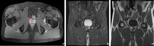
Figure 1: PD-weighted MR image in axial plane (a) and STIR-weighted MR image in coronal (b) plane showing a well-defined hyperintense lesion (red arrow) in the space of Retzius, abbuting the prostate gland and in contiguity with the symphyseal cartilage with a connecting stalk (blue arrowhead). T1-weighted MR image (c) shows the lesion as isointense to muscle and in contiguity with the symphyseal cartilage.
Discussion
Retropubic cysts are rare benign lesions, much more common in women due to its association with pregnancy, vaginal delivery and pelvic trauma.1,2 In fact, our case appears to be one of the few reported cases in male patients.
The pubic symphysis joint is composed by a fibrocartilaginous disc. Retropubic cysts consist of a collagenous capsule containing fibrocartilaginous tissue with extensive mucinous cystic degeneration as a of repeated mechanical stress.1,2,3They are also called supra- or subpubic cysts depending on their relationship to the pubic symphysis.1
Retropubic cysts are primarily located in the space of Retzius between the anteroinferior bladder wall and pubic symphysis and are asymptomatic in the early stages.1 As they grow in size, they may produce symptoms according to their location by compressing adjacent structures.1,2
They appear hypo- to iso-intense on T1-weighted imaging and hyperintense on T2. Smooth rim enhancement may be seen post-contrast images.1
MRI demonstrates the cystic nature of the lesion and its origin from the symphysis pubis allowing differential diagnosis from other entities such as Bartholin’s or Gartner duct cysts specially in symptomatic patients.1,2 It may also be an incidental finding such as in our case, revealing particular value in cases of oncologic surveillance, making distinction of metastatic disease crucial.1,2
As these lesions are benign, patient management should be guided by patient’s symptoms. Surgical resection may be an option for symptomatic patients.1,2
In conclusion, pubic cartilaginous cysts are slow-growing degenerative lesions, more common in post-menopausal women but that may also occur in men.1,2 Its MRI characteristics and origin from the pubic symphysis allow a confident diagnosis and proper patient management.1,2,3

