English (pdf)
 Article in xml format
 Article references
 Send this article by e-mail
 Cited by SciELO
  Similars in
SciELO
Similars in
SciELO 
 Permalink
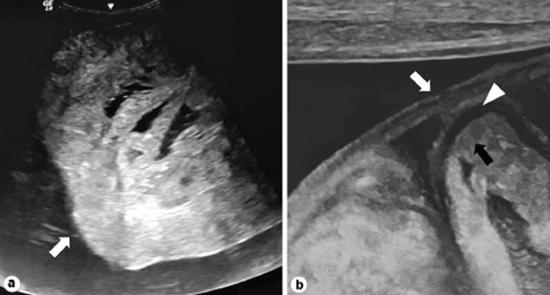
A 50-year-old male presented with vomiting and abdominal pain for 3 days. He is a known case of chronic renal failure for the past 2 years undergoing repeated peritoneal dialysis. On physical examination, his abdomen was distended. Laboratory investigation showed increased serum creatinine levels ∼6 mg/dL. Ultrasound abdomen showed shrunken kidneys with increased cortical echoes and ascites. Apart from that, thickened peritoneum or a membrane overlying the bowel loops was seen on high-frequency ultrasound as a triple-layer appearance, which is called “ultrasound trilaminar sign” (Fig. 1a, b) [1]. The “ultrasound trilaminar sign” is considered characteristic of the abdominal cocoon or encapsulating peritoneal sclerosis (EPS) [2]. EPS is characterized by clustered small bowel loops with narrow base and surrounding thick fibro-collagenous membrane. It may be idiopathic or secondary to repeated peritoneal dialysis, abdominal tuberculosis, abdominal surgeries, and drugs like propranolol [1, 3]. The three layers forming the ultrasound trilaminar appearance are the superficial hyperechoic peritoneal membrane, a middle hypoechoic layer of the bowel wall, and the deep hyperechoic layer produced by the bowel gas or contents [1]. This trilaminar appearance is not to be confused with computed tomography trilaminar sign, which represents submucosal bowel edema [1]. Presence of ascites is usually essential for identifying the trilaminar appearance and cauliflower-like appearance of clustered bowel loops. The patient was advised surgical evaluation for removal of membrane and adhesiolysis.
.