SciELO - Scientific Electronic Library Online

 
vol.82 issue2Disseminated cutaneous leishmaniasis due to Leishmania guyanensis in an infant author indexsubject indexarticles search
Home Pagealphabetic serial listing  

Services on Demand

Journal

Article

Indicators

Related links

  • Have no similar articlesSimilars in SciELO

Share


Portuguese Journal of Dermatology and Venereology

Print version ISSN 2795-501XOn-line version ISSN 2795-5001

Port J Dermatol Venereol. vol.82 no.2 Lisboa June 2024  Epub Dec 20, 2023

https://doi.org/10.24875/pjdv.23000085 

DERMATOLOGY IMAGES

Following the path: an unusual location for cutaneous larva migrans

Seguindo o caminho: uma localização incomum para larva migrans cutânea

Miguel Santos-Coelho1  * 

Joana A. Barbosa1 

Juliana Baptista1 

1Department of Dermatology and Venereology, Hospital de Santo António dos Capuchos, Centro Hospitalar Universitário de Lisboa Central, E.P.E., Alameda Santo António dos Capuchos, Lisbon, Portugal


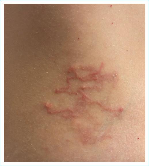
A 28-year-old woman with no relevant past medical history presented to the Dermatology department with pruriginous, slowly growing lesions on her right abdominal flank. The lesions had been first noted a week earlier, after returning home from a trip to Mexico. She denied other symptoms.

Clinical examination revealed several erythematous papules and plaques with a serpiginous distribution affecting the right abdominal flank (Fig. 1). The remaining physical examination was unremarkable. Based on clinical presentation and considering the recent travel history, a diagnosis of cutaneous larva migrans was made.

Figure 1 Lesions of the right abdominal flank. 

The patient was treated with single dose oral ivermectin (200 mcg/kg), with full resolution of lesions at 1-month follow-up.

Cutaneous larva migrans is an infection caused, most frequently, by Ancylostoma braziliense (a hookworm transmitted by soil contaminated with feces of cats or dogs)1,2. Although worldwide distributed (and with some reported autochthonous cases in Europe3), infections are more frequently found in tropical/subtropical countries in Central and South America and Asia1.

After penetrating the skin, the larva undergoes epidermal migration, giving rise to the characteristic clinical appearance. The lower limbs (especially the feet) are the most common locations for lesions, but the trunk, as in our patient, can be affected in up to 7% of cases4. Exceptionally, hematogenous dissemination to the lungs can occur4.

Although self-limited, treatment with oral albendazole or ivermectin relieves pruritus and decreases the chance of bacterial superinfection1. In cases where oral therapy is not possible, patients can be treated with topical treatment using ivermectin 1% or thiabendazole 15% cream3,5.

References

1. Feldmeier H, Schuster A. Mini review:hookworm-related cutaneous larva migrans. Eur J Clin Microbiol Infect Dis. 2012;31:915-8. [ Links ]

2. Purdy KS, Langley RG, Webb AN, Walsh N, Haldane D. Cutaneous larva migrans. Lancet. 2011;377:1948. [ Links ]

3. Blaizot R, Goiset A, Caumes E, Gabriel F, Milpied B. Cutaneous larva migrans:a case in Bordeaux, France and a systematic review of locally acquired cases in Europe. Eur J Dermatol. 2017;27:426-9. [ Links ]

4. French SJ, Lindo JF. Severe cutaneous larva migrans in a traveler to Jamaica, West Indies. J Travel Med. 2003;10:249-50. [ Links ]

5. Gerbig AW, Kempf W. Topical treatment of cutaneous larva migrans with ivermectin 1. Int J Dermatol. 2020;59:e21-2. [ Links ]

FundingNone.

Ethical disclosure

Protection of human and animal subjects. The authors declare that no experiments were performed on humans or animals for this study.

Confidentiality of data. The authors declare that they have followed the protocols of their work center on the publication of patient data.

Right to privacy and informed consent. The authors have obtained the written informed consent of the patients or subjects mentioned in the article. The corresponding author is in possession of this document.

Use of artificial intelligence for generating text. The authors declare that they have not used any type of generative artificial intelligence for the writing of this manuscript, nor for the creation of images, graphics, tables, or their corresponding captions.

Received: October 16, 2023; Accepted: October 30, 2023

*Correspondence: Miguel Santos-Coelho E-mail: mscoelho.derma@gmail.com

Conflicts of interest

None.

Creative Commons License Portuguese Society of Dermatology and Venereology. Published by Permanyer. This is an open access article under the CC BY-NC-ND license