


 English (pdf)
 Article in xml format
 Article references
 Send this article by e-mail
 Cited by SciELO
  Similars in
SciELO
Similars in
SciELO 
 Permalink
Tumors of the terminal ileum are rare, and their diagnosis and treatment standards have not been established. The effectiveness of underwater endoscopic mucosal resection (UEMR) in the duodenum and colon has been reported [1, 2]; however, its effectiveness in the small intestine is rarely reported [3]. We present a case of an ileum adenoma diagnosed using magnifying endoscopy and completely resected using UEMR (online supple-mentary video; for all online suppl. material, see https://doi.org/10.1159/000531774).
A 56-year-old woman with a lesion in the terminal ileum was referred to our hospital for treatment. Colono-scopy revealed a 10 mm slightly depressed lesion with marginal elevation in the terminal ileum (Fig. 1a, b). Magnifying narrow-band imaging showed a tubular sur-face pattern with regular vessels on the slightly elevated marginal area and regular brown vessels on the slightly depressed central area (Fig. 1c). Magnifying chromoendoscopy using crystal violet staining showed a branch-like or gyrus-like pattern on the marginal area and a roundish and tubular structure on the central area (Fig. 1d). The lesion was diagnosed as an adenoma in the terminal ileum, similar to a colonic adenoma; hence, we decided to perform UEMR. The lesion was completely removed using a 10 mm snare (10 mm, Captivator Ⅱ;BostonScientific, Marlborough, MA, USA) with an electric generator (Endocut Q effect 2, interval 1, duration 4; VIO 300D; ERBE, Tübingen, Germany) (Fig. 2). No intra- or post-operative complications occurred. The pathological diagnosis revealed that it was an intestinal-type low grade adenoma with negative margins. At the margins of the lesions, the tumor tended to form a villous structure, while the center appeared tubular with a relatively flat surface (Fig. 3). These histopathological findings were consistent with the magnifying endoscopic findings.
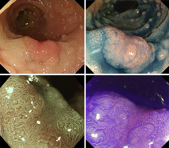
Fig. 1 Endoscopic images showing a 10 mm slightly depressed lesion with mar-ginal elevation in the terminal ileum, sug-gesting type 0-Ⅱa in the Paris classification: in white light (a); chromoendoscopy using indigo carmine (b). c Magnifying narrow-bind imaging: a tubular surface pattern with regular vessels on the marginal elevation area and regular brown vessels on the slightly depressed area, suggesting the Narrow-Band Imaging International Colorectal Endoscopic (NICE) classification type 2. d Magnifying chromoendoscopy image using crystal violet staining showing a pit pattern like type Ⅳ on the marginal area and type ⅢL on the slightly depressed area.
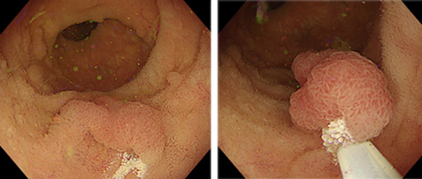
Fig. 2 After immersing the terminal ileum with normal saline and floating the lesion in it, we performed UEMR.
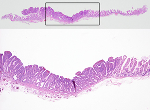
Fig. 3 Histological examination (hematoxylin and eosin stain): A villous structure at the margins of the lesions and a tubular structure with a relatively flat surface at the center. The lesion size was 13 × 9 mm.
Because of the limited luminal space in the terminal ileum, it might be difficult to completely remove a lesion using conventional endoscopic mucosal resection, especially in cases where submucosal injection would prevent snaring. UEMR allows for easier and complete capture of the lesions by suctioning the lumen air and filling it with water, thereby making flat lesions smaller and more polypoid. Therefore, UEMR may be an effective method for terminal ileum tumor resection.

