


 Inglés (pdf)
 Articulo en XML
 Referencias del artículo
 Enviar articulo por email
 Citado por SciELO
  Similares en
SciELO
Similares en
SciELO 
 Permalink
Castleman Disease: atypical cause of pneumonectomy
A previously healthy 24-year-old male, smoker (4 pack- year), presented with a traumatic fracture of the right humerus. Preoperative chest radiography revealed a left perihilar lesion (Figure 1A). He had no respiratory or constitutional symptoms. The physical examination was unremarkable. Laboratory data, including neuron-specific enolase and chromogranin, were normal. Chest computed tomography showed a rounded left perihilar lung mass, well-defined, with 45x40mm and slight contrast uptake (Figure 1B and C). Positron emission tomography revealed increased fluorodeoxyglucose-F18 uptake (SUVmax:5,4) in the left hilar lesion. 68Ga-DOTA-NOC PET-CT also showed an abnormal uptake from the nodular formation in the left pulmonary hilum, suggesting a neuroendocrine tumor. Endobronchial ultrasound-guided transbronchial needle aspiration (EBUS-TBNA) was performed with punction of the left mass; TBNA samples were negative for malignancy. Lung function was normal. He was therefore referred for evaluation of thoracic surgery.
Given the tumor’s central location with major pulmonary vessels and main bronchus involvement, he underwent left pneumonectomy. Hematoxylin and eosin staining showed regressed germinal centers with follicular dendritic cell prominence, surrounded by mantle zones containing small lymphocytes arranged in a concentric pattern (Figure 1D). Microscopic features and immunostaining were consistent with Castleman Disease-Hyaline Vascular Variant. The patient received no further therapy, maintaining regular surveillance.
Unicentric Castleman Disease (UCD) frequently presents as an incidental solitary mediastinal mass, however, intrapulmonary location with the hilum involvement is rare. The preoperative diagnosis can be challenging as clinical and radiological findings are nonspecific.1,2The standard treatment for UCD is complete surgical resection.3 This case emphasizes that although UCD with hilar-presentation is a rare and benign condition, anatomic resection and even a pneumonectomy may be required for diagnostic and therapeutic purposes.
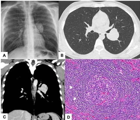
Figure 1: A: PA Chest radiograph showing a left hilar lesion. B and C: Axial (lung window) and coronal (mediastinal window) chest CT, respectively, revealing a rounded and well-defined left perihilar mass with mild contrast uptake. D: Hematoxylin and Eosin (H-E) staining revealing an enlarged follicle with concentric layering of mantle zone lymphocytes (arrows) encircling an atretic germinal center (x200).

