Serviços Personalizados
Journal
Artigo
Indicadores
Links relacionados
Compartilhar
Corrosão e Protecção de Materiais
versão impressa ISSN 0870-1164
Corros. Prot. Mater. vol.32 no.4 Lisboa dez. 2013
ARTIGO
Optimization of an Epifluorescence Microscopy Technique for rapid quantification of bacteria asociated to corrosion products in produced water
Otimização de uma técnica de microscopia de epifluorescência para a quantificação rápida de bactérias associadas a produtos de corrosão em água de produção
D. Silva(1), M. Gonçalves and A. Costa (*)
(1) Universidade do Estado do Rio de Janeiro, PPG-EQ, Instituto de Química, Rua São Francisco Xavier 524, Maracanã, CEP:20550-900, Rio de Janeiro, Brazil, E-mail: d.silv@yahoo.com.br, marciamg@uerj.br
(*) Correspondig author, e-mail: acosta@uerj.br
ABSTRACT
Microbial cells constitute a severe problem for the petroleum industry. Among the microorganisms, the general anaerobic bacteria (GAnB) are the most common, which includes the sulfate-reducing bacteria (SRB). Nowadays, the GAnB are enumerated by the most probable number (MPN), providing the final quantification after 28 days. In this work, a method for the rapid quantification of bacteria in the produced water from oil extraction was optimized using epifluorescence microscopy with acridine orange dye. This method provided a more accurate estimate of the number of GAnB when compared with enumeration by the MPN technique. Furthermore, it is performed in just 30 minutes per sample.
Keywords: Fluorescence Microscopy, Acridine Orange, Anaerobic Bacteria, Microbial Quantification, Produced Water from Oil
RESUMO
As células microbianas constituem um problema sério para a indústria do petróleo. De entre os microrganismos, as bactérias anaeróbias totais (BAnT) são as mais comuns, incluindo as bactérias redutoras de sulfato (BRS). Atualmente, as BAnT são enumeradas pela técnica do número mais provável (NMP), permitindo a quantificação final após 28 dias. Neste trabalho, um método para quantificação rápida de bactérias em água de produção da extração de óleo foi otimizado empregando-se microscopia de epifluorescência com o corante laranja de acridina. Este método permitiu uma estimativa mais precisa do número de BAnT quando comparado com a enumeração de células pela técnica do NMP. Além disso, é uma técnica que pode ser conduzida em apenas 30 minutos por amostra avaliada.
Palavras-chave: Microscopia de Fluorescência, Laranja de Acridina, Bactérias Anaeróbias, Quantificação Microbiana, Água de Produção de Petróleo
1. INTRODUCTION
The extraction of oil and gas at offshore platforms is followed by a significant amount of waste called produced water. The composition of this water is very complex and highly variable. It consists of water, inorganic and organic constituents, microorganisms, corrosion products, oil and gas [1]. Among the microorganisms found in these samples, the general anaerobic bacteria (GAnB) are the most common, which includes the sulfate-reducing bacteria (SRB) [2]. The detrimental effects of the SRB primarily consist of souring the oil and gas deposits and contributing to microbiologically influenced corrosion (MIC) [3]. The cost of controlling the bacterial activity in produced water is significant at an annual estimate of US$150,000.00 per platform when only biocides are used [4]. Thus, there have been considerable efforts to develop techniques for the quantification of primarily anaerobic bacteria to provide early diagnosis of problems and allow the establishment of effective prevention and control measures [5].
Culture methods based on the most probable number (MPN) technique have been extensively used for several decades and remain the standard methods for enumerating GAnB. An alternative method is the direct counting of bacteria by fluorescence microscopy that is commonly used in environmental studies, which enumerates the microbial cells using fluorescent dyes [6,7]. The direct count usually exceeds the counts obtained by the MPN method because the accuracy of the results obtained by direct count does not vary with the nutritional requirements of microorganisms. Another advantage to the direct count against MPN method is that it provides the results in a short period of time.
The aim of this work was to optimize a methodology for rapid quantification of bacterial cells in produced water from oil by epifluorescence microscopy with acridine orange (AO) dye. Additionally, the results obtained were compared with the quantification of GAnB obtained by the MPN method.
2. MATERIALS AND METHODS
Initially, an adjustment of epifluorescence microscopy methodology with AO dye was developed for total microbial count of cells from saline samples. In a second step, the quantifications of GAnB were made using the conventional MPN method.
Ten produced water samples were collected from a single tank of water/oil located in the State of Rio de Janeiro, Brazil over a period of two months at different dates and were stored at 4 oC, during one week, until the tests were performed.
2.1 Optimization of the fluorescence microscopy method
The samples were prepared for fluorescence microscopy by several steps including dilution, fixation, staining and visualization of microorganisms [6]. In all tests, we used a 1:100 dilution, in which a large number of cells could be observed.
Different diluents were tested in order to identify the most suitable diluent to be used in serial dilutions of the sample prior to staining, including ultra-pure water, synthetic sea water [8] and a NaCl solution (0.85 % w/v). All solutions were autoclaved at 121 oC for 15 min and filtered (0.22 µm pore). Glutaraldehyde was tested as a fixative in a range of concentrations (0.1 %, 0.5 %, 1.0 %, 1.5 %, 2.0 % and 2.5 % (v/v)). To determine the optimal volume of acridine orange solution (0.1 % m/v) to use during the staining process, 1 to 10 mL of the dye solution was applied to the sample. Additionally, the optimal volume of the stained sample to use for filtration was determined by testing volumes from 1 to 10 mL. The entire stained sample was filtered through a 0.22 µm black polycarbonate membrane, 25 mm in diameter (Millipore), using a vacuum manifold and it was rinsed with 2.0 mL of ultra-pure water to remove excess AO from the membrane.
Subsequently, the membranes were mounted on glass microscope slides with one drop of immersion oil free self-fluorescence (Olympus, Brazil) below and above the filter, and a coverslip was placed on top of the filter. The slides were stored in the dark at 4 oC and examined the same day that they were prepared to minimize the loss of AO fluorescence. A Carl Zeiss Axioskop epifluorescence microscope was used; it was equipped with filter UV BP 450-490 (excitation) and LP 515 (emission) and an Axiocam MRc 5 camera. The images were processed using the imaging program AxioVision, version 4.8.1 (Carl Zeiss).
2.2 Direct count by fluorescence microscopy
For direct count, all samples were serially diluted 10-fold to 10-3. The final dilution was performed in a 50 mL tube c ontaining 9 mL of solution, and the sample w as then shaken thoroughly on a vortex mixer (IKA Model Lab Dancer). Subsequently, the fixative solution and the AO solution were added to the sample, and the sample was incubated for a minimum of 5 min in the dark f or staining. An aliquot of the stained sample w as filtered through a membrane and rinsed with ultra-pure water. After staining, the membranes were mounted on a glass slide and st ored in the dark at 4 oC. All of the samples were counted on the same day they were prepared, using an ocular grid at a magnification of 1000x. The total bacterial number was based on counts in six randomly selected fields of the membrane surface, but only 25 squar es of the grid in each field were counted to minimize quenching. The equation used to calculate the number of bacteria in the samples has been previously reported by Kepner and Pratt [9].
2.3 Enumeration of GAnB by Most Probable Number (MPN) Method
The GAnB were enumerated by the most probable number (MPN) method (n=3) using culture medium prepared in anaerobic conditions with synthetic sea water (ASTM D1141-98, 2008) as previously described by Luna et al. [5]. Oxygen depletion was achieved by introducing nitrogen gas. This medium had the following composition (per Liter): Glucose, 5.0 g; peptone, 4.0 g; yeast extract, 1.0 g; rezasurin 4 mL (0.025 % m/v). The pH was adjusted to 7.6 using NaOH 1 M and the sterilization of the medium was performed in an autoclave at 121 °C for 20 minutes. After sterilization, the thioglycolate solution (12.4 g/L) was added to the medium in aliquots of 0.1 mL before inoculation.
3. RESULTS AND DISCUSSION
3.1 Optimization of direct count using orange acridine dye (AO)
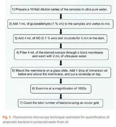
The experiments established an optimal methodology for the rapid quantification of bacterial cells in produced water from oil by epifluorescence microscopy with acridine orange dye. figure 1 shows the optimized method.

The use of ultra-pure water as diluent allowed obtaining images with lower background fluorescence compared to the other diluents tested (synthetic sea water and saline solution 0.85 % m/v). A concentration of 1% v/v of glutaraldehyde yielded the best results for sample fixation; it showed a significant effect on the number of microorganisms fixed in relation to other concentrations studied (0.1 % to 2.5 % v/v). Furthermore, an excess of glutaraldehyde may contribute to the deterioration of the fluorescence emitted by the cells, probably because it is toxic at higher concentrations [10]. A similar glutaraldehyde concentration was identified as optimal for the enumeration of protozoa by Stevik et al. [11].
The best staining results were obtained by using 4 mL of the AO solution (0.1 % w/v) to stain the sample. With this amount of dye, there was no background fluorescence, and the microorganisms were clearly distinguishable over the black membrane, which facilitated subsequent quantification.
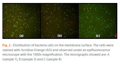
Additionally, the tests showed that a volume of 4 mL of stained sample resulted in an optimal distribution of bacteria on the membrane surface (Figure 2). The time required to directly count the bacteria using the optimized method (Figure 1) was 30 min per sample. However, the operator must be familiar with epifluorescence microscopy to count the samples.

3.2 Direct count versus MPN
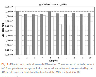
The method optimized in this study was used to determine the number of total bacterial cells in produced water from oil (Figure 3). Statistical analysis applying Grubbs test at P<0.05 was used for identification and rejection of outliers on the results presented in Figure 3. Regarding the results from the direct count method, outliers were not detected. However, for MPN method, the results for sample 2 proved to be an outlier (Grubbs test: G=2.48).

The quantification of the GAnB by MPN (Figure 3) shows that these results are included in the range of the number of bacteria enumerated by the AO direct counting method (108 to 109 cells/mL). It means that approximately 80 to 90 % of total bacterial cells in produced water from oil are anaerobic (GAnB), not considering the sample 2 (outlier). These results are of concern because the SRB are included within GAnB group, and they are considered the most problematic microbial group for the petroleum of industry in economic terms [3].
Unfortunately, the AO fluorochrome is not specific to any type of microorganisms because it binds to both DNA and RNA [12]. For a more specific quantification of different microbial groups, epifluorescence microscopy can be combined with other methods such as fluorescent in situ hybridization (FISH) [13]. In these cases, fluorescent dyes, such as AO or DAPI, are used for the rapid quantification of total bacterial cells, but the quantification and identification of specific microbial groups is performed using specific nucleic acid probe with the dyes to a particular genus [14-16]. Therefore, this method should be employed for samples with previous knowledge of the target microbial community of interest [17,18].
4. CONCLUSION
It was established an optimal methodology for the rapid quantification of bacterial cells in produced water from oil by epifluorescence microscopy with acridine orange dye.
One of the great advantage of this methodology is a short execution time (30 minutes) while the MPN technique takes about 28 days. This time difference of the technique of fluorescence microscopy is of great importance for the oil industry, early diagnosis of problems allow to establish an effective prevention and control measures, avoiding unnecessary expenses.
This optimized method was applied in direct count of total bacterial cells derived from saline samples with success. However, the limitation of this technique was the lack of specificity to distinguish any kind of microorganisms.
REFERENCES
[1] A. Fakhrul-Razi, A. Pendashteha, A. C. Luqman, D. R. A. Biaka, S. S. Madaeni and Z. Z. Abidina, J. Hazard. Mater., 170, 530 (2009). [ Links ]
[2] M. T. Madigan, J. M. Martinko D. A. Stahl and D. P. Clark. Brock Biology of Microorganisms, Thirteenth Ed., San Francisco: Pearson Education - Benjamin Cummings, p. 1152 (2010). [ Links ]
[3] W. A. Hamilton and W. Lee (Biocorrosion) in Biotechnology Handbook 8 - Sulfate Reducing Bacteria (L.L. Barton, ed.), Plenum Press, New York, USA, p. 243 (1995). [ Links ]
[4] S. Maxwell, K. Mutch, P. Charlton, P. Badalek and G. Hellings (In- Field Biocide Optimization for Magnus Water Injection System) in Proceedings of the Corrosion 2002 (Nace Internationals Annual Conference - Paper 02031, 2002), Denver, NACE international, USA, p. 1-12 (2002). [ Links ]
[5] A. S. Luna, A. C. A. da Costa, M. M. M. Gonçalves and K. Y. M. Almeida, Appl. Biochem. Biotechn., 147, 77 (2008). [ Links ]
[6] S. M. McMath, A. Delanove, D. M. Holt, A. H. L. Chamberlain and B. J. Lioyd, Water and Environment Journal,12, 113 (1998). [ Links ]
[7] H. L. Gough and D. A. Stahl, J. Microbiol. Meth., 52, 39 (2003). [ Links ]
[8] ASTM D1141-98. Standard Practice for the Preparation of Substitute Ocean Water, ASTM standards, American Society for Testing and Materials, Pennsylvania, USA (2008). [ Links ]
[9] R. L. Kepner and J. Pratt, Microbiol. Rev., 58, 603 (1994). [ Links ]
[10] M. O. B. O. Pereira (Comparação da eficácia de dois biocidas (carbamato e glutaraldeído) em sistemas de biofilme), dissertação apresentada para a obtenção do grau de doutor, Departamento de Engenharia Biológica, Universidade do Minho, Portugal (2001). [ Links ]
[11] T. K. Stevik, J. F. Hansen and P. D. Jenssen, J. Microbiol. Meth., 33, 13 (1998). [ Links ]
[12] G. A. McFeters, F. P. Yu, B. H. Pyle and P. S. Stewart, J. Microbiol. Meth., 21, 1 (1995). [ Links ]
[13] S. Lucker, D. Steger, K. U. Kjeldsen, B. J. MacGregor, M. Wagner and A. Loy, J. Microbiol. Meth., 69, 523 (2007). [ Links ]
[14] T. Hug, W. Gujer and H. Siegrist, Water Res., 39, 3837 (2005). [ Links ]
[15] B. Icgen and S. Harrison, Res. Microbiol., 157, 922 (2006). [ Links ]
[16] A. F. J. Santos, L. L. F. Batista, J. B. T. Lima, R. C. C. Almeida, M. R. A. Roque and P. F. Almeida, Chemical Engineering Transactions, 20, 139 (2010). [ Links ]
[17] J. S. Hirasawa (Avaliação da metanogênese e sulfatogênese na presença de oxigênio, sob diferentes relações etanol/sulfato, utilizando técnicas de biologia molecular), dissertação apresentada para a obtenção do grau de doutor, Escola de Engenharia São Carlos, Universidade de São Paulo, Brasil (2007). [ Links ]
[18] D. M. Levine, M. L. Berenson, T. C. Krehbiel and D. F. Stephan (Statistics for Managers using MS Excel), 6th Ed., Prentice Hall, New Jersey, USA, p.840 (2010). [ Links ]
ACKNOWLEDGEMENTS
The authors thank Igor de Almeida Campos Lima for his help with the statistical analysis. This study was supported by grants from the Brazilian Government (CNPq and Faperj).
Artigo submetido em Maio de 2013 e aceite em Outubro de 2013


