Serviços Personalizados
Journal
Artigo
Indicadores
-
 Citado por SciELO
 -
 Acessos
Links relacionados
-
 Similares em
SciELO
Compartilhar
Portuguese Journal of Nephrology & Hypertension
versão impressa ISSN 0872-0169
Port J Nephrol Hypert vol.26 no.4 Lisboa out. 2012
C4d detection in renal allograft biopsies: immunohistochemistry vs. immunofluorescence
Ana Santos1, Helena Viana1, Maria João Galvão1, Fernanda Carvalho1, Fernando Nolasco2
1 Laboratório de Morfologia Renal, Lisbon, Portugal.
2 Serviço de Nefrologia, Hospital Curry Cabral, Lisbon, Portugal.
ABSTRACT
Introduction. Peritubular capillary complement 4d staining is one of the criteria for the diagnosis of antibody-mediated rejection, and research into this is essential to kidney allograft evaluation. The immunofluorescence technique applied to frozen sections is the present gold-standard method for complement 4d staining and is used routinely in our laboratory.
The immunohistochemistry technique applied to paraffin-embedded tissue may be used when no frozen tissue is available.
Material and Methods.The aim of this study is to evaluate the sensitivity and specificity of immunohistochemistry compared with immunofluorescence. We describe the advantages and disadvantages of the immunohistochemistry vs. the immunofluorescence technique. For this purpose complement 4d staining was performed retrospectively by the two methods in indication biopsies (n=143) and graded using the Banff 07 classification.
Results.There was total classification agreement between methods in 87.4% (125/143) of cases. However, immunohistochemistry staining caused more difficulties in interpretation, due to nonspecific staining in tubular cells and surrounding interstitium. All cases negative by immunofluorescence were also negative by immunohistochemistry. The biopsies were classified as positive in 44.7% (64/143) of cases performed byimmunofluorescence vs. 36.4% (52/143) performed by immunohistochemistry. Fewer biopsies were classified as positive diffuse in the immunohistochemistry group (25.1% vs. 31.4%) and more as positive focal (13.2% vs. 11.1%). More cases were classified as negative by immunohistochemistry (63.6% vs. 55.2%). Study by ROC curve showed immunohistochemistry has a specificity of 100% and a sensitivity of 81.2% in relation to immunofluorescence (AUC: 0.906; 95% confidence interval: 0.846-0.949; p=0.0001).
Conclusions.The immunohistochemistry method presents an excellent specificity but lower sensitivity to C4d detection in allograft dysfunction. The evaluation is more difficult, requiring a more experienced observer than the immunofluorescence method.
Based on these results, we conclude that the immunohistochemistry technique can safely be used when immunofluorescence is not available.
Key-Words: Antibody-mediated rejection; C4d; Immunofluorescence; Immunohistochemistry; kidney allograft.
INTRODUCTION
Complement 4d (C4d) is a fragment of the classical complement pathway component C4, which is activated by antigen-antibody complexes. C4 is activated and proteolytically cleaved into C4a and C4b which have a reactive thiolester group that binds to nearby molecules covalently. C4b is later inactivated by cleavage into C4c and C4d. This fragment contains the covalent bond to the tissue and remains at the site of complement activation for longer periods, in contrast with other complement fragments1.
In renal transplantation the presence of specific antidonor antibodies reacting with the graft endothelium activates the classical complement pathway which leads to the deposition of C4d in peritubular capillaries (PTC)2. The knowledge that C4d in peritubular capillary persist for prolonged periods allowed the recognition of antibody-mediated process in acute and chronic renal transplant dysfunction.
Antibody-mediated renal transplant rejection can be divided into four different types: hyperacute rejection, acute humoral rejection (AHR), chronic humoral rejection (CHR) and, according to some authors, accommodation. Hyperacute rejection presents with immediate graft failure, minutes or hours after reperfusion, while in accommodation there is antibody and complement deposition in the PTC, but the graft function and histology are not affected. Also AHR and CHR present PTC C4d deposits but have different clinical and histological characteristics. AHR is characterized by acute injury lesions, such as the presence of neutrophils and macrophages in glomeruli and PTC, acute tubular injury and sometimes fibrinoid arterial necrosis. CHR is characterised by chronic injury lesions such as glomerular basement membrane duplication, mononuclear cells in glomeruli and PTC, intimal fibrosis, tubular atrophy and interstitial fibrosis, peritubular basement membrane reduplication3.
In both AHR and CHR the presence of C4d deposits in PTC is crucial to distinguish them from other nonantibody-mediated rejections2,4.
Renal grafts with C4d deposits in PTC have lower survival rates than grafts without C4d, especially when C4d deposition occurs early after transplant.
C4d staining score in renal grafts has been divided into four categories according to the percentage of staining area: C4d negative (0%); C4d positive minimal (1<10%); C4d positive focal (10<50%); C4d positive diffuse (>50%). This score was established by the patterns observed by immunofluorescence (IF). The C4d deposition evaluation on PTC can be made in both cortex and medulla without scarring or infarction5.
Minimal staining is considered as negative. The diffuse staining is defined as positive, but the clinical and histological relevance of focal staining is not yet established. Some studies suggested that focal staining may be associated with glomerulitis and capillaritis6 while others stated that focal C4d cases may have an intermediate prognosis between diffuse and negative cases7. Some studies had been made comparing staining of C4d by IF and immunohistochemistry (IHC).
Adasdy et al.8 compared a three-step IF method on frozen sections, a two-step IF method on frozen sections and an IHC method in formalin-fixed, paraffinembedded (FFPE) tissue using different antibodies in twenty biopsies. They concluded that the three-step IF method appeared to be the most sensitive and IHC the less sensitive.
Troxell et al.9 compared indirect IF with the monoclonal antibody anti-C4d from Quidel (ref. A213) and IHC with the polyclonal antibody anti-C4d from Biomedica Gruppe Austria in 107 biopsies (26 positive) and, establishing the IF method as the gold standard, found that the IHC specificity was 98% and sensitivity 87.5%. They also referred to the difficult interpretation of IHC, which is due to unspecific background staining. This study also confirmed the observation of Regele et al.10 that normal glomeruli show mesangial C4d staining with an IF method in frozen tissue but not with an IHC method in FFPE tissue.
Seemayer et al.11 compared the same antibodies and methods as the previous study in 64 biopsies. They concluded that, on average, the degree of C4d staining with an IHC method was lower by about a degree, which means that many diffusely staining cases in IF method turned focally positive in the IHC method. They also observed that in normal renal tissue C4d was detected in the mesangium of the glomeruli in frozen tissues but not in FFPE tissues. In the presence of glomerular damage a strong C4d staining of the glomerular basement membrane was detected by both methods. The endothelia of arteries and arterioles were sometimes positive.
All three studies recommended the use of the IF method in frozen tissue. However, a biopsy does not always have enough material for conventional processing (FFPE) and to be frozen for IF.
MATERIAL AND METHODS
The aim of our study was to evaluate the sensibility and specificity of IHC compared to the goldstandard method and to establish the safety of IHC use when IF is not available.
The study was performed retrospectively in 143 renal biopsies made for graft dysfunction with frozen and formalin-fixed, paraffin-embedded (FFPE) tissue available.
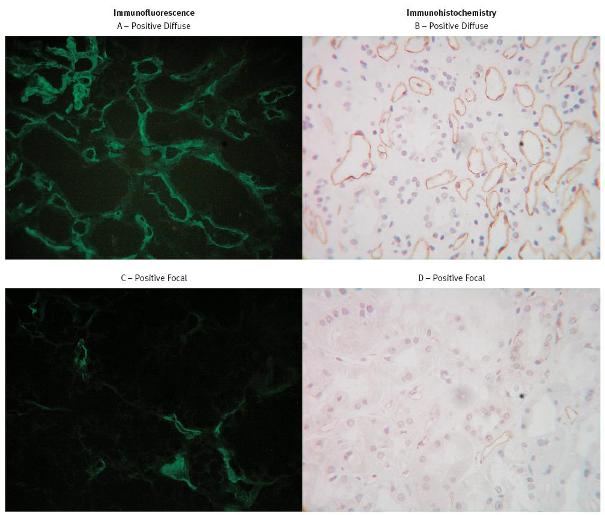
Immunofluorescence technique (Figures 1A and 1C) was performed in 3 μ m frozen tissue, acetonefixed sections, using monoclonal antibody. The primary antibody used was Ms/Hm anti-C4d (clone 033II-317.1.3.X by Quidel, ref. A213) in dilution of 1:30 over 30 minutes of incubation. The secondary antibody was Rb/Ms IgG-FITC (Dako, ref. F261) in dilution of 1:20 over 30 minutes of incubation.
The indirect IHC technique (Figures 1B and 1D) was performed in paraffin-embedded tissue with polyclonal antiserum. The epitope retrieval was heat-induced at 95-99ºC, over 20 minutes, at pH6, with the Target Retrieval Solution (Dako, ref. S2369). The primary antibody used was polyclonal antibody Rb/Hm anti-C4d (Serotec, ref. 0030-0230) in a dilution of 1:30, over 45 minutes. Others reagents used in this technique were Hydrogen Peroxide Block (Thermo Fisher Scientific , ref. TA-125-HP); Ultra V Block (Thermo Fisher Scientific, ref. TA-125-UB); UltraVision ONE HRP Polymer (Thermo Fisher Scientific, ref. TL-125-PHJ) and DAB+ (Dako, ref. K3468).

Figure 1
Positive C4d detection in peritubular capillaries according to technique and Banff classification.
The calculate cost was EUR 20.70 per biopsy by IF vs. EUR 38.63 per biopsy by IHC.
Biopsies were independently evaluated by two nephropathologists using the Banff 07 classification and scored negative (0-10%), positive focal (10-50%) and positive diffuse (>50%). In cases of non-concordance the biopsies were reviewed together and classified according to consensus.
RESULTS
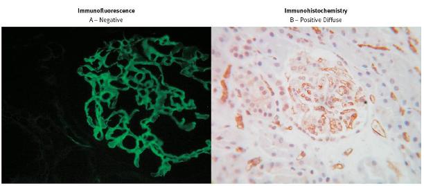
By IF, C4d was always present in glomerulus; i.e. positive and negative cases for PTC C4d showed mesangial C4d staining (Figure 2A). The same was demonstrated for artery and arteriole endothelium.

By IF, positive and negative cases for PTC C4d showed mesangial C4d staining (A). By IHC, only PTC C4d positive cases presented staining in the glomerular basement membrane (B).
Using IHC, only PTC C4d positive cases presented staining in the glomerular basement membrane.
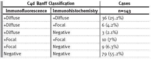
The results were summarised in Table I. There was total agreement on Banff classification in 87.4% (125/143) cases. All negative cases by IF were also negative by IHC. The biopsies were classified as positive in 44.7% (64/143) of cases performed by IF vs. 36.4% (52/143) performed by IHC. Fewer biopsies were classified as positive diffuse in the IHC group (25.1% vs. 31.4%) and more as positive focal (13.2% vs. 11.1%). More cases were classified as negative by the IHC method (63.6% vs. 55.2%).
Table I
C4d detection in peritubular capillaries according to technique and Banff classification

Study by ROC curve showed that IHC has a specificity of 100% and a sensitivity of 81.2% in relation to IF (AUC: 0.906; 95% confidence interval: 0.846-0.949; p=0.0001).
DISCUSSION
IHC ADVANTAGE
1 – Excellent specificity. The results obtained in this study confirm and extend the results from other authors with a lower number of cases.
No false positive cases were registered in our study.
2 – Feasible in formalin-fixed, paraffin-embedded tissue FFPE is the usual tissue processing for kidney biopsies, so this material is always available. The IHC technique could be used when no frozen tissue is accessible.
IHC DISADVANTAGE
1 – Lower sensibility. Although IHC presents an excellent specificity, it has a lower sensitivity than IF. Some false negative were registered.
The positive staining area by IHC is in some cases lower than by IF cases. That explains some discrepancies between classifications: positive diffuse by IF was graded as positive focal by IHC. Results obtained with IHC must be interpreted with some reserve since clinical relevance has only been established for IF results, and a focal staining in IHC can correspond to a diffuse staining in IF, for example.
2 – Nonspecific staining. Both nephropathologists stated that IHC staining causes more difficulties in interpretation, due to the existence of unspecific staining in tubular cells and surrounding interstitium. This was also referred to in other studies9. To overcome this issue, transplantation centres and laboratories must establish protocols to minimise the impact of preanalytical factors in IHC, and laboratories must follow strict protocols both in analytical and postanalytical procedures12.
3 – Cost and time preparation. The IHC technique was approximately EUR 18 per biopsy, more expensive than IF. The IHC method takes over one hour more to complete than the IF technique.
The IF technique is applied to frozen sections, does not need previous processing and can be finished in one hour. By IF method the results can be available to medical staff less than two hours after tissue reception in the laboratory.
4 – Need for external controls. Since mesangium and artery endothelium in frozen sections are always C4d positive (Figure 2A), there is no need for an external positive control. In IHC (Figure 2B) we recommend the use of two external positive controls – one with focal and the other with diffuse staining. When that is not possible, human tonsil specimens from acute tonsillitis can also be used as a positive control as C4d presents strong staining in germinal centres of secondary lymphoid follicles13.
CONCLUSIONS
The IHC method presents an excellent specificity but lower sensitivity to C4d detection in allograft dysfunction. The evaluation is more difficult, requiring a more experienced observer than the IF method.
In our laboratory, the use of IHC for C4d detection seems to us acceptable and safe when frozen tissue is unavailable. Extensive studies should be made to determine the real correspondence between results of both techniques and to establish guidelines for the interpretation of IHC results.
References
1. Chakravarti DN, Campbell RD, Porter RR. The chemical structure of the C4d fragment of the human complement component C4. Mol Immun 1987;24:1187-1197 [ Links ]
2. Mauiyyedi S, Crespo M, Collins AB, et al. Acute humoral rejection in kidney transplantation: II, morphology, immunopathology, and pathological classification. J Am Soc Nephrol 2002;13:779-787 [ Links ]
3. Colvin RB. Antibody-mediated renal allograft rejection: diagnosis and pathogenesis. J Am Soc Nephrol 2007;18:1046-1056 [ Links ]
4. Mauiyyedi S, Pelle PD, Saidman S, et al. Chronic humoral rejection: identification of antibody-mediated chronic renal allograft rejection by C4d deposits in peritubular capillaries. J Am Soc Nephrol 2001;12:574-582 [ Links ]
5. Solez K, Colvin RB, Racusen LC, et al. Banff classification of renal allograft pathology: updates and future directions. Am J Transplant 2008;8:1-8 [ Links ]
6. Mengel M, Borgers J, Bosmans JL, Serón D, Moreso F, Carrera M. Incidence of C4d stain in protocol biopsies from renal allografts: results from a multicenter trial. Am J Transplant 2005;5:1050-1056 [ Links ]
7. Magil AB, Tinckam KJ. Focal peritubular capillary C4d deposition in acute rejection. Nephrol Dial Transplant 2006;21:1382-1388 [ Links ]
8. Nadasdy GM, Bott C, Cowden D, Pelletier R, Fergunson R, Nadasdy T. Comparative study for the detection of peritubular capillary C4d deposition in human renal allograft using different methodologies. Hum Pathol 2005;36:1178-1185 [ Links ]
9. Troxell ML, Weintraub LA, Higgings JP, Kambham N. Comparison of C4d immunostaining methods in renal allograft biopsies. Clin J Am Soc Nephrol 2006;1:583-591 [ Links ]
10. Regele H, Exner M, Watschinger B, et al. Endothelial C4d deposition is associated with inferior kidney allograft outcome independently of cellular rejection. Nephrol Dial Transplant 2001;16:2058-2066 [ Links ]
11. Seemayer CA, Gaspert A, Nickeleit V, Mihastsch MJ. C4d staining of renal allograft biopsies: a comparative analysis of different staining techniques. Nephrol Dial Transplant 2007;22:568-576 [ Links ]
12. Goldstein NS, Hewitt SM, Taylor CR, et al. Recommendations for improved standardization of immunohistochemistry. Appl Immunohistochem Mol Morphol 2007;15:124-133 [ Links ]
13. Zwirner J, Felber E, Schmidt P, Riethmuller G, Feucht E. Complement activation in human lymphoid germinal centres. Immunology 1989;66:270-277 [ Links ]
Dr Helena Viana
Laboratório de Morfologia Renal
Serviço de Nefrologia
Hospital Curry Cabral
Rua da Beneficência, 8
1069-166 Lisbon, Portugal
E-mail: viana.helena@gmail.com
Conflict of interest statement. None declared.
Received for publication: 10/09/2012
Accepted in revised form: 08/11/2012


