Serviços Personalizados
Journal
Artigo
Indicadores
-
 Citado por SciELO
 -
 Acessos
Links relacionados
-
 Similares em
SciELO
Compartilhar
Portuguese Journal of Nephrology & Hypertension
versão impressa ISSN 0872-0169
Port J Nephrol Hypert vol.30 no.3 Lisboa set. 2016
MINI‑REVIEW
Oxalate nephropathy following Roux‑en‑Y gastric bypass surgery – Mini‑Review
Miguel Verdelho, Marco Mendes, Francisco Ribeiro, Helena Sousa Viana, Fernanda Carvalho, Fernando Nolasco
Nephrology Department, Hospital Curry Cabral, Centro Hospitalar de Lisboa Central, Lisboa, Portugal
ABSTRACT
Oxalate Nephropathy is characterised by the presence of tubular crystalline deposits of calcium oxalate, which can lead to both acute and chronic tubular injury and progressive renal failure. Enteric hyperoxaluria is the most common cause of moderate hyperoxaluria; it occurs in conditions associated with fat or bile acid malabsorption, which include jejunoileal bypass and other bariatric procedures such as Roux‑en‑Y gastric bypass surgery.
We present the clinical case of a 69‑year‑old man who was hospitalised for non‑oliguric renal dysfunction, with a serum creatinine of 10 mg/dl and normocytic normochromic anaemia. There was no prior history of renal disease. Twenty months before admission the patient was diagnosed with a gastro‑oesophageal junction adenocarcinoma and was treated with pre‑operative chemotherapy, followed by total gastrectomy, with a Roux‑en‑Y gastric bypass reconstruction. On discharge from gastric surgery, renal function was normal.
On the first day of hospital stay haemodialysis was initiated. Over the following days, the rapid unexplained renal impairment was investigated, and this workup [2] included a kidney biopsy. Histological examination of the biopsy specimen revealed a predominantly interstitial nephropathy with tubular atrophy and interstitial fibrosis, with bright intra‑tubular calcium oxalate crystals in over 50% of the tubules and so the histological diagnosis was of oxalate nephropathy. Subsequently, no recovery of renal function was observed, so the patient is currently undergoing regular haemodialysis.
Oxalate nephropathy is a rare but severe complication of Roux‑en‑Y gastric bypass surgery that can lead to a rapid progression to kidney failure. Although the treatment of obesity is the main indication for this surgery, this is also the preferred approach for gastrointestinal reconstruction after total gastrectomy for treatment of gastric carcinoma. Considering the rapid progression of oxalate nephropathy to kidney failure, patients who undergo Roux‑en‑Y gastric bypass surgery should have regular follow‑up of renal function.
Keyword: bariatric surgery; hyperoxaluria; malabsorption, oxalate nephropathy.
INTRODUCTION
Oxalate nephropathy is an entity characterised by the presence of tubular crystalline deposits of calcium oxalate, which can lead to both acute and chronic tubular injury1.
Dietary oxalate accounts for only 10 to 20% of the daily urinary oxalate excretion, with the remainder of its urinary excretion of endogenous origin. However, intake of even a small amount of oxalate from major dietary sources can substantially increase daily urinary oxalate excretion2.
Known causes of oxalate nephropathy include primary (hereditary) hyperoxaluria; excessive ingestion of dietary sources of oxalate such as spinach, rhubarb, tea, parsley, nuts and cocoa; ethylene glycol intoxication; vitamin B6 deficiency and enteric hyperoxaluria1‑3.
Enteric hyperoxaluria is the most common cause of moderate hyperoxaluria. It occurs in conditions associated with fat or bile acid malabsorption, such as inflammatory bowel disease, pancreatic insufficiency, bowel resection, blind loop syndrome, jejunoileal bypass and other bariatric procedures such as Roux‑en‑Y gastric bypass surgery (RYGB). All these gastrointestinal disorders are characterised by malabsorption of bile salts and/or fatty acids. As fat is malabsorbed, excessive intraluminal free fatty acids bind to and saponify calcium ions and fat‑soluble vitamins, leading to steatorrhoea.
In the normal state, calcium and oxalate within the intestinal lumen combine to form calcium oxalate complexes that are insoluble and excreted in faeces.
However, in the presence of malabsorptive conditions, there is decreased calcium‑oxalate binding due to decreased calcium availability in the intestinal lumen, which will lead to increased quantity of soluble free oxalate. This increased unbound oxalate can be actively or passively absorbed in the colonic mucosa. Concurrently, permeability of the colonic mucosa to oxalate can be enhanced by exposure to free long‑chain fatty acids and unconjugated bile salts, both increased in the gastrointestinal tract after RYGB1,3.
Patients with intestinal malabsorption may have additional factors predisposing to stone formation other than increased oxalate excretion (which can be as high as 80 to 200 mg [0.9 to 2.2 mmol] per day). The diarrhoeal fluid losses can lead both to a reduction in urine volume and, if the patient has a metabolic acidosis, to a low urine pH and a marked decrease in citrate excretion. These changes can promote uric acid as well as calcium oxalate stone formation4.
CASE SUMMARY
A 69‑year‑old man was referred to our hospital with complaints of anorexia and fatigue of recent onset. He denied other symptoms, such as oedema, worsening of hypertension, nausea, vomiting, diarrhoea, decreased urine output, or significant weight loss. Manifestations of malabsorption, such as diarrhoea with pale, voluminous, foul‑smelling stools, were absent and patient had no signs of dehydration, hypovolaemia or infection and there was no recent history of exposure to contrast media or other nephrotoxic agents. The patient had been diagnosed with hypertension 15 years ago and had a 5‑year history of type 2 diabetes mellitus. There was no prior history of renal disease. The prescribed medication included metformin, sitagliptin, ramipril and hydrochlorothiazide3, with a good metabolic and blood pressure control. Twenty months before the current admission, a diagnosis of gastro‑oesophageal junction adenocarcinoma was made. At that time, the patient was admitted to the hospital and was submitted to pre‑operative combination chemotherapy with epirubicin, oxaliplatin and capecitabine, followed by total gastrectomy, with a Roux‑en‑Y gastric bypass reconstruction (RYGB). On discharge from surgery, 8 months before current hospitalization, the patient presented a normal renal function (serum creatinine of 0.8 mg/ dl). There was no evidence of cancer recurrence during follow‑up.
Laboratory investigation showed a normocytic normochromic anaemia with a haemoglobin of 6.5 g/dl, and renal insufficiency with a serum creatinine of 10 mg/dl. There was no evidence of blood loss or haemolysis (bilirubin and LDH levels were within the normal range), and a bone marrow biopsy performed prior to the admission found no evidence of neoplastic infiltration.
A urinalysis showed 30 mg/dl of proteinuria, without erythrocytes or leucocytes, and a 24‑hour urine collection revealed hypocalciuria, hypophosphaturia and hypomagnesuria; urinary oxalate was within the normal range and urinary citrate wasnt determined.
Antinuclear antibodies (ANA) were positive (1:160), while all other immunological tests were negative (Anti‑dsDNA antibody, ANCA‑MPO and ANCA‑PR3 antibodies, Anti‑GBM antibody, Anti‑Cardiolipin antibodies, Anti‑SSA antibody, Anti‑Ro52kD antibody, Anti‑SSB antibody, and Anti‑RNP and Anti‑Sm antibodies). Complement levels were within the normal range. Serology for HIV, HCV, and HBV was negative.
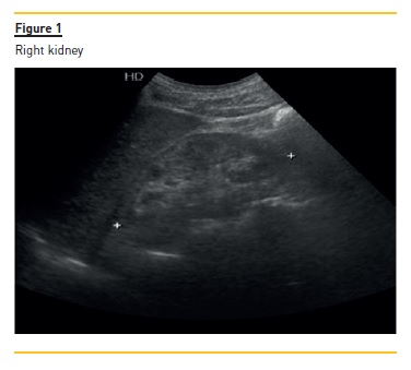
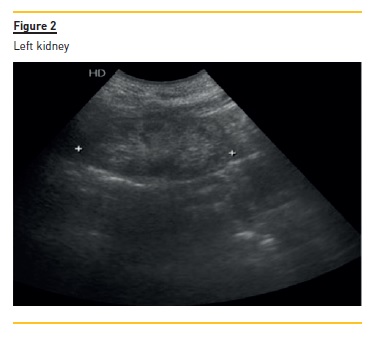
A kidney ultrasound (longitudinal view) revealed a right kidney measuring 9.9 cm in length (figure 1) and a left kidney of 8.8cm in length (figure 2), with hyperechoic cortex and loss of demarcation between the outer cortex and middle pyramids and columns of Bertin.


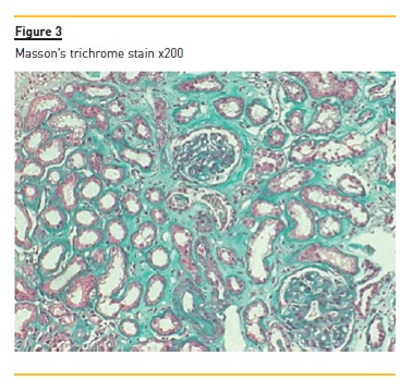
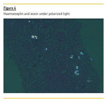
Three days after admission, a kidney biopsy was performed. Histological examination of the biopsy specimen revealed, predominantly, interstitial nephropathy with irreversible tubular atrophy and interstitial fibrosis. The glomerulus visualized presented ischaemic aspects (figure 3). Observation with Haematoxylin and Eosin (figure 4) showed bright intra‑tubular calcium oxalate crystals under polarized light in over 50% of the tubules and so a histological diagnosis of oxalate nephropathy was made.


On the first day of hospital stay haemodialysis was initiated. During follow‑up, no recovery of renal function was observed, so the patient is currently undergoing regular haemodialysis. No specific treatment was prescribed, apart from the avoidance of food rich in oxalates, mostly due to the advanced stage of renal failure and the irreversibility of the lesions found on kidney biopsy specimen.
MINI‑REVIEW
RYGB has not only proved to be an effective long‑term weight reduction treatment option, which concomitantly can also treat diabetes and hypertension in the obese population3, but has also become the preferred mode for intestinal reconstruction after total gastrectomy for treatment of gastric carcinoma. This is because of its feasibility and capability to prevent biliary and pancreatic secretions from reaching the esophageal mucosa. Some patients undergoing RYGB may develop weight loss, diarrhoea and steatorrhoea due to rapid intestinal transit, bacterial overgrowth and pancreatic under stimulation with consequent fat malabsorption1.
The development of bariatric surgery began in the 1950s with the appearance of jejunoileal bypass5, with nephrolithiasis and oxalate nephropathy widely recognized complications of this procedure1,6. Jejunoileal bypass was largely abandoned in 1979 due to the high rate of serious complications6. Several newer bariatric procedures emerged, of which RYGB surgery has become the most widely used7.
Despite the benefits inherent in the RYGB procedure, emerging data suggests that RYGB is associated with increased risk factors for the development of calcium oxalate nephrolithiasis and oxalate nephropathy.
Most of these are studies with small sample sizes. A prospective longitudinal study by Duffey et al. of 24 morbidly obese adults observed a significant 3‑month postoperative increase in daily oxalate excretion (p=0.026)2. These findings were confirmed in a 2‑year prospective longitudinal study with a cohort of 21 patients. Oxalate excretion increased significantly 2 years after RYGB in both male and female patients (31 ± 9 mg/day versus 59 ± 28 mg/day; p=0.001)8. Sinha et al. described a study including 60 patients who underwent RYGB and subsequently developed nephrolithiasis. Hyperoxaluria was a common risk factor in these stone‑ forming patients, although low urinary volumes and reduced 24‑hour urine excretions of citrate and magnesium appeared to be important factors as well7. A recent prospective study was performed to determine when the increased risk for calcium oxalate stone formation occurs after RYGB. A total of 13 patients were enrolled, with mean age 41.1 ± 7.2 years. It was noted that the calcium oxalate relative saturation ratio significantly increased as early as 2 months after RYGB and continued to remain significantly elevated at 4 and 6 months. An increase in urinary oxalate excretion was also noted at 2 months9. Nasr et al. reported an association of RYGB with renal insufficiency as a result of oxalate nephropathy, identifying a total of eleven cases. The cohort consisted of six women and five men with a mean age of 61.3 years (range 45‑79).
The indication for RYGB was morbid obesity for eight patients (72.7%) and gastric adenocarcinoma for the remaining three (27.3%). The three patients with gastric adenocarcinoma underwent total gastrectomy and Roux‑en‑Y oesophageal jejunostomy. Urine oxalate levels were available in two patients and serum oxalate levels in one patient and both parameters were markedly increased. Oxalate crystals were noted in the urine of two patients and three patients developed nephrolithiasis.
Patients presented with acute renal failure (ARF), often superimposed on chronic kidney disease (CKD). The mean and median times from surgery to discovery of ARF were 33 and 12 months, respectively (range 4‑96 months). All patients were followed‑up post‑biopsy, for a mean and median time of 19.4 and 11 months, respectively (range 2.5 to 58.0 months).
Within this period, eight (72.7%) patients rapidly progressed to kidney failure at a mean of 3.2 weeks (range 0.0 to 12.0 weeks). Regarding renal biopsy findings, the distinctive histological feature was an acute and chronic tubulointerstitial nephropathy with acute tubular injury diffusively involving non‑atrophic tubules, associated with varying degrees of tubular atrophy and interstitial fibrosis and abundant tubular calcium oxalate deposits, with this findings very similar to those described in this case report. These deposits were mainly seen in the renal cortex and involved both proximal and distal tubules. Immunofluorescence was performed in all cases and confirmed the absence of immune‑type deposits1.
Treatment of oxalate nephropathy aims to reduce intestinal oxalate absorption. This can be achieved by limiting fat intake to reduce the binding of calcium by free fatty acids, reduction in oxalate rich‑food, increased hydration in order to raise total urine volume and decrease urine supersaturation3.
Calcium supplementation using calcium citrate2,3 can be used to increase oxalate excretion by forming an insoluble calcium‑oxalate salt that is excreted in the faeces2. However, calcium supplementation can be limited by its saponification with increased intestinal free fatty acids and subsequent decreased availability to bind with oxalate2.
Supplementation with citric salts, such as potassium citrate, may also be beneficial3. The use of cholestyramine has been advocated as the presence of bile salts in the colon increase colonic permeability to organic acids such as oxalate10. Probiotics may also have a beneficial effect, although compelling data is lacking3.
Surgical reversion may be attempted, although it is not clear whether this could lead to an improvement in renal function once oxalate nephropathy is established1,11.
Oxalate nephropathy after RYGB, albeit rare, has a poor prognosis, with a rapid progression to kidney failure1.
In this particular case, oxalate nephropathy occurred after RYGB reconstruction following total gastrectomy, and the patient developed kidney failure eight months after surgery. It is predictable that a larger number of cases will be observed due to the increasing use of bariatric surgery1,9. Therefore, patients who undergo RYGB would benefit from regular follow‑up of renal function and rapid implementation of dietary restrictions. It has been suggested that post‑surgery administration of oral calcium may be beneficial. However studies and research will be needed for an evidence‑based approach11.
References
1. Nasr SH, DAgati VD, Said SM, et al. Oxalate nephropathy complicating Roux‑en‑Y Gastric Bypass: an underrecognized cause of irreversible renal failure. Clin J Am Soc Nephrol. 2008 Nov; 3(6):1676–1683. [ Links ]
2. Duffey BG, Pedro RN, Makhlouf A, Kriedberg C, Stessman M, Hinck B, et al. Roux‑en‑Y Gastric Bypass is associated with early increased risk factors for development of calcium oxalate nephrolithiasis. J Am Coll Surg. 2008 Jun; 206(3):1145‑1153. [ Links ]
3. Canales BK, Gonzalez RD. Kidney stone risk following Roux‑en‑Y gastric bypass surgery. Transl Androl Urol. 2014 Sep; 3(3):242–249. [ Links ]
4. Nelson WK, Houghton SG, Milliner DS, Lieske JC, Sarr MG. Enteric hyperoxaluria, nephrolithiasis, and oxalate nephropathy: potentially serious and unappreciated complications of Roux‑en‑Y gastric bypass. Surg Obes Relat Dis. 2005 Sep‑Oct; 1(5):481–485. [ Links ]
5. Park AM, Storm DW, Fulmer BR, Still CD, Wood GC, Hartle JE. A prospective study of risk factors for nephrolithiasis after Roux‑en‑Y gastric bypass surgery. J Urol. 2009 Nov; 182(5):2334–2239. [ Links ]
6. Mole DR, Tomson CR, Mortensen N, Winearls CG. Renal complications of jejuno ileal bypass for obesity. QJM. 2001 Feb; 94(2):69–77. [ Links ]
7. Sinha MK, Collazo‑Clavell ML, Rule A, Milliner DS, Nelson W, Sarr MG, et al. Hyperoxaluric nephrolithiasis is a complication of Roux‑en‑Y gastric bypass surgery. Kidney Int. 2007 Jul; 72(1):100–107. [ Links ]
8. Duffey BG, Alanee S, Pedro RN, Hinck B, Kriedberg C, Ikramuddin S, et al. Hyperoxaluria is a long‑term consequence of Roux‑en‑Y Gastric bypass: a 2‑year prospective longitudinal study. J Am Coll Surg. 2010 Jul; 211(1):8‑15. [ Links ]
9. Agrawal V, Liu XJ, Campfield T, Romanelli J, Enrique Silva J, Braden GL. Calcium oxalate supersaturation increases early after Roux‑en‑Y gastric bypass. SurgObesRelat Dis. 2014 Jan‑Feb; 10(1):88–94. [ Links ]
10. Nagaraju SP, Gupta A, McCormick B. Oxalate nephropathy: an important cause of renal failure after bariatric surgery. Indian J Nephrol. 2013 Jul‑Aug; 23(4):316–318. [ Links ]
11. Ahmed MH, Byrne CD. Bariatric surgery and renal function: a precarious balance between benefit and harm. Nephrol Dial Transplant. 2010 Oct; 25(10):3142-7 [ Links ]
Miguel Verdelho
Hospital Curry Cabral, Rua da Beneficência,nº8, 1069‑166,
Lisbon, Portugal
E‑mail: miguelverdelhocosta@gmail.com
Disclosures of Potential Conflicts of Interest: None declared
Received for publication: Jan 8, 2016
Accepted in revised form: June 1, 2016


