Serviços Personalizados
Journal
Artigo
Indicadores
-
 Citado por SciELO
 -
 Acessos
Links relacionados
-
 Similares em
SciELO
Compartilhar
Portuguese Journal of Nephrology & Hypertension
versão impressa ISSN 0872-0169
Port J Nephrol Hypert vol.30 no.4 Lisboa dez. 2016
MINI-REVIEW
BK virus nephropathy in kidney transplantation – A literature review following a clinical case
Patrícia Barreto1,2, Manuela Almeida1, Leonídio Dias1, Pedro Vieira1,3, Sofia Pedroso1, La Salete Martins1, António Castro Henriques1, António Cabrita1
1 Department of Nephrology, Centro Hospitalar do Porto. Porto, Portugal
2 Department of Nephrology, Centro Hospitalar de Vila Nova de Gaia/Espinho. Vila Nova de Gaia, Portugal
3 Department of Nephrology, Centro Hospitalar do Funchal. Funchal, Portugal
ABSTRACT
Over the last 15 years, better immunosuppressive drugs have decreased acute rejection rates in kidney transplantation but have also led to an increase in the incidence and impact of BK virus nephropathy. The authors report the case of a 62--year-old man submitted to a renal transplant of a deceased donor with an immunosuppression regimen free of rabbit anti -thymocyte globulin and tacrolimus, in whom BK nephropathy was diagnosed at seven weeks post-transplant. Intravenous human immunoglobulin (IVIG) was administered after immunosuppression reduction. Instituted treatment was successful. This clinical case highlights the importance of a high index of suspicion for an atypical presentation of BK nephropathy in renal transplant recipients and strengthens the need for other therapeutic interventions beyond the reduction of immunosuppression. It was the starting point for a review of BK virus nephropathy in kidney transplantation with a focus on risk factors, diagnosis and treatment.
Key-Words: Atypical presentation, BK virus, early diagnosis, kidney transplant, nephritis, therapeutic interventions.
CASE REPORT
The authors report the case of a 62-year-old Caucasian male with a personal history of hypertension, diabetes, and chronic kidney disease of unknown aetiology, who began haemodialysis in February 2009.
He was submitted to renal transplantation of a 60-year-old deceased donor (death from cerebrovascular accident) on September 29, 2014. Both the donor and the recipient had the same ABO and Rhesus blood type (O+). The recipient had 2 HLA -mismatches (1 in AB and 1 in DR), a panel reactive antibody (PRA) of 0%, negative CDC crossmatch for B and T lymphocytes and anti-HLA alloantibody class I and II research using Luminex® technology was negative.
The immunosuppression protocol was basiliximab (first and fourth post-transplant days), methylprednisolone (500-250-125-80mg on 1-4 post-transplant days, respectively), prednisolone (35 mg at 5 post–transplant day, followed by progressive tapering until nearly 5mg at 30 post-transplant day), cyclosporine (175 mg twice daily with target levels achieved) and mycophenolate mofetil (1000mg twice daily).
After the surgery the patient presented delayed graft function (DGF). The patient needed one dialysis session due to hyperkalaemia. At discharge date, eleven days post-transplant, his serum creatinine (SCr) was 2.53 mg/dL. Progressive reduction of immunosuppression was made because of pancytopaenia, and even prednisolone was quickly tapered to the maintenance dose due to uncontrolled glycaemia.
Eighteen days post -transplant he presented BK virus positivity in urine (identification of decoy cells on analysis of urine cytology). The patient presented progressive improvement of graft function and two weeks later had a SCr of 1.02 mg/dL.
He was admitted seven weeks post-transplant for acute graft dysfunction (with SCr 2.56 mg/dL). On that date his maintenance immunosuppression was cyclosporine 150mg twice daily, mycophenolate mofetil 250mg twice daily and prednisolone 7.5mg daily. A positive BK viraemia of 197250 copies/mL was detected [by quantitative polymerase chain reaction (PCR) in plasma]. Immunosuppression was immediately reduced (cyclosporine was reduced to 100 mg twice daily and mycophenolate mofetil was suspended) and a ciprofloxacin course was initiated (250mg 12/12h). Obstruction was excluded and the patient was hydrated. Graft pyelonephritis by Enterococos faecalis was diagnosed.
He was treated with amoxicillin-clavulanic acid and the pigtail was removed. He had partial graft function recovery (SCr 1.9 mg/dL). Graft biopsy was performed one week after admission, which was compatible with polyomavirus associated interstitial nephritis (BK virus nephropathy). Non -specific intravenous human immunoglobulin 1 g/Kg was administered in 2 divided doses one month apart. One month and two weeks after the mentioned treatment, graft function improved (SCr 1.5mg/dL) and viraemia disappeared.
REVIEW
Over the last 15 years, more potent immunosuppression regimens have decreased the rates of acute rejection in kidney transplantation but simultaneously have led to the emergence of BK virus nephropathy1.
BK is a DNA virus from the polyomavirus family, which includes JC virus. Based on DNA sequence variations, BK can be divided into six genotypes. Genotype I is the most frequent worldwide (80%), followed by genotype IV (15%)2. The BK genome encodes three capsid structural proteins, called viral capsid protein 1 (VP1), VP2 and VP3, as well as the large T and small t antigens.
Primary infection is often subclinical or manifests as mild respiratory illness and is acquired in childhood; BK virus is almost ubiquitous in adults with seroprevalence of 80%3. After primary infection, the virus establishes latency in the uroepithelium renal and particularly in tubular epithelial cells. In the setting of immunosuppression, the virus reactivates and begins to replicate, triggering a cascade of events starting with tubular cell lysis and consequent viruria. The BK virus then multiplies in the interstitium and crosses into the peritubular capillaries, causing viraemia and may also invade the allograft interstitium, leading to the tubulointerstitial lesions that characterize BK virus nephropathy4.
Approximately 20 to 60% of renal transplant recipients present viruria; one third of patients with viruria will develop BK viraemia and BK virus nephropathy develops in 2 to 10% of recipients5.
The most consistent risk factor identified across studies for the development of BK virus nephropathy is the overall level of immunosuppression6. Other risk factors identified for BK virus nephropathy which have not been uniformly observed in all studies include male gender, older and younger age, white ethnicity, diabetes mellitus, DGF, rejection episodes, degree of human leukocyte antigen (HLA) mismatching, prolonged cold ischaemia, BK serostatus, ureteral trauma/ureteral stent placement, cytomegalovirus infection and glucocorticoid maintenance therapy5-12.
Induction and also maintenance immunosuppression seems to influence BK virus nephropathy risk.
Dharnidharka et al demonstrated that the use of rabbit anti-thymocyte globulin induction and tacrolimus or mycophenolate mofetil based immunosuppression increased the risk for the development of BK viraemia13.
Hirsch et al performed a secondary analysis of the data of the DIRECT trial which was a prospective, randomized trial of cyclosporine versus tacrolimus in conjunction with basiliximab induction14. They showed that patients in the cyclosporine arm had a lower rate of BK virus nephropathy at 6 and 12 months post -transplant, compared with the tacrolimus group. This result supports an effect of more potent maintenance immunosuppression on BK reactivation. It has been shown that the risk of BK virus nephropathy is higher in recipients that received maintenance immunosuppression with tacrolimus-mycophenolate mofetil, or prednisone versus no prednisone, tacrolimus versus cyclosporine, high versus low levels of tacrolimus, and high versus low doses of prednisone15. Prospective studies designed to address the effect of induction and maintenance immunosuppression on BK virus nephropathy are needed4.
Rejection and/or ischaemia of the kidney allograft may increase the risk of BK nephropathy16. This statement is based on the fact that BK virus nephropathy rarely occurs in the native kidneys of patients who undergo non -renal solid organ transplantation, despite immunosuppression17,18.
BK virus nephropathy may be higher among human leukocyte antigen (HLA) - and ABO -mismatched patients, which may be due to the increased rate of rejection and increased immunosuppression among such patients19. The risk among ABO -incompatible patients seems to be higher than among HLA-mismatched patients. ABO incompatibility was inclusively assumed as an independent predictor of BK virus nephropathy in the study of Sharif A et al11.
Currently, pre-transplant screening of donors and recipients for BK seropositivity is neither mandatory nor routinely performed. Nevertheless, although pre-transplant BK seropositivity in adult recipients has not been shown to influence the development of BK virus nephropathy after transplantation20,21, Bohl et al identified BK virus antibody-positive donors as a risk for post-transplant BK viraemia in recipients20. In this study, recipients of BK seropositive donor kidneys developed BK viraemia at earlier post -transplant timepoints; had higher viral titers and were slower to clear the virus.
This data suggests a role for donor pre-transplant serotyping as a means of BK risk assessment and immunosuppression management.
The induction of immunosuppression in our patient was not based on rabbit anti-thymocyte globulin but on basiliximab; and in spite of tacrolimus he had cyclosporine as maintenance immunosuppression. According to collected data concerning induction and maintenance immunosuppression regimens, this patient did not belong to a recipients group at a higher risk of developing BK virus nephropathy. However, he presented several risk factors that contributed to the increase in his overall degree of immunosuppression: history of diabetes mellitus and the development of pancytopaenia. In addition, other risk factors not universally accepted were also present, such as male gender, older age, white ethnicity and DGF. Therefore the diagnosis hypothesis of BK virus nephropathy would be more plausible than it first seemed.
Among kidney transplant recipients, BK virus causes tubulointerstitial nephritis and seldom ureteral stenosis22.
Haemorrhagic and non -haemorrhagic cystitis is rarely observed among this subgroup of patients, although it may be a complication in bone marrow transplantation23,24.
Patients with BK virus nephropathy most commonly present with an asymptomatic acute or slowly progressive rise in serum creatinine concentration25. Routine urinalysis most commonly reveals pyuria, haematuria and/or cellular casts consisting of renal tubular cells and inflammatory cells, which are suggestive findings of interstitial nephritis.
Prospective screening studies have demonstrated that BK virus nephropathy is predominantly an early complication of renal transplantation, with most cases occurring within the first year post -transplantion4, with disease onset occurring at mean 10 to 13 months post-transplantation. However, the onset of nephritis may occur as early as six days post -transplantation, or as late as five years23,26–28.
Literature supports that such an early BK virus nephropathy presentation, as occurred with our patient, is rare but it has already been described. We emphasize that, in our case, BK virus nephropathy presented nearly seven weeks post-transplant, much earlier than three months which is when several centers begin screening for BK virus infection.
BK virus nephropathy is first suspected in the patient with clinical findings suggestive of interstitial nephritis19.
The suspicion of the diagnosis is supported by the identification of decoy cells on analysis of urine cytology and/or by quantitative polymerase chain reaction (PCR) of urine and/or plasma19. A definitive diagnosis of BK virus nephropathy is made by analysis of tissue obtained by renal biopsy19. However the diagnosis may be missed on biopsy since definitive lesions have a focal distribution. As BK virus nephropathy has limited treatment options, the goal of screening is to facilitate early diagnosis of patients when viruric or viraemic, and to intervene prior to the development of overt nephropathy4.
BK viral infections progress through recognized stages. Viral replication in the urine precedes BK viraemia by nearly four weeks and BK virus nephropathy occurs nearly 12 weeks after BK viruria5. Renal transplant recipients with higher urine DNA levels are more likely to develop detectable DNA in plasma, and prolonged DNAemia usually precedes clinically overt nepropathy19.
The approach to the evaluation of patients with suspected BK nephritis varies by institution and their surveillance protocols. One possible approach is first to screen by using urine cytology and, if positive on two successive occasions over four weeks and plasma PCR is positive (DNA PCR load > 104 copies/ml), an allograft biopsy is obtained regardless of renal function4,19.
Based on the available literature4, one responsible approach is to routinely screen all renal transplant patients starting at 3 months until 24 months post-transplantation. Screening is not recommended out of that period once BK virus nephropathy is rare, unless renal dysfunction is present.
BK virus is detectable in both blood and urine4. BK viral loads are measured by PCR. Viruria is defined as presence of >2000 copies/mL of urine and viraemia is defined as presence of >2000 copies/mL of whole blood. The sensitivity and specificity of the PCR appears to be institution dependent and must be interpreted according to clinical setting19.
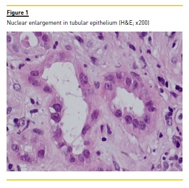
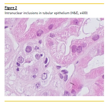
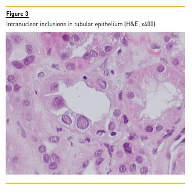
Urine can be screened for BK virus by cytology or by quantification of urine BK DNA by PCR4. Tubular epithelial cells infected with BK virus are shed in the urine and are called decoy cells4 (due to their resemblance to renal carcinoma cells). Cytological examination of urine may reveal such BK -infected cells. Their most characteristic abnormality is an enlarged nucleus with a single large basophilic intranuclear inclusion.
The presence of characteristic cytopathology changes in infected cells is strongly suggestive of BK virus infection19.
Thus, urine cytology showing characteristic cytological abnormalities is suggestive, but not definitive, for BK virus nephropathy. Their absence does not exclude the disease. Urine BK PCR is more sensitive and specific than urine cytology for the detection and diagnosis of BK virus nephropathy29 (100% sensitivity and 78% specificity for urine PCR vs 25% and 85%, respectively, for the presence of decoy cells in urine cytology). If only urine BK virus screening is to be performed, urine BK PCR is recommended as the superior assay, considering the threshold of >1x10⁷ copies/mL as suggestive of BK virus nephropathy4. Given the widespread availability of the plasma PCR assay and its greater predictive value for BK virus nephropathy, it may be unnecessary to perform urine BK screening first before plasma testing4.
BK detection by PCR in plasma is very sensitive and specific for the development of BK virus nephropathy.
Depending on the study, sensitivity can achieve 100% and specificity is almost 90%20,29. A definitive viral load cut-off associated with BK virus nephropathy has not been established yet, but retrospective studies have suggested that a BK viral load >104 copies/mL is strongly associated with the presence of BK virus nephropathy5.
The patient previously reported presented decoy cells on urine cytology eighteen days post-transplant.
It is important to mention that, in this case, urine cytology was requested sooner than usual because this patient had an elevated creatinine level (SCr 2.53mg/dL) at discharge. Even though he had achieved a normal graft function (Scr 1.02mg/dL) transiently, seven weeks post-transplant he presented acute graft dysfunction associated to a much higher BK viraemia than the threshold above which renal biopsy is mandatory. This case is in accordance with the progression of BK virus infections through detectable stages advocated by literature (Table I). This patient probably, developed BK virus infection in the first fewdays post-transplant.
Renal biopsy remains the gold standard for the diagnosis of BK virus nephropathy. It is recommended in patients with a high level of BK viraemia (>104 copies/mL) with or without an elevation in serum creatinine4.
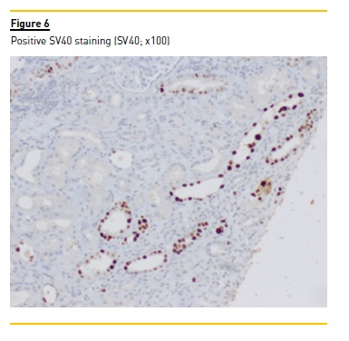
A definitive diagnosis of BK virus induced nephropathy requires the following findings on renal biopsy: characteristic cytopathic changes plus positive immunohistochemistry tests using antibodies directed specifically against BK or against the cross-reacting SV40 large T antigen. Positive SV40 staining is pathognomonic as it is associated with a specificity of almost 100 percent19.
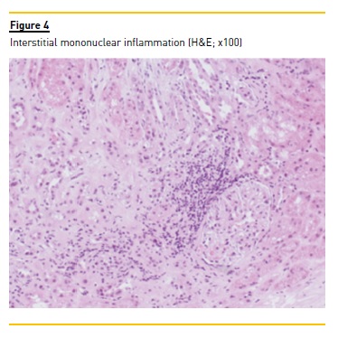
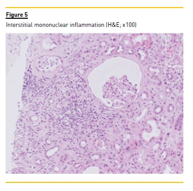
The characteristic cytopathic changes mentioned above include: intranuclear basophilic viral inclusions without a surrounding halo; interstitial mononuclear or polymorphonuclear cell infiltrates in the areas of tubular damage; tubular injury which is characterized by tubular cell apoptosis, cell drop out, desquamation, and flattened epithelial lining, and tubulitis which is manifested by lymphocyte permeation of the tubular basement membrane that can be mistaken for acute cellular rejection23,26,30,31.
Three patterns have been proposed to describe the histological findings in patients with BK virus nephropathy19: pattern A consists of cytopathic/cytolytic changes with absent or minimal inflammation; pattern B consists of cytopathic/cytolytic changes associated with patchy or diffuse tubulointerstitial inflammation and atrophy, and pattern C consists of graft sclerosis. Therefore the degree of fibrosis and tubular atrophy are assumed to be the most predictive of allograft outcome4.
Because of the focal nature of early BK nephropathy, the diagnosis may be missed in one -third of biopsies32,33, making diagnosis on occasion challenging.
Owing to these difficulties, a negative biopsy cannot rule out early BKVN with 100% certainty. A minimum of two biopsy cores, preferably including medulla, should be examined34. Medullary tissue should be included because the virus is more likely to be present in the medulla. If the initial biopsy does not confirm BK, a biopsy repetition should be considered.
A presumptive diagnosis of BK virus nephropathy may be made in the absence of definitive findings on biopsy. Findings of sustained (more than two weeks of duration) urinary viral shedding and significant BK replication plasma DNA PCR load > 104 copies/mL) with or without kidney dysfunction defines presumptive BK nephropathy5,32.
Our patient´s renal biopsy showed the characteristic cytopathic changes of BK virus nephropathy, namely the intranuclear basophilic viral inclusions in tubular cells and interstitial mononuclear cell infiltrate accompanied by positive SV40 staining. It allowed the definitive diagnosis of BK virus nephropathy. This result was unexpected in view of the timing of the development and the immunosuppressive regimen used. Thus, the other possible diagnosis hypothesis of cellular acute rejection became less probable, although the coexistence of both entities at the date the renal biopsy was performed could not be completely excluded. As cellular acute rejection shares similar features, such as interstitial mononuclear cell infiltrate and tubulitis, with BK virus nephropathy the distinction between them is not always easy but is crucial as the treatment of rejection is the opposite of that required for BK virus nephropathy. In view of the patient´s clinical response to immunosuppression reduction, the possibility of rejection was afterward definitely excluded.
Cellular adaptative immunity is determinant for the control and resolution of BK virus nephropathy. CD4+ and CD8+ cells are implicated in BK clearance. In kidney transplant recipients, a strong CD8 response was associated with lower BK viral loads in blood and urine4,15.
There is no objective data concerning BK virus nephropathy treatment, as current therapeutic recommendations are largely based on small case series5,23,25,35. This is a worrisome problem since established BK virus nephropathy is associated with allograft failure in 15 to 50 percent of cases.
Evidence suggests that prevention consisting of a screening and pre-emptive approach is more effective than a strategy based on therapy of established disease35.
Several studies have evaluated the efficacy of screening with subsequent immunosuppression reduction20,34,36,37.
They concluded that BK viruria without viraemia poses minimal risk; that the minimization of immunosuppression upon detection of BK viraemia is associated with excellent graft survival, low rejection rates and preserved renal function at five years, and that such an approach may prevent clinically evident BK virus nephropathy. These outcomes have been strengthened by more recent studies38-40. Sawinski et al4 recommend that patients who have their immunosuppression reduced for BK viraemia should be monitored carefully, with serum creatinine checked every 1-2 weeks and BK viral loads repeated at 2-4 week intervals. Few observational studies have reported successful clearance of viraemia of > 85 percent with immunosuppression reduction15.
When BK virus nephropathy is established, immunosuppression reduction remains the cornerstone of therapy25,41. However, in this stage, treatment becomes more complex and immunosuppression reduction alone is often not sufficient to stabilize renal function. The main objective of immunosuppression reduction is to restrain viral replication without triggering rejection, but the optimal method to achieve it is unclear. The particular regimen is frequently center specific42. One approach that is used for patients who are on a maintenance regimen consisting of triple immunosuppression therapy with a calcineurin inhibitor (cyclosporine or tacrolimus), an antimetabolite (azathioprine, mycophenolate mofetil) and prednisone is to discontinue completely the anti-metabolite (usually mycophenolate) and decrease the dose of the calcineurin inhibitor.
Additional interventions are based upon the clinical response to this initial strategy as measured by the reduction in BK viraemia load after a period of one to two months. The resolution of viraemia may take several months. An alternative approach that is used by others is to decrease the mycophenolate dose by 50 percent, followed by a 50 percent decrease in the calcineurin inhibitor at three months if decoy cells persist.
Using this approach, the target serum tacrolimus trough level is 4 to 6 ng/mL, and the target serum cyclosporine trough level is 60 to 100 ng/mL. Mycophenolate may be discontinued completely if viral activation persists.
Maintenance immunosuppression then consists of tacrolimus and low-dose prednisone.
Discontinuation of the anti-metabolite is the most common approach and a recent study43 suggested that both tacrolimus and cyclosporine can inhibit anti-BK T-cell responses in vitro, strengthening this practice4.
For patients who have progressive allograft dysfunction, despite a maximal decrease in immunosuppressive therapy for a period of several weeks to months, agents with hypothetical antiviral activity may be tried. However the efficacy of this approach has not been proven44.
Antiviral agents that have been associated with anecdotal success include intravenous immune globulin (IVIG) and cidofovir42. Regardless of the treatment strategy employed, rapid viral reduction has been associated with stable or improving glomerular filtration rate (GFR)4. Treatment is based on biopsy findings: if the kidney biopsy is compatible with BK virus nephropathy and simultaneously has an important interstitial inflammation, treatment options include administration of IVIG, immunosuppression reduction and antiviral therapy; if the kidney shows findings of BK virus nephropathy but without significant interstitial inflammation, treatment shall include immunosuppression reduction and antiviral therapy; and if the biopsy does not show evidence of BK nephropathy, immunosuppression reduction and continued blood BK viral load monitoring every two weeks until the assay becomes negative is recommended.
The reported efficacy of adjuvant antiviral agents is difficult to interpret because they have been used in combination with immunosuppression reduction and, occasionally, in combination42. So there are no randomized controlled trials showing superiority of these drugs over the timely reduction of immunosuppression; additionally, the United States Food and Drug Administration has not approved any specific drug for the treatment of BKV15.
We will specifically focus on intravenous human immunoglobulin, cidofovir, leflunomide, fluoroquinolones and mTOR inhibitors.
Intravenous human immunoglobulin (IVIG) therapy has immunomodulatory effects on inflammation and autoimmune diseases through formation of immunoglobulin complexes and it has also shown potential anti -BK virus properties45. It has been administered in doses of 0.2-2.0g/Kg together with reduction of the immunosuppression42. A recent study46 assessed the impact of treatment with IVIG in 30 renal transplant recipients with BK virus nephropathy previously submitted to 8 weeks immunosuppression reduction and leflunomide without response. Ninety percent of patients presented a positive response in clearing viraemia.
It was concluded that IVIG administration appeared to be safe and effective in treating BK virus nephropathy and preventing graft loss in patients who had inadequate response to immunosuppression reduction and leflunomide therapy. The authors argued that the administration of IVIG for treating BK virus nephropathy not only reduces the BK viral load but also seems to offer additional protection against acute rejection during the reduction of immunosuppression. This study had the largest sample size using IVIG treatment in patients with persistent BK virus nephropathy.
Cidofovir is a cytosine nucleotide analogue that is active against various DNA viruses and is approved for both human immunodeficiency virus (HIV) associated cytomegalovirus retinitis and the topical treatment of genital warts42. Its mechanism of action in BK virus infection is not yet clear. It has in vitro activity against BK virus, but the results of in vivo activity are contradictory15.
However, cidofovir is highly nephrotoxic, resulting in proteinuria and renal failure in 20 percent of patients42. To treat BK nephropathy, cidofovir is administered at low doses of 0.25 -1mg/Kg every 1 to 3 weeks15. Monitoring should include creatinine, leukocyte count, visual symptoms, and plasma viral load every 2 weeks. Among the most important adverse effects reported is anterior uveitis in up to 12 to 35 percent of cases. Some studies report stabilization of renal function47,48, whereas others report no demonstrable benefit49,50.
Brincidofovir (CMX001), administrated orally, is a lipid conjugate of the acyclic nucleotide phosphonate cidofovir. Unlike cidofovir, CMX001 is not concentrated in the proximal tubules of the kidney and is unlikely to produce renal toxicity51. It has been proven effective against polyomavirus52.
Leflunomide is a prodrug whose active metabolite A771726 (teriflunomide) has both immunosuppressive and antiviral activity53. Although currently approved for treatment of rheumatoid arthritis, several reports describe its use in treatment of BK virus nephropathy54-56.
Despite there being limited experience with this drug, leflunomide in combination with a low-dose calcineurin inhibitor and prednisone seems to facilitate virus clearance and to stabilize graft function without increasing the risk of graft rejection53. Leflunomide has a modest activity in vitro by blocking replication of BKV, but its mechanism of action against BK virus is unknown15. It is also active in vivo against other DNA viruses, such as CMV and herpes simplex virus. It is administered orally, with a loading dose of 100mg/day during 5 days followed by a maintenance dose of 40mg/day. Patients undergoing this treatment must have blood counts and liver function once a month and BKV loads every 2 weeks. Reported side effects with leflunomide are hepatitis, haemolysis, thrombotic microangiopathy, aplastic anaemia, and fungal pneumonia. In some but not all studies therapeutic response is associated with levels of between 40 and 100 ug/mL57-59. However, the wide interpatient level variability, the limited availability of A771726 levels assays and the potential for haematologic and hepatic toxicity preclude routine use of leflunomide for BK virus infections42.
FK778 is a derivative of the active metabolite of leflunomide and inhibits pyrimidine biosynthesis to prevent lymphocyte proliferation. One study60 has assessed the efficacy of FK778 for the treatment of BK virus nephropathy. FK778 did decrease BK viraemia and BK viruria in patients treated with it, but acute rejection rates and incidence of allograft loss in the FK778 treatment group were much higher than in the immunosuppression reduction group. No further studies using FK778 are planned.
A recent systematic review61 suggested that the addition of leflunomide or cidofovir to immunosuppression reduction does not result in a decrease in the rate of allograft failure. However, the evidence base is poor and highlights the urgent need for randomized trials to define the adequate adjunctive therapy for cases of BK virus nephropathy with no response to immunosuppression reduction alone.
Fluoroquinolones have shown in vitro activity against BKV42. They inhibit bacterial replication by inhibiting type II topoisomerases and have activity against BKV helicase, which is encoded by the long T antigen, although with low selectivity index62,63. However, two recent randomized trials64,65 showed no benefit of levofloxacin given either prophylactically immediately following transplantation, or as treatment for active BK viraemia.
Sirolimus and the newer mTOR inhibitor, everolimus, reduce BKV large T antigen expression in vitro66. Therefore, the potential effect of mTOR inhibitors may be not only from lowering the intensity of immunosuppression but also by a more direct effect. A mTOR inhibitor based regimen is associated with lower rates of BKV infection in clinical studies. Furthermore it has the advantages of a lower viraemia peak and a faster resolution of BK viraemia when infection occurs66. Its use in this setting deserves further investigation. A recently published study67 concluded that everolimus conversion for the treatment of BKV infection is promising but did not reach statistical significance. Currently a randomized trial comparing the efficacy of reduction of immunosuppression versus substitution of tacrolimus for sirolimus to the treatment of BK viraemia or BK virus nephropathy is ongoing.
Prospective randomized studies with a longer follow-up are still needed to evaluate different treatment strategies while assessing the possibility of chronic allograft dysfunction due to systematic reduction of immunosuppression.
In what concerns our patient´s treatment, despite being submitted to progressive immunosuppression reduction since the first days post.transplant, he developed BKV nephropathy one month and three weeks post-transplant. In this setting we decided to associate IVIG as an antiviral agent at diagnosis date. Beyond current available literature that reports anecdotal success with IVIG, the finding of an important interstitial inflammation in his renal biopsy contributed to our option. Six weeks after treatment with IVIG our patients viraemia had resolved and graft function improved (SCr 1.9 -> 1,5mg/dL). Our favorable outcome with IVIG is supported by the recent study of Vu D et al52 which concluded that IVIG is effective in treatment of BK virus nephropathy in recipients unresponsive to the combination therapy of immunosuppression reduction and leflunomide therapy. Even though we did not wait 8 weeks for the absence of response after maximal immunosuppressive reduction nor used leflunomide, we observed a significant decrease of BK viral load within 1 month of IVIG treatment, accompanied by improvement of graft function, as reported in this study.
Although IVIG therapy is expensive, this cost may be justified in selected patients in light of the fact that BK virus nephropathy may compromise graft survival.
Retransplantation after allograft loss due to BK virus nephropathy is a reasonable option, although it has been recommended that the absence of BK replication should be confirmed prior to retransplantation5.
Because BK nephropathy appears to be donor derived and cytotoxicity mediated, this usually necessitates intense screening for the activation of BK virus in the new allograft42. Transplant nephrectomy in patients with failed graft due to BK virus nephropathy has not been found protective after retransplantation4.
CONCLUSION
This case illustrates the importance of a high index of suspicion to an atypical presentation of BK virus nephropathy in renal transplant recipients, since this entity is currently a real threat to allograft survival. In spite of an early presentation (before 3 months post-transplantation) and the absence of immunosuppression regimen based on rabbit anti-thymocyte globulin and tacrolimus (typically associated with BK virus nephropathy), this patient presented several risk factors that contributed to increasing his overall degree of immunosuppression, including history of diabetes mellitus and the development of pancytopaenia. Therefore the diagnosis hypothesis of BK virus nephropathy was more plausible than it first seemed.
A tight screening is determinant in order to detect the early stages of BK virus infection (when patients are still viruric or viraemic) and to intervene prior to the development of overt nephropathy. This timely intervention is crucial, as when BK virus nephropathy is established, treatment becomes more complex and immunosuppression reduction is often not sufficient to stabilize renal function. Furthermore, the efficacy of adjuvant treatment with agents with antiviral activity remains to be proven.
A recent study revealed efficacy and safety of IVIG as adjuvant treatment of BK virus nephropathy. The case presented here may strengthens these outcomes. However, randomized trials are needed to define the adequate adjunctive therapy for cases of BK virus nephropathy with no response to immunosuppression alone.
References
1. Ramos E, Drachenberg CB, Wali R et al. The decade of Polyomavirus BK –Associated Nephropathy: state of affairs. Transplantation 2009;87:621 -630 [ Links ]
2. Takasaka T, Goya N, Tokumoto T et al. Subtypes of BK virus prevalent in Japan and variation in their transcriptional control region. J Gen Virol 2004;85:2821 -2827
3. Stolt A, Sasnauskas K, Koskela P et al. Seroepidemiology of the human polyomaviruses. J Gen Virol 2003;84:1499 -1504 [ Links ]
4. Sawinski D, Goral S. BK virus infection: an update on diagnosis and treatment. Nephrol Dial Transplant 2015;30:209 -217 [ Links ]
5. Hirsch HH, Brennan DC, Drachenberg CB et al. Polyomavirus-associated nephropathy in renal transplantation: interdisciplinary analyses and recommendations. Transplantation 2005;79:1277-1286 [ Links ]
6. Wiseman AC. Polyomvirus Nephropathy: a current perspective and clinical considerations. Am J Kidney Dis 2009;54:131-142 [ Links ]
7. Hirsch HH, Randhawa P, and The AST Infectious Diseases Community of Practice. BK polyomavirus in solid organ transplantation. Am J Transplant 2013;13:179 -188 [ Links ]
8. Schold JD, Rehman S, Kayle LK et al. Treatment for BK virus: incidence, risk factors and outcomes for kidney transplant recipients in the United States. Transpl Int 2009;22:626-634 [ Links ]
9. Thomas A, Dropulic LK, Tahman MH et al. Ureteral stents: a novel risk factor for polyomavirus nephropathy. Transplantation 2007;84:433-436 [ Links ]
10. Randhawa P, Ramos E. BK viral nephropathy: an overview. Transplantation Reviews 2007;21:77-85 [ Links ]
11. Sharif A, Alachkar N, Bagnasco S et al. Incidence and outcomes of BK virus allograft nephropathy among ABO - and HLA -incompatible kidney transplant recipients. Clin J Am Soc Nephrol 2012;7:1320 -1327 [ Links ]
12. Knight RJ, Gaber LW, Patel SJ et al. Screening for BK viremia reduces but does not eliminate the risk of BK nephropathy: a single -center retrospective analysis. Transplantation 2013;95:949-954 [ Links ]
13. Dharnidharka VR, Cherikh WS, Abbot KC. An OPTN analysis of national registry data on treatment of BK virus allograft nephropathyin the United States. Transplantation 2009;87:1019-1026 [ Links ]
14. Hirsch HH, Vicenti F, Friman S et al. Polyomavirus BK replication in the novo kidney transplant recipients receiving tacrolimus or cyclosporine: a prospective, randomized, multicenter study. Am J Transplant 2013;13:136-145 [ Links ]
15. Gonzalez S, Escobar-Serna DP, Suarez O et al. BK virus nephropathy in kidney transplantation: an approach proposal and update on risk factors, diagnosis, and treatment. Transplant Proc 2015;47:1777-1785 [ Links ]
16. Bentall A, Neil D, Sharif A et al. ABO-incompatible kidney transplantation is a novel risk factor for BK nephropathy. Transplantation 2015;99:e8-e9 [ Links ]
17. Bohl DL, Brennan DC. BK virus nephropathy anda kidney transplantation. Clin J Am Soc Nephrol 2007;2(Suppl 1):S36 [ Links ]
18. Limaye AP, Smith KD, Cook L et al. Polyomavirus nephropathy in native kidneys of non-renal transplant recipients. Am J Transplant 2005;5:614 -620 [ Links ]
19. Brennan DC, Ramos E. Clinical manifestations and diagnosis of BK virus –induced (polyomavirus -induced) nephropathy in kidney transplantation. Available at http://www.uptodate.com. Accessed January 29,2015 [ Links ]
20. Hirsch HH, Knowles W, Dickenmann M et al. Prospective study of polyomavirus type BK replication and nephropathy in renal transplant recipients. N Engl J Med 2002;347:488-496 [ Links ]
21. Bohl DL, Brennan DC, Ryschkewitsch C et al. BK virus antibody titers and intensity of infections after renal transplantation. J Clin Virol 2008;43:184 -189 [ Links ]
22. Nickeleit V, Hirsch HH, Zeiler M et al. BK -virus nephropathy in renal transplant –tubular necrosis, MHC -class II expression and rejection in a puzzling game. Nephrol Dial transplant 2000;15:324 -332 [ Links ]
23. Randhawa PS, Finkelstein S, Scantlebury V et al. Human polyoma virus –associated interstitial nephritis in the allograft kidney. Transplantation 1999;67:103 -109 [ Links ]
24. Peinemann F, de Villiers EM, Dorries K et al. Clinical course and treatment of haemorrhagic cystitis associated with BK type of human polyomavirus in nine paediatric recipients of allogeneic bone marrow transplants. Eur J Pediatr 2000;159:182-188 [ Links ]
25. Vasudev B, Hariharan S, Hussain SA et al. BK virus nephritis: risk factors, timing, and outcome in renal transplant recipients. Kidney Int 2005;68:1834 -1839 [ Links ]
26. Howell DN, Smith SR, Butterly DW et al. Diagnosis and management of BK polyomavirus interstitial nephritis in renal transplant recipients. Transplantation 1999;68;1279-1288 [ Links ]
27. Sachdeva MS, Nada R, Jha V et al. The high incidence of BK polyoma virus infection among renal transplant recipients in India. Transplantation 2004;77:429-431 [ Links ]
28. Dall A, Hariharan S. BK virus nephritis after renal transplantation. Clin J Am Soc Nephrol 2008;3(Suppl 2):S68 [ Links ]
29. Viscount HB, Eid AJ, Espy MJ et al. Polyomavirus polymerase chain reaction as a surrogate marker of polyomavirus associated nephropathy. Transplantation 2007;84:340-345 [ Links ]
30. Drachenberg CB, Beskow CO, Cangro CB et al. Human polyoma virus in renal allograft biopsies: morphological findings and correlation with urine cytology. Hum Pathol 1999;30:970 [ Links ]
31. Nickeleit V, Hirsch HH, Zeiler M et al. BK -virus nephropathy in renal transplants-tubular necrosis, MHC-class II expression and rejection in a puzzling game. Nephrol Dial Transplant 2000;15:324 -332 [ Links ]
32. Ramos E, Drachenberg CB, Papadimitriou JC et al. Clinical course of polyoma virus nephropathy in 67 renal transplant recipients. J Am Soc Nephrol 2002;13(8):2145 -2151 [ Links ]
33. Wiseman AC. Polyomavirus nephropathy: a current perspective and clinical considerations. Am J Kidney Dis 2009;54:131 -142 [ Links ]
34. Drachenberg CB, Papadimitriou JC, Hirsch HH et al. Histological patterns of polyomavirus nephropathy: correlation with graft outcome and viral load. Am J Transplant 2004;4:2082-2092 [ Links ]
35. Buehrig CK, Lager DJ, Stegall MD et al. Influence of surveillance renal allograft biopsy on diagnosis and prognosis of polyomavirus -associated nephropathy. Kidney Int 2003;64:665-673 [ Links ]
36. Wadei HM, Rule AD, Lewin M et al. Kidney transplant function and histological clearance of virus following diagnosis of polyomavirus -associated nephropathy (PVAN). Am J Transplant 2006;6:1025 -1032 [ Links ]
37. Brennan DC, Agha I, Bohl DL et al. Incidence of BK with tacrolimus versus cyclosporine and impact of preemptive immunosuppression reduction. Am J Transplant 2005;5:582-594 [ Links ]
38. Sood P, Senanayake S, Sujeet K et al. Management and outcome of BK viremia in renal transplant recipients: a prospective single center study. Transplantation 2012;94:814-821 [ Links ]
39. Schaub S, Hirsch HH, Dickenmann M et al. Reducing immunosuppression preserves allograft function in presumptive and definitive polyomavirus associated nephropathy. Am J Transplant 2010;10;2615 -2623 [ Links ]
40. Hardinger KL, Koch MJ, Bohl DJ et al. BK virus and the impact of preemptive immunosuppression reduction: 5 years results. Am J Transplant 2010;10:407-415 [ Links ]
41. The American Society of Transplantation Infectious Diseases Guidelines. Am J Transplant 2009;9(Suppl 4):S92 [ Links ]
42. Brennan DC, Ramos E. Management of BK virus-induced (polyomavirus -induced) nephropathy in kidney transplantation. Available at http://www.uptodate.com. Accessed January 29,2015 [ Links ]
43. Elgi A, Kohli S, Dickenmann M et al. Inhibition of polyomavirus BK specific T cell responses by immunosuppressive drugs. Transplantation 2009;88:1161-1168 [ Links ]
44. Randhawa P, Brennan DC. BK virus infection in transplant recipients: an overview and update. Am J Transplant 2006;6:2000 -2005 [ Links ]
45. Blanckaert K, De Vriese An S. Current recommendations for diagnosis and management of polyoma BK virus nephropathy in renal transplant recipients. Nephrol Dial Transplant 2006;21:3364-3367 [ Links ]
46. Vu D, Shah T, Ansari J et al. Efficacy of intravenous immunoglobulin in the treatment of persistent BK viremia and BK virus nephropathy in renal transplant recipients. Transplant Proc 2015;47:394 -398 [ Links ]
47. Vats A, Shapiro R, Singh RP et al. Quantitative viral load monitoring and cidofovir therapy for the management of BK virus -associated nephropathy in children and adults. Transplantation 2003;75:105 -112 [ Links ]
48. Araya CE, Lew JF, Fennel RS et al. Intermediate dose cidofovir does not cause additive nephrotoxicity in BK virus allograft nephropathy. Pediatr Transplant 2008;12:790-795 [ Links ]
49. Wu SW, Chang HR, Lian JD. The effect of low-dose cidofovir on the long-term outcome of polyomavirus -associated nephropathy in renal transplant recipients. Nephrol Dial Transplant 2009;24:1034 -1038 [ Links ]
50. Kuypers DR, Vandooren AK, Lerut E et al. Adjuvant low -dose cidofovir theraphy for BK polyomavirus interstitial nephritis in renal transplant recipients. Am J Transplant 2005;5:1997-2004 [ Links ]
51. Hostetler KY. Alkoxyalkyl prodrugs of acyclic nucleoside phosphonates enhance oral antiviral activity and reduce toxicity:current state of the art. Antiviral Res 2009;82:A84-A98 [ Links ]
52. Randhawa P, Farasati NA, Shapiro R et al. Ether lipid ester derivatives of cidofovir inhibit polyomavirus BK replication in vitro. Antimicrob Agents Chemother 2006;50:1565-1566 [ Links ]
53. Teschner S, Gerke P, Geyer M et al. Leflunomide therapy for polyomavirus –induced allograft nephropathy: efficient BK virus elimination without increased risk of rejection. Transplant Proc 2009;41:2533-2538 [ Links ]
54. William JW, Javaid B, Kadambi PV et al. Leflunomide for polyomavirus type BK nephropathy. N Engl J Med 2005;352:1157-1158 [ Links ]
55. Teschner S, Geyer M, Wilpert J et al. Remission of polyomavirus-induced graft nephropathy treated with low-dose leflunomide. Nephrol Dial Transplant 2006;21:2039-2040 [ Links ]
56. Josephson MA, Gillen D, Javaid B et al. Treatment of renal allograft polyoma BK virus infection with leflunomide. Transplantation 2006;81:704 -710 [ Links ]
57. Josephson MA, Williams JW, Chandraker A et al. Polyomavirus-associated nephropathy: update on antiviral strategies. Transplant Infect Dis 2006:8:95 -101 [ Links ]
58. Williams JW, Javaid B, Kadambi PV et al. Leflunomide for polyomavirus type BK nephropathy. N Engl J Med 2005;352:1157-1158 [ Links ]
59. Josephson MA, Gillen D, Javaid B et al. Treatment of renal allograft polyoma BK virus infection with leflunomide. Transplantation 2006;81:704-710 [ Links ]
60. Gausch A, Roy -Chaudhury P, Woodle ES et al. Assessment of efficacy and safety of FK778 in comparison with standard care in renal transplant recipients with untreated BK nephropathy. Transplantation 2010;90:891-897 [ Links ]
61. Johnston O, Jaswal D, Gill JS et al. Treatment of polyomavirus infection In kidney transplant recipients: a systematic review. Transplantation 2010;89(9):1057-1070 [ Links ]
62. Ali SH, Chandraker A, DeCaprio JA. Inhibition of simian virus 40 large T antigen helicase activity by fluroquinolones. Antivir Ther 2007;12:1-6 [ Links ]
63. Sharma BN, Li R, Bernhoff E et al. Fluoroquinolones inhibit human polyomavirus BK (BKV) replication in primary human kidney cells. Antiviral Res 2011;92:115-123 [ Links ]
64. Lee BT, Gabardi S, Grafals M et al. Efficacy of levofloxacin in the treatment of BK viremia: a multicenter, double-blinded, randomized, placebo-controlled trial. Clin J Am Soc Nephrol 2014;9:583-589 [ Links ]
65. Knoll GA, Humar A, Fergusson D et al. Levofloxacin for BK virus prophylaxis following kidney transplantation: a randomized clinical trial. JAMA 2014;312:2106 -2114 [ Links ]
66. Tohme FA, Kalil RS, Thomas CP et al. Conversion to a sirolimus -based regimen is associated with lower incidence of BK viremia in low-risk kidney transplant recipients. Transpl Infect Dis 2015;17:66-72 [ Links ]
67. Wojciechowski, Webber A, Chandran S et al. Everolimus conversion to treat BK virus infection in kidney transplant recipients (2015 American Transplant Congress abstract 1548)
Dra. Patrícia Barreto
Department of Nephrology
Centro Hospitalar de Vila Nova de Gaia/Espinho, Rua Conceição
Fernandes, 4434 -502 Vila Nova de Gaia, Portugal
Disclosure of potential conflict of interest: None declared.
Received for publication: Mar 14, 2016
Accepted in revised form: Jul 22, 2016


