Serviços Personalizados
Journal
Artigo
Indicadores
-
 Citado por SciELO
 -
 Acessos
Links relacionados
-
 Similares em
SciELO
Compartilhar
Portuguese Journal of Nephrology & Hypertension
versão impressa ISSN 0872-0169
Port J Nephrol Hypert vol.31 no.1 Lisboa mar. 2017
CASE REPORT
Membranous nephropathy: A diagnostic and therapeutic challenge?
Joana Gameiro, MD1, Sofia Jorge, MD1, José António Lopes, MD1, PhD, Lurdes Correia, MD2, António Gomes da Costa, MD1
1Department of Nephrology and Renal Transplantation
2Anatomic Pathology Department, Hospital de Santa Maria, Centro Hospitalar Lisboa Norte, EPE
ABSTRACT
Membranous nephropathy (MN) is the most common cause of nephrotic syndrome in Caucasian adults aged over 60 years old. Approximately two thirds of cases of membranous nephropathy (MN) in adults are primary (idiopathic), and the remaining are secondary forms which have been attributed to a variety of agents or conditions. Recent advances in pathophysiology and the availability of new laboratory diagnostic methods reinforce the importance of differentiating idiopathic from secondary MN, given the repercussions on therapy.
We present the case of a 71-year-old Caucasian male referred to Nephrology due to nephrotic syndrome.
He had a history of gastric adenocarcinoma and of parathyroid cancer, but was considered cured. Investigation excluded immunologic disease, dysproteinaemia, chronic infection and active malignancy. Kidney biopsy was performed and confirmed MN. Histological characterization of IgG subtypes was not available, nor was biopsy study for the reactivity for PLA2 receptor. The patient was started on conservative treatment, but, after a one-year follow-up, presented persistent nephrotic proteinuria (5g/day) and renal function decline (serum creatinine of 1.6mg/dL). Serological determination of phospholipase A2 receptor antibody was positive, but with borderline titer. Considering his history of malignancy as well as kidney function deterioration, Rituximab was prescribed. At one year of follow-up the patient has proteinuria of 500 mg/day, stable renal function (serum creatinine of 1.2 mg/dL) and negative serum anti-PLA2 receptor antibody.
Despite the recent availability of serological determination of phospholipase A2 receptor antibody and histological characterization of IgG subtypes, in this case they did not allow us to safely distinguish between idiopathic and secondary MN. Therefore the past medical history played a significant part in the therapeutic option. Recent studies suggest that Rituximab has similar results to non-specific immunosuppression, promoting remission in patients with severe and persistent MN, with less adverse effects, and thus can be considered as first-line therapy.
Keywords: Membranous nephropathy, Nephrotic syndrome, PLA2 receptor antibodies, Rituximab
INTRODUCTION
Membranous nephropathy (MN) is the most common cause of nephrotic syndrome in Caucasian adults (aged over 60 years old), and the third most common cause of end stage kidney disease due to glomerulonephritis.
The majority of cases are idiopathic, with only one third being caused by viral infections, autoimmune diseases or malignancy. Nephrotic syndrome is the most common presentation, and these patients have an increased risk of thromboembolic and cardiovascular complications.1-3
In its pathogenesis, there is an accumulation of immune complexes in the subepithelial side of the glomerular basement membrane.3 Recent advances in pathology, with the discovery of phospholipase A2 receptor antibody and thrombospondin type 1 domaincontaining 7A, have allowed for a more comprehensive investigation to distinguish idiopathic from secondary forms, with significant impact on therapeutic management.1-7
Immunosuppressive therapy is a crucial part of the treatment of patients with idiopathic membranous nephropathy. Current guidelines support first-line therapy with corticosteroids and alkylating agents or calcineurin inhibitors, although associated with limited response in some cases and significant side effects.2,3,8-10
Recent studies include Rituximab as first-line therapy, but it is still a cause of controversy.11-13 Nevertheless, given the key role that B cells play in the pathogenesis of membranous nephropathy, Rituximab is a promising and more specific immunosuppressive therapy.
CASE REPORT
A 71-year-old Caucasian male was referred to Nephrology due to nephrotic syndrome. He had a history of gastric adenocarcinoma with partial gastrectomy five years earlier, and of parathyroid cancer with right thigh metastasis, submitted to subtotal parathyroidectomy and metastasis resection three years earlier. There was no relapse of either carcinoma on follow-up. Genetic susceptibility tests for MEN (multiple endocrine neoplasia) were negative.
At the time of presentation, he had oedema, hypoalbuminaemia (2.2 g/dL), hyperlipidaemia (total cholesterol: 208 mg/dL), proteinuria of 7g/day and serum creatinine ranging from 1.0 to 1.4 mg/dL. Urine examination presented no erythrocytes, or cylinders.
Renal echography revealed normal kidneys. Laboratory investigation including dysproteinaemia study, serum complement levels, cryoglobulin determination, anti-neutrophil cytoplasmic antibodies, antinuclear antibody, hepatitis B and C and HIV serology, was negative or in the normal range. Thyroid function was normal and syphilis serology (VDRL) was negative.
Abdominal fat biopsy was performed and was negative for amyloid substance. Active malignancy was excluded with echo-endoscopy of the stomach, body-CT scan, leg MRI and PET scan.
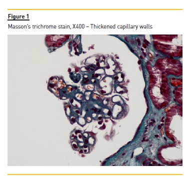
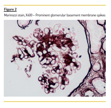
Kidney biopsy revealed a total of 11 glomeruli, absence of crescents and proliferation, glomerular membrane basement thickening and glomerular membrane basement spikes, evident on Massons trichrome (Fig. 1) and Marinozzi staining (Fig. 2). Immunofluorescence was positive only for IgG with granular subepithelial deposits. Histological characterization of IgG subtypes (1 to 4) was not available and neither was biopsy study for the reactivity for PLA2 receptor in the deposits. The final diagnosis was of MN.


He was started on low-sodium and low-protein diet, ACE inhibitor, statin, diuretic and pentoxifylline. After a one-year follow-up, the patient maintained proteinuria of 5g/day, hypoalbuminemia (serum albumin of 3.1mg/dL), and presented with hypertension and a decline in renal function (serum creatinine of 1.6mg/dL), with periorbital and lower limb oedema. Due to lack of responsiveness to conservative therapy, before starting immunosuppressive therapy, active malignancy was once again excluded and serological phospholipase A2 receptor antibody was determined, and positive, but at a borderline level (result: 10, positivity cutoff³10) by indirect immunofluorescence method.
Despite discarding active malignancy and other secondary causes, patients past history and renal function deterioration had to be taken into account when deciding on immunosuppressive therapy. Additionally, the borderline level of antiPLA2 receptor antibody did not support a diagnosis of Idiopathic MN (iMN). Accordingly, immunosuppressive therapy was started with Rituximab.
Patient received two doses of intravenous (IV) rituximab, of 1g each, with a fourteen-day interval, with IV methylprednisolone 500mg. No adverse effects of Rituximab occurred. At 6 month follow-up after rituximab, there were no adverse effects of therapy; proteinuria decreased at three months to 800 mg/day with oedema resolution, serum albumin correction to 3.8 g/dL and serum creatinine recovery to 1.2 mg/dL.
At one year of follow-up the patient remains asymptomatic and without oedema, with proteinuria of 500 mg/day, stable renal function (serum creatinine of 1.2 mg/dL, normoalbuminaemia, normal lipid profile. The titration of phospholipase A2 receptor antibody was repeated at 11 months follow-up and was negative. His CD19+ lymphocytes remained depleted at the 6th, 8th and 12th month after rituximab.
DISCUSSION
MN is the most common cause of nephrotic syndrome in Caucasian adults, presenting primarily as nephrotic syndrome, usually without hypertension, and normal kidney function. Idiopathic and secondary forms have similar clinical presentations. iMN is diagnosed after excluding all secondary causes.1-3
Some histological patterns are suggestive of iMN. These include granular IgG4 deposits1-3, without other immunoglobulin subtypes; no cell proliferation; and only subepithelial deposits. Mesangial or tubular deposits of IgG1, IgG2, IgA or IgM suggest a secondary cause for MN1-3.
Recent advances have demonstrated that iMN is an autoimmune disease and the establishment of its pathogenic mechanism is presently under investigation.
In fact, most of our knowledge of the pathogenesis of iMN has been based on experimental models such as the Heyman nephritis model, which have shown that immune deposits are formed in situ by the reaction of autoantibodies against specific podocyte antigens.1-3,6
Podocyte proteins can also act as autoantigens, as seems to be the case of M-type phospholipase A2 receptor (PLA2R), which can be found in approximately 70% of iMN patients (but only rarely in other glomerulonephritides).1-6,14 There are other antigenic targets, such as Thrombospondin Type-1 Domain-Containing 7A, under investigation7. But there are still many unanswered questions; the podocyte damage in experimental models is complement-mediated, but in humans, although the presence of complement within the subepithelial deposits is well established, IgG4, which represents the predominant subclass of IgG anti-PLA2R, does not activate complement by classical or alternative pathways. Some evidence suggests that IgG4 anti-PLA2R autoantibodies can bind mannan-binding lectin and activate the lectin complement pathway.4,5
Presently, antibodies against phospholipase A2 receptor may represent a specific marker for iMN1,14. The determination of the serum titer of these antibodies has diagnostic and prognostic value1,14. The serological levels of anti-PLA2R correlate with the disease activity and proteinuria; they become undetectable in disease remission and elevate before relapse. Thus, serological testing before and during immunosuppression can be used to predict remission and monitor treatment response.1,6,14
In our case, MN diagnosis was established according to the histological finding of thickened basement membrane with granular subepithelial IgG deposits.
All secondary causes for MN were excluded. The biopsy was consistent with the diagnosis of an idiopathic form of MN as there were no deposits of other types of immunoglobulins, no mesangial or subendothelial deposits. Although we were unable to characterize the subtypes of IgG, the positive PLA2R antibodies suggest iMN, despite their low titer.
This distinction was especially important in this case, with a previous history of two malignancies which could be correlated with MN. The identification of an active malignancy would have prognostic and therapeutic impact on the medical approach.
MN is a chronic disease, with spontaneous remission and relapses. Approximately 30% of patients will experience spontaneous remission that can occur up to 2 years after presentation. The other two thirds divide equally into those who maintain proteinuria, and those who progress to end stage kidney disease.1-3,14
Evaluating the prognosis is critical. Older age, male gender, hypertension, low albumin, high creatinine, persistent nephrotic range proteinuria, high IgG, beta-2-microglobulin and C5b9 urinary excretion, focal glomerular sclerosis and tubulointerstitial disease are predictors of poor outcome and higher risk for renal disease progression.2
Initial therapy should be supportive and involves restricting dietary protein and sodium intake, controlling blood pressure, hyperlipidaemia, and oedema.
Pentoxifylline may be used in MN due to its anti-inflammatory and protective effects and it has been demonstrated to decrease proteinuria in patients with chronic kidney disease.3 Immunosuppressive therapy is started if at 6-months the patient maintains proteinuria, or if patient has higher risk for disease progression. First-line immunosuppression is a combination of corticosteroids and alkylating agents, or calcineurin inhibitors (CNIs)2,3,8,10. The treatment of iMN remains controversial because of the high rate of spontaneous remission and side effects from the alkylating agents and renal toxicity of CNIs10.
As alternative therapies, Rituximab has generally demonstrated beneficial outcomes with limited toxicity and has been proposed as a first-line therapy11,13. In treatment-resistant patients, mycophenolate mofetil (MMF) plus steroids has been demonstrated to induce modest reduction in proteinuria and increased remission15,16 with risk of severe infection and malignancy, and ACTH has demonstrated antiproteinuric and renoprotective effects without significantly increasing the risk of infections17,18.
This 71-year-old male patient had a one-year followup with conservative therapy maintaining proteinuria, hypoalbuminaemia, oedema, hypertension and a decline in renal function. As the patient was at high risk of disease progression, a decision was made to start immunosuppression. Considering the patients history of malignancy, alkylating agents and MMF were excluded as a therapeutic option; the past malignancies and the deteriorated renal function precluded CNI use; consequently, rituximab was considered as first-line immunosuppressive therapy.
Rituximab is a monoclonal antibody against the cellsurface protein CD20, a molecule selectively expressed in cells of B lymphocyte lineage. Recent data suggests that B lymphocyte depletion with Rituximab is an effective and safe treatment approach for first-line therapy of MN, with less adverse effects and relapses demonstrated10-12,14,15. The MENTOR trial12 will provide some insight into the comparison between rituximab and cyclosporine before widespread use of this agent, given its high cost and unknown long-term toxicity.
A recently presented randomized controlled trial11,14 demonstrated that the addition of rituximab to nonimmunosuppressive antiproteinuric therapy leads to significant improvements in immunologic and clinical outcomes, not only at 6 months, but extending to a 24-month observational period. Although the remission rate and time to remission were similar for both groups at month 6, during the observation phase the rates of remission were significantly higher in the rituximab group. In this study, the remission was associated with the titer of anti-PLA2R, and rituximab showed a dramatic effect on the anti-PLA2R titer. Moroni et al19 demonstrated partial response in a small number of patients and complete remission in a minority of patients treated with low-dose rituximab, which might mean that perhaps higher doses and longer treatments are necessary to treat MN, particularly in patients with high titers of anti-PLA2R antibodies. The GEMRITUX trial20 also demonstrated rituximab efficacy on proteinuria remission after 6 months in patients treated with rituximab and non-immunosuppressive antiproteinuric therapy, without increasing adverse effects.
Thus, rituximab seems a promising therapy for MN not only in those patients with contraindication for alkylating agents or calcineurin inhibitors, but in other subgroups of patients, namely those maintaining high titers of anti-PLA2R. Nevertheless, the optimal dosing of rituximab is yet to be established.
Given the risks of CKD progression and nephrotic syndrome complications, we believe that treating this patient with rituximab was the best decision. Despite the histological, laboratory and complementary exam data previously mentioned favouring the hypothesis of an iMN, only long term follow-up will allow us to conclude if this was indeed the case.
References
1. Ronco P, Debiec H. Pathophysiological advances in membranous nephropathy: time for a shift in patients care. Lancet 2015;385:1983-1992 [ Links ]
2. Fervenza F, Sethi S, Specks U. Idiopathic membranous nephropathy: diagnosis and treatment. Clin J Am Soc Nephrol 2008;3:3905-3919 [ Links ]
3. Lai WL, Yeh TH, Chen PM, et al. Membranous nephropathy: a review on the pathogenesis, diagnosis, and treatment. J Formosan Med Association 2015;114:102-111 [ Links ]
4. Sinico RA, Mezzina N, Trezzi B, et al. Immunology of membranous nephropathy: from animal models to humans. Clin Exp Immunol 2016 Feb;183(2):157-165 [ Links ]
5. Bally S, Debiec H, Ponard D, et al. Phospholipase A2 receptor-related membranous nephropathy and mannan-binding lectin deficiency. J Am Soc Nephrol 2016; epub ahead of print [ Links ]
6. Francis JM, Beck LH Jr, Salant DJ. Membranous nephropathy: a journey from bench to bedside. Am J Kidney Dis 2016; epub ahead of print [ Links ]
7. Tomas N, Beck LH Jr, Meyer-Schwesinger C, et al. Thrombospondin type-1 domaincontaining 7A in idiopathic membranous nephropathy. N Engl J Med 2014;371(24):2277-2287 [ Links ]
8. Kidney disease: Improving global outcomes (KDIGO) glomerulonephritis work group. KDIGO clinical practice guideline for glomerulonephritis. Kidney IntSuppl 2012;2:124-138 [ Links ]
9. Laudari S. Membranous glomerulonephritis – A case report. J Coll Med Sciences-Nepal 2010;(6)3:64-67 [ Links ]
10. Tran T, J Hughes G, Greenfeld C, et al. Overview of current and alternative therapies for idiopathic membranous nephropathy. Pharmacotherapy 2015;35(4):396-411 [ Links ]
11. Cravedi P, Remuzzi G, Ruggenenti P. Rituximab in primary membranous nephropathy: first-line therapy, why not? Nephron Clin Pract 2014;128:261-269 [ Links ]
12. Fervenza F, Canetta PA, Barbour SJ, et al. A multicenter randomized controlled trial of rituximab versus cyclosporine in the treatment of idiopathic membranous nephropathy (MENTOR). Nephron 2015;130(3):159-168 [ Links ]
13. Bomback A, Derebail V, McGregor J, et al. Rituximab therapy for membranous nephropathy: a systematic review. Clin J Am Soc Nephrol 2009;4:734 –744 [ Links ]
14. Ruggenenti P, Debiec H, Ruggiero B, et al. Anti-phospholipase A2 receptor antibody titer predicts post-rituximab outcome of membranous nephropathy. J Am Soc Nephrol 2015;26(10):2545-2558 [ Links ]
15. Branten AJ, du Buf-Vereijken PW, Vervloet M, et al. Mycophenolate mofetil in idiopathic membranous nephropathy: a clinical trial with comparison to a historic control group treated with cyclophosphamide. Am J Kidney Dis 2007;50:248 [ Links ]
16. Miller G, Zimmerman R 3rd, Radhakrishnan J, et al. Use of mycophenolate mofetil in resistant membranous nephropathy. Am J Kidney Dis 2000;36:250 [ Links ]
17. Kittanamongkolchai W, Cheungpasitporn W, Zand L. Efficacy and safety of adrenocorticotropic hormone treatment in glomerular diseases: a systematic review and metaanalysis. Clin Kidney J 2016;9(3):387-396 [ Links ]
18. van de Logt AE, Beerenhout CH, Brink HS, et al. Synthetic ACTH in high risk patients with idiopathic membranous nephropathy: a prospective, open label cohort study. PLoS ONE 2015;10(11):e0142033 [ Links ]
19. Moroni G, Depetri F, Vecchio L, et al. Low-dose rituximab is poorly effective in patients with primary membranous nephropathy. Nephrol Dial Transplant 2016;0:1-6 [ Links ]
20. Dahan K, Debiec H, Plaisier E, et al. Rituximab for severe membranous nephropathy: a 6-month trial with extended follow-up. J Am Soc Nephrol 2016; epub ahead of print [ Links ]
Joana Gameiro
Service of Nephrology and Renal Transplantation,
Department of Medicine
Hospital de Santa Maria, Centro Hospitalar Lisboa Norte, EPE
Av. Prof. Egas Moniz, 1649-035 Lisboa, Portugal
Telephone +351939811447
E-mail: joana.estrelagameiro@gmail.com
ACKNOWLEDGEMENTS
Professor M. Praga (Department of Nephrology, Hospital Universitario Doce de Octubre, Madrid, Spain) for the kind discussion of the case and Professor João Martin Martins (Department of Endocrinology, HSM, CHLN, Lisbon) for the medical co-work.
Received for publication: May 14, 2016
Accepted in revised form: Jan 30, 2017


