Serviços Personalizados
Journal
Artigo
Indicadores
-
 Citado por SciELO
 -
 Acessos
Links relacionados
-
 Similares em
SciELO
Compartilhar
Portuguese Journal of Nephrology & Hypertension
versão impressa ISSN 0872-0169
Port J Nephrol Hypert vol.31 no.2 Lisboa jun. 2017
ORIGINAL ARTICLE
Renal pathology in HCV infected patients – Report of 148 patients and review of the literature
Isabel Mesquita, Helena Sousa, Fernanda Carvalho, Fernando Nolasco
Nephrology Department, Hospital Curry Cabral, Centro Hospitalar de Lisboa Central, Lisboa, Portugal
ABSTRACT
Background: Hepatitis C virus infection (HCV) is a major public health problem with a reported incidence of 3‑4 million cases per year. Renal injury secondary to HCV was initially observed in autopsy studies and later in kidney biopsies. Several types of renal disease have been recognized in association with HCV patients. Objectives: Characterize the type of renal disease found in HCV‑infected patients and established as possible relation with clinical presentation. Methods: Unicentric retrospective study of HCV patients with a renal biopsy from January 1988 to December 2015. The clinical data at biopsy time was analyzed according to histological diagnosis. Results: HCV infection was present in 148 cases. Male gender was predominant (76.7%), as was Caucasian race (79.1%). Mean age was 41.46±11.47years. Histological study of renal biopsies revealed membranoproliferative glomerulonephritis (MPGN) type 1 to be the commonest lesion encountered (37.2%), followed by proliferative glomerulonephritis (16.9%), focal segmental glomerulosclerosis (FSGS) (10.1%), and tubulointerstitial nephropathy (10.1%). Other patterns (amyloidosis, diabetic nephropathy, membranous nephropathy, IgA nephropathy) were observed. Hypocomplementaemia and cryoglobulinaemia showed correlation with MPGN diagnosis. A statistically significant correlation was observed with human immunodeficiency virus (HIV) infection and FSGS diagnosis. Amyloidosis diagnosis was associated with advanced age. No other significant correlations were found. Conclusions: Renal disease in HCV patients has a broad spectrum. No strong correlations between clinical data and pattern of renal disease have been established and it seems that is not possible to predict the renal disease based on clinical criteria alone. Renal biopsy remains the gold standard for diagnosis.
Key Words: hepatitis C virus infection; renal biopsy; renal disease
INTRODUCTION
Hepatitis C virus (HCV) infection is a major public health problem with a reported incidence of 3‑4 million cases per year1. It is known that 3% of the worlds population is currently infected with HCV2. Hepatitis C is a major cause of liver‑related morbidity3 and accounts for over 1 million deaths as a result of cirrhosis and liver cancer4,5.
HCV infection leads to chronic liver disease6, but also should be considered as a systemic disorder which is often associated with a number of extrahepatic manifestations7,8. These include porphyria cutanea tarda, type 2 diabetes mellitus, lymphoproliferative disorders, cryglobulinaemia and renal diseases9.
Almost 40% of patients with HCV develop at least one extrahepatic manifestation during the course of the disease2.
Several types of renal disease have been recognized in association with HCV infection10,11 Membranoproliferative glomerulonephritis (MPGN), is thought to be the most common histological pattern, whether cryoglobulinaemic or noncryoglobulinemic12,13. An association between HCV infection and FSGS13,14, IgA nephropathy15,16 and membranous nephropathy17 has also been reported. Other forms of renal disease have been previously linked to chronic HCV infection, including proliferative glomerulonephritis18, fibrillary and immunotactoid glomerulopathy, thrombotic microangiopathies and postinfectious glomerulonephritis9,19. However, glomerular disease is not the only type of renal manifestation of chronic viral hepatitis. Tubular injury may also be present20.
This retrospective study aims to characterize the type of renal disease found in Portuguese HCV‑infected patients and establish possible relationships between clinical and laboratory data.
PATIENTS AND METHODS
We performed a retrospective study, reviewing all native biopsies of patients with positive HCV serology (n=148) analyzed in our Nephropathology Department, between January 1988 and December 2015. All had renal dysfunction and/or abnormal findings on urinalysis.
Proteinuria was defined as protein urinary excretion of more than 300mg/24h and haematuria was defined as urine sedimentary red blood cell counts of more than five in microscope x400 visual field.
Tissues for optical microscopy were stained with haematoxylin‑eosin, periodic acid Schiff, Massons trichrome, methenamine silver and Congo red. Immunofluorescence (IF) was performed in frozen sections, using labeled human immunoglobulin (IgA, IgG, IgM), light chains (λ e κ), complement (C1q, C3), and fibrinogen.
When no frozen fragment was available, indirect immunoperoxidase using formalin‑ fixed paraffin embedded section was performed. Amyloid substance was characterized using immunofluorescence or immunochemistry staining for SAA or κ and λ light chains.
When required, electron microscopy was performed. Clinical and laboratory data at the time of biopsy were obtained: demographic (age, gender), clinical (presence of renal failure, nephrotic syndrome, hypertension, microscopic haematuria, nephritic syndrome, purpura, other viral co‑infections, chronic hepatic disease), laboratory (serum creatinine, proteinuria, haematuria, cryoglobulins, hypocomplementaemia).
We searched for relationships between renal histological findings and clinical data.
Mean and standard deviation were used to characterize quantitative variables. Qualitative variables were analyzed using absolute and relative frequencies. Background factors were compared with the different histological patterns by using the qui‑square test for categorical variables and the Kruskal‑Wallis test for comparison among the groups. A p value <0.05 was considered significant. All tests were performed using SPSS, version 22.
RESULTS
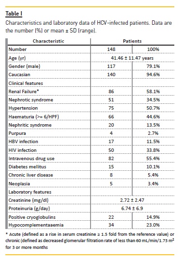
From January 1988 to December 2015, 148 biopsies of native kidney of HCV‑infected patients were analyzed in our department. The baseline clinical characteristics of HCV patients are summarized in Table I.

Male gender (79.1%) and Caucasian race (94.6%) were predominant. Mean age was 41.46 ± 11.47 years. 10.1% (n=15) of the patients had diabetes mellitus and 5.4% (n=8) had chronic liver disease. Co‑infections with HIV and hepatitis B virus (HBV) were present in 33.8% (n=50) and 53.3 (n=17), respectively. 82 patients (55.4%) had past or present history of intravenous drug use.
The mean serum creatinine level was 2.72 ± 2.47mg/ dl (0.4 – 12.0), 24‑hour protein excretion was 6.74 ± 6.9 g (0.30 – 30.6), and haematuria was positive in 66 of the 148 patients (44.6%). Cryoglobulinaemia was detected in 22 patients (14.9%) and hypocomplementaemia in 34 patients (23.0%).
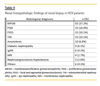
Ten different histological findings were recognized (Table II).

The predominant lesion was MPGN (n=55, 37.2%), followed by PGN (n=25, 16.9%), FSGS (n=15, 10.1%) and tubulointerstitial nephropathy (n=15, 10.1%). Amyloidosis was the histological diagnosis in 10 patients and diabetic nephropathy in nine.
The histological and clinical features and the major cause for performing renal biopsy are summarized in Table III and Table IV, respectively.
Histological feature
1. MPGN
MPGN was diagnosed in 55 of the 148 patients. All patients were Caucasian and eighteen patients were co‑infected: 12 had HIV and six had HBV. Intravenous drug use was identified in 37 of 55 patients. No patients had clinical or serological signs for lupus. Mean Scr was 2.0±1.02 mg/dl, and mean proteinuria was high (6.5±5.38g). Haematuria was present in 29 (53%) patients.
Cryoglobulinaemia was present in 35% (n=19 patients) of the group and hypocomplementaemia in 38% (n=21 patients). Three patients presented purpura, all with cryoglobulinaemia and one with hypocomplementaemia.
2. Proliferative glomerulonephritis
Caucasian race was predominant (92%). Seven patients were co‑infected with HIV and two with HBV.
Sixteen patients (64%) were intravenous drug users.Mean Scr was 2.2±1.50 mg/dl, mean proteinuria was high (5.0±5.46g), and two thirds had haematuria. No patients had clinical or serological signs for lupus. Cryoglobulinaemia was present in two patients and hypocomplementaemia in eight patients.
Renal histology was characterized by two different findings. In 23 patients, diffuse proliferative endocapillary glomerulonephritis, with hypercellularity of all glomeruli, no significant interstitial infiltration or tubular atrophy, and positive IMF for C3, IgG and IgM were found. In two patients, crescentic glomerulonephritis, and positive IMF for C3 and IgM was observed. ANCA was negative.
3. FSGS
Fifteen patients had a histological pattern of focal and segmental glomerulosclerosis (FSGS). Mean age of these patients was 37±9.69 years and Caucasian race was predominant (93.3%). We found significant proteinuria (6.1±4.91g), with 80% of patients having nephrotic proteinuria. HIV co‑infection was present in ten patientS and HBV in five (all intravenous drug users).
Sclerotic lesion location was extremely heterogeneous, without specific histologic variant. IMF was positive with segmental deposits of C3, IgM and IgG in 8/15 patients.
Collapsing FSGS was found in one HCV‑ infected patient. This biopsy was obtained from a 28‑year‑old black male, intravenous drug user and HIV co‑infected.
He presented with heavy proteinuria (20g/day) and rapidly progressive renal insufficiency (Scr 8.6 mg/dl).
Renal histology was characterized by microcystic distortion of the tubules. IMF was not available.
4. Tubulointerstitial nephropathy
A predominant interstitial pathology was present in 15 patients. All patients had normal glomeruli and marked interstitial nephritis. Eight biopsies had features of chronicity.
The proteinuria was non‑nephrotic in 54% of the cases (1.3±0.61 g/24h), 5 patients had haematoproteinuria and in all patients renal function was significantly deteriorated (3.90±2.52 mg/dl).
5. Amyloidosis
Amyloidosis was found in ten HCV‑infected Caucasian patients. All presented with nephrotic proteinuria (mean proteinuria 10.5±8.29g), including 8 with nephrotic syndrome. The majority of these patients were male (n=9) and this diagnosis mainly occurred in older patients (51.80±12.76 years). Renal function was deteriorated: Scr 2.4±1.55mg/dl. Two patients had primary amyloidosis.
In the patients with AA amyloidosis (n=8), six had pulmonary tuberculosis or chronic respiratory disease (obstructive pulmonary disease and bronchiectasis). Six patients were intravenous drug users, all with amyloidosis AA. Glomeruli were sclerosed and had mesangial nodules. Fibrosis and interstitial infiltrates were moderate.
6. Diabetic nephropathy
This lesion was present in nine HCV‑infected patients. Caucasian race was pre‑dominant (78%). All patients had renal insufficiency (mean Scr 2.5±2.42mg/dl). Mean proteinuria was 6.3±2.88g and HBV co‑infection was present in one patient.
7. IgA nephropathy
Six of the 148 patients were diagnosed as having IgAN. A small amount of C3 was detected in 4 of the 6 patients. All patients had proteinuria (4.1±3.41 g/24h), 50% in the nephrotic range and one had normal renal function.
Haematuria was detected in three patients. Co‑infection with HIV was identified in two patients, one of which had severe renal impairment (Scr 8.0 mg/dl).
8. Membranous nephropathy
Four patients were diagnosed as having membranous nephropathy. All patients had proteinuria (3 in nephrotic range and with nephrotic syndrome) and 1 had also microhaematuria and presented with nephritic syndrome. Renal failure (Scr >1.5mg/dl) was present in all patients (2.9±1.55 mg/dl). Two patients were co‑infected with HIV. IF showed diffuse granular deposits of IgG and C3 in the capillary walls.
9. Other renal diseases
Nefroangiosclerosis: three male patients, one presented with anuric kidney injury due to malignant nephroangiosclerosis
Minimal change lesion: two HCV‑infected patients, one with a Hodgkin lymphoma
Microscopic polyarteritis: two HCV‑infected females, without any viral co‑infections. Patients had crescent formation and IF showed no deposition of immunoglobulins or complement. All cases had positive anti‑myeloperoxidase‑neutrophil cytoplasmic antibodies (MPO‑ANCA).
Anti‑GBM disease: one 43‑year‑old Caucasian male, without any viral co‑infections
Acute tubular necrosis (ATN): one HCV‑HIV co>‑infected male, with chronic liver disease
Comparison between histological and clinical data
The clinical findings within distinct histological types were compared.
Statistically significant results were found between patients ages and histological diagnosis (p=0.018). The data suggested that older patients had a higher frequency of amyloidosis (M=51.80, SD=12.76), while younger patients had a higher frequency of FSGS (M = 36.93, SD = 9.69), PGN (M = 37.92, SD = 10.92), or even MPGN (M = 40.05, SD = 11.67).
The incidence of hypocomplementaemia was twice as high in the MPGN group (61.8%), a statistically significant difference (p=0.013). No patient with FSGS or MN presented hypocomplementaemia.
A statistically significant association was found between the histological diagnosis and the presence of cryoglobulinaemia (p<0.001). Patients with this condition had a much higher prevalence of MPGN (86.4%) than patients without cryoglobulinaemia (28.6%).
HIV co‑infected patients had a lower prevalence of MPGN diagnose (24%) than non‑HIV patients (43.9%).
On the other hand, HIV infection seems to be related to the diagnosis of FSGS (20.0%) and TIN (18.0%). The association between histological diagnosis and HIV infection was statistically significant (p=0.004).
Intravenous drug use presented a statistically significant relationship with histological diagnosis (p=0.003). Patients with drug addiction had a higher prevalence of MPGN diagnosis (45.1%) than patients without drug dependence (27.3%).
The diagnosis of type 2 diabetes mellitus presented a statistically significant relationship with histological diagnosis (p<0.001). Diabetic nephropathy represented 60% of the total diagnosis in diabetic patients.
Regarding gender there was a predominance of males in the MPGN group (40.2%) although statistically non‑significant (p=0.408).
The serum creatinine level was not significantly different between the groups. Nevertheless all patients with TIN diagnose had plasma creatinine levels >1.5 mg/dl.
There was statistically no significant difference regarding incidence of chronic liver disease, HBV infection and proteinuria level (p=0.561, 0.889, and 0.119 respectively).
DISCUSSION
Chronic HCV viraemia is present in 150–170 million people worldwide21. Kidney disease is regarded as one of the most important of the extrahepatic manifestations of HCV infection22.
With the current availability of direct‑acting antivirals (DAA), which can achieve a viral remission of >95% for most HCV genotypes, the prevalence of glomerular disease in this population should progressively decline in the coming decade23.
HCV‑associated glomerular disease is primarily a consequence of viral antigen – immune complex formation with glomerular basement membrane deposition21. The expansion of HCV‑reactive B‑cells leads to antibody production, including cryoprecipitating antibodies24. The classic pathologic hallmark of HCV renal disease is type 1 MPGN as a consequence of type 2 mixed cryoglobulinaemia (MC), although the same renal lesion may also develop from noncryoglobulinaemic immune complexes25.
In addition, HCV is also an important cause of polyarteritis nodosa (PAN) from immune complex deposition in medium size blood vessels leading to renal ischaemia and infarction similar to HBV PAN and idiopathic PAN21.
The pattern of glomerular injury will depend on the physic‑chemical characteristics of the immune‑complexes such as their size and charge, their rate of production and factors such as phagocytic clearance and quantity of complexes reaching the kidney11.
The typical renal manifestations in HCV‑infected patients include proteinuria, microscopic haematuria, hypertension, acute nephritis and nephrotic syndrome. However, mostly the renal disease is asymptomatic, and thus patients with HCV infection should be screened for proteinuria, haematuria, hypertension and cryoglobulinaemia9.
Various other forms of histological types of renal diseases have been described in HCV patients, mesangial proliferative and focal proliferative GN, IgA nephropathy, membranous nephropathy (MN), focal segmental glomerulosclerosis (FSGS), fibrillary glomerulonephritis, immunotactoid glomerulopathy, renal thrombotic microangiopathy, vasculitic renal involvement and interstitial nephritis albeit far less commonly than MPGN9,17,19,21.
It is important to note that renal dysfunction in a patient with HCV is rarely a result of GN (<10%) and hospital admissions for MPGN from HCV have been decreasing for the past decade26. The majority of renal diseases in HCV patients with liver disease are a consequence of acute tubular necrosis, hepatorenal syndrome, prerenal azotaemia, and CKD and therefore it is essential to be familiar with the typical presentation and characteristics of acute GN in HCV patients and how to differentiate them from the more common diagnoses causing AKI in this population21,26.
In our review, the histological lesions associated with HCV infection were MPGN (55 cases), followed by PGN (25 cases), FSGS (15 cases) and tubulointerstitial nephropathy (15 cases). Amyloidosis was the histological diagnose in 10 patients and diabetic nephropathy in nine. IgA nephropathy was found in six cases and membranous nephropathy in two.
The prevalence of HCV infection in MPGN varies between 15% and 60% in different series and rises to 96% when cryoglobulinaemia is also present11,19.
In our patients with HCV‑associated MPGN, hypertension and microscopic haematuria were the main clinical features followed by renal failure and nephrotic syndrome. These clinical findings are consistent with those described in the literature27.
In cases of cryoglobulinemic glomerulonephritis, one‑third of these patients may have vasculitis of small renal arteries however extracapillary crescents are rarely seen19. IF microscopy may reveal C3, IgM and IgG depositions on the capillary wall and mesangium.
Intraluminal and subendothelial deposits may have a fibrillary pattern on electron microscopy probably representing cryoglobulin deposition1,19.
In our study, cryoglobulinaemia was present in 35% of all HCV‑positive patients. The clinical presentation was not different from that of patients without cryoglobulinaemia, and there were no differences in levels of plasma creatinine and proteinuria. A statistically significant correlation was observed between cryoglobulinaemia and the diagnosis of MPGN. Johnson et al.28 found similar results in a group of 35 patients with MPGN, 31.4% having cryoglobulinaemia.
Interestingly, classical signs/symptoms of cryoglobulinaemia such as palpable purpura, arthritis, or neuropathy, are present in only 30–50% of patients with HCV and a MPGN19,29. In our patients palpable purpura was identified only in four patients, three of them with diagnosis of MPGN and positive cryoglobulins. Many questions remain unanswered on the relationship between the occurrence of cryoglobulinaemia and the duration of HCV infection, viral load, viral genotype, the number of mutations and cytokine profile19,21.
Hypocomplementaemia is a distinguishing feature of MPGN, with C4 being low in 93% of patients, whereas C3 is depressed in only 53%6,21. In our study, hypocomplementaemia was present in 38% of all HCV‑positive MPGN patients, a feature statistically significant.
Proliferative glomerulonephritis was the second most frequent histological diagnosis. About one‑third of patients had hypocomplementaemia but only a small percentage had positive cryoglobulins. Reports about the association between HCV and FSGS have been published30‑32.
Confounding factors must be stressed for FSGS. For example, intravenous drug abuse and HIV infection are common causes of both FSGS and HCV infection19,31. In this study, FSGS was the histological diagnosis in 10% of patients and nephrotic proteinuria was the main clinical feature. Two thirds were co‑infected with HIV and 80% were intravenous drug user. These concurrent predisposing conditions could explain a much higher prevalence of the FSGS diagnosis in our patients. Furthermore, HIV infection showed a significant association with the diagnosis of FSGS.
Association of IgA nephropathy with HCV infection has also been described in several reports15,16,33. Fabrizi et al.34 reviewed the association of HCV infection and glomerulonephritis and found that 6% of patients with IgA nephropathy were HCV‑positive.
On the other hand, Ström et al.35 analysed the association between IgA and HCV infection from an autopsy series in cirrhotic patients: of 27 patients, 13 had IgA nephropathy and only two were HCV infected, suggesting that HCV does not play a role in the pathogenesis of IgA nephropathy associated with cirrhosis. These contradictory findings suggest that confounding factors must be taken into account, such as HBV infection and alcohol consumption19.
Regarding IgA nephropathy, our prevalence in HCV infection was lower than in other reported series. Several authors have suggested that the cases of IgA nephropathy were probably secondary to chronic hepatic disease and not directly related with the HCV infection11. However, in our study no association was found between the presence of chronic liver disease and the diagnosis of IgAN.
Several cases of MN have been described in HCV‑infected patients36,37. The clinical presentation and the histological findings are usually similar from that occurring in HCV‑negative patients. Nevertheless, atypical features, such as glomerular hypercelullarity and/or mesangial and subendothelial immuno‑complexes, are more often associated with the secondary form of membranous nephropathy related to HCV‑infection than with the idiopathic disease11. Published series (38, 39) support a possible pathogenic role of HCV in the development of MN. However, there was no increased prevalence of MN associated with HCV in the cohort of renal transplants in the Saint Etienne study40, as well as in the Maiza et al. series41. As with IgA nephropathy, the possibility of confounding factors, such as HBV infection and causes of secondary membranous glomerulopathies must be taken into consideration11,19.
In our patients a much lower percentage of MN (2.7%) was found. No association with HBV infection was established.
The prevalence of HCV positivity in type 2 diabetic population ranges between 1.7% and 12.1%42. A high prevalence of HCV infection has also been observed in patients with diabetic nephropathy43. After kidney transplantation, HCV infection has been identified as a predictive factor for DM44. HCV infection might also alter the progression of diabetic‑related nephropathy45,46.
In our study, 10% of patients had diabetes mellitus and 9 were diagnosed as having diabetic nephropathy.
The high prevalence of HCV‑positive patients with diabetes mellitus and diabetic nephropathy may reflect the association between HCV and diabetes.
There are reports of renal amyloidosis in HCV infected patients. AA amyloid was described in patients with hepatosplenic schistosomiasis and HCV infection47,48.
Renal AA amyloidosis is most seen in intravenous drug users with a history of skin‑popping and suppurative skin infections49. Intravenous drug abusers are also a population at risk for developing HCV infection and multiple others risk factors for renal disease (HBV and HIV infection). The state of immunosuppression of this population makes it also more susceptible to serious infections that may also contribute to the development of secondary amyloidosis50.
HCV is a well‑known risk factor for lymphoproliferative disorders51,52. In AL amyloidosis, it is possible that the proliferation of monoclonal B cells resulted from HCV infection associated with chromosomal rearrangement, which led to the production of amyloid proteins.
In our study, ten patients were diagnosed with amyloidosis, two with primary amyloidosis. In all patients with AA amyloidosis risk factors were identified for development of secondary amyloid deposition.
Advanced age was related to amyloidosis diagnosis. Regardless of the tubulointerstitial injury associated with different glomerular lesions, HCV may lead to tubular injury in its own right48. Kasuno et al.20 report that tubulointerstitial changes are observed frequently in HCV‑infected patients, and the viral antigen and RNA of HCV were detected in the tubulointerstitium of these patients.
TIN was identified in 10% of our patients. It is interesting to note that all patients presented serum creatinine levels > 1.5 mg/dl, though without statistical significance. These results support the literature findings that HCV infection is a potent pathogenic factor of tubulointerstitial injury.
Although current findings suggest that HCV infection is a potent pathogenic factor of tubulointerstitial injury, further prospective studies are required to prove the causal roles of HCV in the initiation or progression of TIN.
Other types of glomerulopathy in association with HCV have been described, but they are mainly case reports. Six cases of fibrillary‑immunotactoid Glomerulopathies have been described in association with HCV infection53‑55.
Thrombotic microangiopathy has been found associated with cryoglobulinaemic MPGN and hepatitis C in two patients56. Antineutrophil cytoplasmic antibodies (ANCA) are frequently found patients HCV infected57,58. No correlation has been found between ANCA positivity and the presence of clinically active vasculitis in these patients59 and only a paucity of cases of ANCA‑associated vasculitis in patients with HCV have been reported60. The rarity of these glomerulopathies makes the causal relationship with HCV infection difficult to demonstrate and would require multicenter studies19.
CONCLUSION
Patients infected with HCV can develop diverse renal pathologies. No strong correlations between clinical data and pattern of renal disease have been seen and it seems that is not possible to predict the renal disease based on clinical criteria alone. Renal biopsy remains the gold standard for establishing the diagnosis and can help predict renal prognosis.
References
1. Ozkok A, Yildiz A. Hepatitis C virus associated glomerulopathies. World J Gastroenterol 2014; 20: 7544–7554 [ Links ]
2. Cacoub P, Renou C, Rosenthal E, Cohen P, Loury I, Loustaud‑Ratti V, Yamamoto AM, Camproux AC, Hausfater P, Musset L, Veyssier P, Raguin G, Piette JC. Extrahepatic manifestations associated with hepatitis C virus infection. A prospective multicenter study of 321 patients. The GERMIVIC. Groupe dEtude et de Recherche en MedecineInterne et Maladies Infectieuses sur le Virus de lHepatite C. Medicine 2000; 79: 47–56 [ Links ]
3. Sandri AM, Elewa U, Poterucha JJ, Fervenza FC. Treatment of hepatitis C‑mediated glomerular disease Nephron Clin Pract 2001; 199: 121–130 [ Links ]
4. Williams R. Global challenges in liver disease. Hepatology 2006; 44: 521–526 [ Links ]
5. Alter MJ. Epidemiology of hepatitis C virus infection. World J Gastroenterol 2007; 13:2436–2441 [ Links ]
6. Rostaing L, Izopet J, Kamar N. Hepatitis C virus infection in nephrology patients. J Nephropathol 2013; 2: 217–233 [ Links ]
7. Cacoub, P, Costedoat‑Chalumeau, N, Lidove, O, Alric, L. Cryoglobulinemia vasculitis. Curr Opin Rheumatol 2002; 14: 29 [ Links ]
8. Galossi A, Guarisco R, Bellis L, Puoti C. Extrahepatic manifestations of chronic HCV infection. J Gastrointestin Liver Dis 2007; 16: 65–73 [ Links ]
9. Perico N, Cattaneo D, Bikbov B, Remuzzi G. Hepatitis C infection and chronic renal diseases. Clin J Am Soc Nephrol 2009; 4: 207–220 [ Links ]
10. Sabry, AA, Sobh, MA, Irving, WL et al. A comprehensive study of the association between hepatitis C virus and glomerulopathy. Nephrol Dial Transplant 2002; 17: 239 [ Links ]
11. Frutuoso M, Eduardo Vazquez‑Martul, E. Glomerular disease and hepatitis C. Fifteen years experience. Port J Nephrol Hypert 2011; 25: 263–268 [ Links ]
12. Ohsawa I, Ohi H, Endo M et al. High prevalence of hepatitis C virus antibodies in elder patients with membranoproliferative glomerulonephritis. Nephron 1999; 82: 366–367 [ Links ] 13. Johnson RJ, Gretch DR, Couser WG et al. Hepatitis C virus associated glomerulonephritis. Effect of alpha‑interferon therapy. Kid Int 1994; 46: 1700–1704 [ Links ] 14. Altraif IH, Abdulla AS, Alsebayl MI et al. Hepatitis C associated glomerulonephritis. Am J Nephrol 1995; 15: 407–410 [ Links ] 15. Gonzalo A, Navarro J, Bárcena R, Quereda C, Ortuño J. IgA nephropathy associated with hepatitis C virus infection. Nephron 1995; 69: 354 [ Links ] 16. Dey AK, Bhattacharya A, Majumdar A. Hepatitis C as a potential cause of IgA nephropathy. Indian J Nephrol 2013; 23: 143–145 [ Links ] 17. Stehman‑Breen C, Alpres CE, Couser WG et al. Hepatitis C virus associated membranous glomerulonephritis. Clinincal Nephrology 1995; 44: 141–147 [ Links ] 18. Kamatsoda A, Imai H, Wahui H et al. Clinicopathological analysis and therapy in HCV associated nephropathy. Int J Med 1996; 35: 529–533 [ Links ] 19. Pouteil‑Noble C, Maiza H, Dijoud F, McGregor B. Glomerular disease associated with Hepatitis C virus infection in native kidneys. Nephrol Dial Transplant 2000; 15: 28–33 [ Links ] 20. Kasuno K, Ono T, Matsumori A et al. Hepatitis C virus–associated tubulointerstitial injury. Am J Kidney Dis 2003; 41767– 41775 [ Links ] 21. Kupin, W. Glomerular Diseases: Viral>‑Associated GN: hepatitis C and HIV. CJASN CJN.04320416; published ahead of print October 24, 2016 [ Links ] 22. Johnson R, Gretch D, Yamabe H, Hart J, Bacchi C, Hartwell P, Couser W, Corey L, Wener M, Alpers C, Willson R. Membranoproliferative glomerulonephritis‑associated hepatitis C virus infection. N Engl J Med 1993; 328: 465–470 [ Links ] 23. Yau AH, Yoshida EM: Hepatitis C drugs: the end of the pegylated interferon era and the emergence of all‑oral interferon‑free antiviral regimens: a concise review. Can J Gastroenterol Hepatol 2014; 28: 445–451 [ Links ] 24. Oyedele AA. Vascular and glomerular manifestations of viral hepatitis B and C: a review. Semin Diag Pathology 2009; 26: 116–121 [ Links ] 25. Gill K, Ghazinian H, Manch R, Gish R. Hepatitis C virus as a systemic disease: reaching beyond the liver. Hepatol Int 2016; 10: 415–423 [ Links ] 26. Tong X, Spradling PR. Increase in nonhepatic diagnoses among persons with hepatitis C hospitalized for any cause, United States, 2004>‑2011. J Viral Hepat 2015; 22: 906–913 [ Links ] 27. McGuire BM, Julian BA, Bynon JS, Cook WJ, King SJ, Curtis JJ, Accortt NA, Eckhoff DE. Brief communication: glomerulonephritis in patients with hepatitis C cirrhosis undergoing liver transplantation. Ann Intern Med 2006; 144: 735–741 [ Links ] 28. Johnson RJ, Wilson R, Yamabe H et al. Renal manifestations of hepatitis C virus infection. Kidney Int 1994; 46: 1255–1263 [ Links ] 29. Monti G, Galli M, Invernizzi F, Pioltelli P, Saccardo F, Monteverde A, Pietrogrande M, Renoldi P, Bombardieri S, Bordin G. Cryoglobulinaemias: a multi centre study of the early clinical and laboratory manifestations of primary and secondary disease. GISC. Italian Group for the Study of Cryoglobulinaemias. QJM 1995; 88: 115–126 [ Links ] 30. Alpers C, Kowalewska J. Emerging paradigms in the renal pathology of viral diseases. Clin J Am Soc Nephrol 2007; 2: S6–S12 [ Links ] 31. Stehman‑Breen C, Alpers CE, Fleet WP, Johnson RJ. Focal segmental glomerular sclerosis among patients infected with hepatitis C virus. Nephron 1999; 81: 37–40 [ Links ] 32. Shah HH, Patel C. Long‑term response to peginterferon in hepatitis C virus associated nephrotic syndrome from focal segmental glomerulosclerosis. Ren Fail 2013; 35: 1182–1185 [ Links ] 33. Ji F, Li Z, Ge H, Deng H. Successful interferon‑α treatment in a patient with IgA nephropathy associated with hepatitis C virus infection. Intern Med 2010; 49: 2531–2532 [ Links ] 34. Fabrizi F, Pozzi C, Farina M, et al. Hepatitis C virus infection and acute or chronic glomerulonephritis: an epidemiological and clinical appraisal. Nephrol Dial Transplant 1998; 13: 1991–1997 [ Links ] 35. Ström EH, Du ̈rmu ̈ller U, Gudat F, Mihatsch MJ. Hepatitis C virus plays no role in the pathogenesis of immunoglobulin A nephropathy in liver cirrhosis. Nephron 1994; 67: 370 [ Links ] 36. Rollino C, Roccatello D, Giachino O et al. Hepatitis C virus infection and membranous glomerulonephritis. Nephron 1991; 59: 319 [ Links ] 37. Davda R, Peterson J, Weiner R et al. Membranous glomerulonephritis in association with hepatitis C virus infection. Am J Kidney Dis 1993; 22: 452. [ Links ] 38. Yamabe H, Johnson RJ, Gretch DR, Fukushi K, Osawa H, Miyata M, Inuma H, Sasaki T, Kaizuka M, Tamura N. Hepatitis C virus infection and membranoproliferative glomerulonephritis in Japan. J Am Soc Nephrol 1995; 6: 220–223 [ Links ] 39. Morales JM, Pascual‑Capdevila J, Campistol JM et al. Membranous glomerulonephritis associated with hepatitis C virus infection in renal transplant patients. Transplantation 1997; 63: 1634 [ Links ] 40. Hammoud H, Haem J, Laurent B et al. Glomerular disease during HCV infection in renal transplantation. Nephrol Dial Transplant 1996; 11: 54–55 [ Links ] 41. Maiza H, MacGregor B, Pouteil‑Noble C. De novo glomerulonephritis in renal transplant patients infected by hepatitis C virus and/or hepatitis B virus. J Am Soc Nephrol 1996; 7: 1935 [ Links ] 42. Okan V, Araz M, Aktaran S, Karsligil T, Meram I, Bayraktaroglu Z, Demirci F. Increased frequency of HCV but not HBV infection in type 2 diabetic patients in Turkey. Int J Clin Pract 2002; 56: 175–177 [ Links ] 43. Soma J, Saito T, Taguma Y, Chiba S, Sato H, Sugimura K, Ogawa S, Ito S. High prevalence and adverse effect of hepatitis C virus infection in type II diabetic‑related nephropathy. J Am Soc Nephrol 2000; 11: 690–699 [ Links ] 44. Yildiz A, Tütüncü Y, Yazici H, Akkaya V, Kayacan SM, Sever MS, Carin M, Karşidağ K. Association between hepatitis C virus infection and development of posttransplantation diabetes mellitus in renal transplant recipients. Transplantation 2002; 74: 1109–1113 [ Links ] 45. Crook ED, Penumalee S, Gavini B, Filippova K. Hepatitis C is a predictor of poorer renal survival in diabetic patients. Diabetes Care 2005; 28: 2187–2191 [ Links ] 46. Sumida K, Ubara Y, Hoshino J et al. Hepatitis C virus‑related kidney disease: various histological patterns. Clin Nephrol. 2010; 74: 446. [ Links ] 47. Barsoum R. The changing face of schistosomal glomerulopathy. Kidney Int 2004; 66: 2472–2484 [ Links ] 48. Barsoum R. Hepatitis C virus: from entry to renal injury–facts and potentials. Nephrol Dial Transplant 2007; 22: 1840–1848 [ Links ] 49. Neugarten J, Gallo GR, Buxbaum J et al: Amyloidosis in subcutaneous heroin abusers (skin poppers amyloidosis). Am J Med 1988; 81: 635–640 [ Links ] 50. Mohan S, Jaitly M, Cheng JT, DAgati VD, Pogue VA. Unusual biopsy findings in a hepatitis C–infected white man with cryoglobulinemia, purpuric rash, and renal failure. Am J Kidney Dis 2006; 48: 513–517 [ Links ] 51. Khoury T, Chen S, Adar T, Ollech JE, Mizrahi M. Hepatitis C infection and lymphoproliferative disease: Accidental comorbidities? World J Gastroenterol 2014; 20: 16197–16202 [ Links ] 52. Cao Y, Zhang Y, Wang S, Zhao M, Zou W. Simultaneous occurrence of hepatitis C virus‑associated glomerulonephritis and AL amyloidosis. Nephrol Dial Transplant 2009; 24:2943‑2945 [ Links ] 53. Markowitz GS, Cheng JT, Colvin RB, Trebbin WM, DAgati VD. Hepatitis C viral infection is associated with fibrillary glomerulonephritis and immunotactoid glomerulopathy. J Am Soc Nephrol 1998; 9: 2244–2252 [ Links ] 54. Coroneos E, Truong L, Olivero J. Fibrillary glomerulonephritis associated with hepatitis C viral infection. Am J Kidney Dis 1997; 29: 132–135 [ Links ] 55. Guerra G, Narayan G, Rennke HG, Jaber BL. Crescentic fibrillary glomerulonephritis associated with hepatitis C viral infection. Clin Nephrol 2003; 60: 364–368 [ Links ] 56. Herzenberg AM, Telford JJ, De Luca LG, Holden JK, Magil AB. Thrombotic microangiopathy associated with cryoglobulinemic membranoproliferative glomerulonephritis and hepatitis C. Am J Kidney Dis 1998; 31: 521–526 [ Links ] 57. Lamprecht P, Gutzeit O, Csernok E et al. Prevalence of ANCA in mixed cryoglobulinemia and chronic hepatitis C virus infection. Clin Exp Rheumatol 2003; 21: S89–S94 [ Links ] 58. Bonaci>‑Nikolic B, Andrejevic S, Pavlovic M et al. Prolonged infections associated with antineutrophil cytoplasmic antibodies specific to proteinase 3 and myeloperoxidase: diagnostic and therapeutic challenge. Clin Rheumatol 2010; 29: 893–904 [ Links ] 59. Cojocaru M, Cojocaru IM, Iacob SA. Prevalence of anti‑neutrophil cytoplasmic antibodies in patients with chronic hepatitis C infection‑associated mixed cryoglobulinemia. Rom J Intern Med 2006; 44: 427–431 [ Links ] 60. Asai O, Nakatani K, Yoshimoto S et al. A case of MPO‑ANCA‑ related microscopic polyangiitis with mixed cryoglobulinemia. Nippon Jinzo Gakkai Shi 2006; 48: 377–384 [ Links ] Isabel Mesquita, Nephrology Department – Hospital Curry Cabral Centro Hospitalar de Lisboa Central Rua da Beneficiência n. 8 1600‑166 Lisboa, Portugal. E-mail: imesquita@sapo.pt Disclosure of potential conflicts of interest: none declared Received for publication: Feb 06, 2017 Accepted in revised form: Jun 14, 2017


