Serviços Personalizados
Journal
Artigo
Indicadores
-
 Citado por SciELO
 -
 Acessos
Links relacionados
-
 Similares em
SciELO
Compartilhar
Portuguese Journal of Nephrology & Hypertension
versão impressa ISSN 0872-0169
Port J Nephrol Hypert vol.32 no.4 Lisboa dez. 2018
CASE REPORT
Urinary schistosomiasis: a forgotten and challenging Diagnosis
Ana C. Castro1, André Garrido1, Maria J. Brito2, Sara Pinto3, Vanda Bento3
1 Pediatrics Department, Hospital Prof. Doutor Fernando Fonseca E.P.E.
2 Pediatric Infectious Diseases Department, Hospital Prof. Doutor Fernando Fonseca E.P.E.
3 Pediatric Nephrology Department, Hospital Prof. Doutor Fernando Fonseca E.P.E.
ABSTRACT
Urinary schistosomiasis is a parasitic disease with a high global burden, especially in poor communities.
Although rare in industrialized countries, schistosomiasis is expected to be seen with increasing frequency and poses a diagnostic challenge. We describe three cases of children presenting with gross hematuria caused by Schistosoma haematobium with the purpose of bringing awareness to this uncommon and treatable cause of hematuria in developed countries.
All three patients were African male adolescents and presented with terminal hematuria. The long incubation period (as long as 2 years in one of our patients), the differential diagnosis with urinary tract infection prompting prescription of inadequate broad-spectrum antibiotics and the low sensitivity of urine standard examination for the presence of infecting schistosomes were the main challenges in these three patients. To increase the detection rate, urine samples can be taken after a mid-day session of physical exercise and a cystoscopy with tissue biopsies may be required if there are solid masses in the bladder wall on ultrasound.
The presented cases aim to show the importance of interdisciplinary collaboration between clinical, surgical specialties and pathologists.
Key-words: Children; Hematuria; Schistosoma haematobium.
INTRODUCTION
Schistosomiasis is a parasitic disease acquired by contact with cercariae (larval forms) in contaminated fresh water. It is estimated that schistosomiasis particularly affects poor communities without potable water and inadequate public sanitation in tropical and sub-tropical areas.1,2 After malaria, schistosomiasis is the second most common devastating parasitic disease. According to the World Health Organization (WHO), at least 206 million people in 2016 required treatment for schistosomiasis and 90% of those lived in Africa.2 In Portugal it is a rare, usually imported, disease.
However, schistosomome infections are being increasingly recognized due to the influx of immigrants from endemic countries (particularly from the Community of Portuguese Language Countries) as well as the tourists returning from those areas.3
Peak incidence of infection with Schistosoma haematobium occurs in early adolescence as a result of frequent exposure. Incidence and prevalence of disease generally decrease in adulthood because, apart from lower exposure, the capacity to resist new infection by eosinophil secretion of antigen-specific immunoglobulin E is age dependent.2
Three Schistosoma major species can cause infection in human beings: Schistosoma haematobium, Schistosoma mansoni and Schistosoma japonicum. Each of these species has a tropism for different body organs, presenting with genitourinary, intestinal or systemic disease.4-7 The two minor species – Schistosoma mekongi and Schistosoma intercalatum – are tropic for the intestines and liver.5 Schistosoma haematobium infection occurs in sub- Saharan Africa, the Middle East and Corsica (France), and is the main cause of genitourinary disease.4
The life cycle of the parasite is complex and requires specific intermediate-host freshwater snails. Schistosoma haematobiumeggs reach freshwater through urine from infected humans. The eggs hatch and release free-living miracidia that infect susceptible snail host where the parasite undergoes asexual replication, shedding thousands of cercariae into the water.5,6,8 The parasite returns to the human via cercarial invasion of skin or mucosa. As schistosomes do not replicate in human host, it is the process of repeated exposure that results in progressive acquisition of higher worm loads.8
Schistosome eggs, and not adult worms, are responsible for the morbidity associated with schistosome infections. Eggs induce a granulomatous host immune response and chronic inflammation that leads to the disease manifestations of schistosomiasis. Clinical manifestations present in four phases:5,6,9
1. Swimmers itch is a localized dermatitis at the site of larval entry. It usually goes unnoticed.
2. Four to eight weeks after infection there is a period of rapid increase in antigen burden with the beginning of egg production. It can be asymptomatic or present as acute schistosomiasis syndrome (Katayama fever) especially among nonimmune hosts such as tourists returning from endemic countries.
3. Genitourinary symptoms begin insidiously months to years after infection. The defining symptom of urogenital schistosomiasis is intermittent terminal hematuria, often presenting with dysuria, urinary frequency or fever. In endemic regions hematuria is so common that it is thought a natural sign of puberty for boys as menses in girls.
4. In chronic infection, the deposition of Schistosomaeggs in the bladder submucosa causesgranulomatous inflammation, ulcerations anddevelopment of pseudopolyps that can mimicmalignancy.
Pathogenic schistosomes can survive and replicate in human hosts for years and even decades and most affected individuals do not develop symptomatic disease.5,10 The natural course of the infection depends on the age and intensity of ongoing exposure, development of immunity and genetic susceptibility.2,5
Severe urinary schistosomiasis leads to chronic fibrosis of the urinary tract, resulting in obstructive uropathy (hydroureter and hydronephrosis) and predisposes to squamous-cell carcinoma of the bladder. Other potential complications of disease include urinary tract infection, schistosomal glomerulopathy, genital disease with sexual dysfunction, infertility and increased transmission of human immunodeficiency virus.2,6 However it is recognized that the occurrence of these end-organ complications is low relative to more subtle but disabling chronic morbidities especially important in children, such as anemia, growth stunting, decreased physical capacity and cognitive impairment.8
Diagnosis of urinary schistosomiasis is made by detection of eggs in urine or on biopsy specimens of the bladder.2,6 The presence of infecting schistosomes cannot be ruled out definitively only based on standard urine examination because of the latters low sensitivity.
To increase detection rate, urine samples should take place after a midday session of physical exercise, the peak period of urine output.7,10,11 Serologic testing has proven useful in the diagnosis but it is unable to discriminate between acute infection and past exposure.6,11 The recent addition of schistosome antigen detection in serum or urine has overcome this difficulty.8
Bladder ultrasonography is a noninvasive screening tool useful in detecting advanced disease in people from endemic regions. The assessment of bladder pathology includes identification of surface irregularities, thickening of the wall, masses and pseudopolyps.11,12 Cystoscopy with bladder biopsy may be needed if the diagnosis is suspected and eggs are not found in urine.2
The approach to diagnosis of returned travelers differs from the approach in endemic settings. Due to the low parasite burden, serology is often required for the diagnosis of infected travelers to endemic zones. It is a less useful diagnostic tool among individuals living in endemic areas, in which cases the parasite burden should be determined by microscopy for egg detection.6,11
Praziquantel is regarded as the gold standard of therapy against all forms schistosomiasis.4 The goal of treatment is reduction of egg production via reduction of worm load. Treatment results in cure in 80% of cases; among those individuals who are not cured, there is a reduction in parasite burden by more than 90%, minimizing the risk of disease progression.10
We present three cases of children presenting with gross hematuria caused by Schistosoma haematobium with the purpose of raising awareness of this uncommon and treatable cause of hematuria in developed countries.
CASE PRESENTATIONS
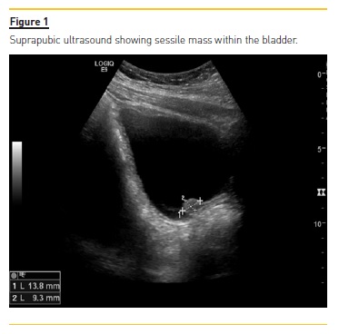
Case 1: An 11-year-old boy from Guinea-Bissau (where he used to swim in local rivers and lakes), resident in Portugal for two years, presented to the emergency room with a 1-month history of dysuria and terminal hematuria without fever. Physical examination showed no abnormal signs. Laboratory testing revealed normocytic normochromic anemia (hemoglobin 10.7g/dL; mean corpuscular volume 88fL; mean corpuscular hemoglobin concentration 21pg) and peripheral eosinophilia (1600/uL) with normal blood urea nitrogen, creatinine and electrolytes. Urinalysis showed 2+ hemoglobin. It was negative for leukocyte esterase, nitrites, glucose and ketones. Urine culture test was negative. Suprapubic ultrasound showed multiple sessile masses within the bladder, not mobile (Figure 1). Renal ultrasound demonstrated normal-sized kidneys bilaterally without hydronephrosis. Urinary microscopic examination for eggs of Schistosoma haematobium and serum detection of Schistosomaimmunoglobulin (Ig)G antibody were negative. Cystoscopy was performed and histopathological examination of biopsy specimens of the bladder lesions revealed moderate inflammatory infiltration, mainly with eosinophils and numerous calcified and active eggs of Schistosomawith terminal spine, suggestive of Schistosoma haematobium (Figure 2).

After exclusion of coinfection with other parasitic diseases (toxocariasis, cysticercosis, strongyloidiasis) the patient was treated with praziquantel (two doses of 20mg/Kg/dose) with resolution of hematuria within 1 week. Because of persistence of dysuria he also received treatment with flavoxate hydrochloride for two months. On follow-up examination 6 months later the patient was asymptomatic and had normal urinalysis and ultrasonography.
Case 2: A 14-year-old African boy from Guinea-Bissau, resident in Portugal for 4 months, presented to his family physician with a 3-week history of intermittent terminal macroscopic hematuria without fever. He had noticed similar episodes of hematuria while living in Guinea-Bissau where he used to bathe in a lake near his house.
Urinalysis was performed and showed hemoglobinuria, leukocyturia and proteinuria. He received antibiotics; because of persistence of hematuria he was referred to a pediatric nephrology appointment. At our hospital, results of investigation revealed normocytic normochromic anemia (hemoglobin 11.7g/dL; mean corpuscular volume 81.5fL; mean corpuscular hemoglobin concentration 26.4pg), peripheral eosinophilia (2400/uL), normal blood urea nitrogen, creatinine and electrolytes; urinary microscopic examination showed presence of red blood cells, white blood cells and eggs of Schistosoma haematobium. Coinfection with other parasitic diseases was excluded. He was treated with praziquantel with two 20mg/Kg doses. At 8 months of follow-up he had no symptoms; urinary microscopic examination was negative for eggs of Schistosoma haematobium; bladder and kidney ultrasound was normal.
Case 3: A 14-year-old African male adolescent, from Angola, living in Portugal for three months, presented in the emergency room with a 1-month history of terminal hematuria without dysuria or fever. He used to swim in a lake near his home and mentioned similar episodes of intermittent hematuria occurring in the last year. Blood testing revealed peripheral eosinophilia (2100/uL). Urinalysis performed in the emergency room was negative for hemoglobin, leucocyte esterase and nitrites. Urine culture test was negative and urinary microscopic examination for eggs of Schistosoma haematobium was negative.
Suprapubic ultrasound showed solid, not mobile, hyperecogenic nodular bladder wall thickening with a maximum of 2cm of length. Renal ultrasound was normal.
In outpatient setting, urinary microscopic examination was repeated after 15 minutes of exercise and eggs of Schistosoma haematobium were seen. Other parasitic coinfections were excluded. The patient was treated with two doses of praziquantel 20mg/Kg. At three months of follow-up urinary microscopic examination was negative for eggs of Schistosoma haematobium. Bladder ultrasound performed 1 year after diagnosis revealed complete resolution of wall thickening.
DISCUSSION
The cases reported illustrate the main challenges in the diagnostic approach of urinary schistosomiasis in a developed non-endemic country.
All three patients were early adolescents and presented with terminal intermittent hematuria without fever. Only one presented with concomitant dysuria. All three were born in endemic countries and were exposed to contaminated waters. Because genitourinary symptoms may begin years after infection, schistosomiasis is a diagnosis to bear in mind in every patient immigrant from endemic countries even if the child is living in an industrialized country and has not been exposed to parasites for many years, such as in the first case described. In the second case described, the main presenting symptoms of urinary schistosomiasis resembled those of urinary tract infection, prompting prescription of inadequate broad-spectrum antibiotics.
Diagnosis of urinary schistosomiasis was based on the identification of Schistosomaeggs on urine microscopic examination in cases 2 and 3. To increase the detection rate, urine samples were taken after a midday session of physical exercise. Cystoscopy with tissue biopsies was performed in case 1 to rule out malignancy as urine samples and serology were both negative. All three cases were treated with two doses of praziquantel 20mg/Kg with complete resolution of hematuria.
In conclusion, schistosomiasis can be a difficult diagnosis since genitourinary symptoms may appear later in the course of the disease and because of low sensitivity of standard urine examination. In chronic infection, eggs cause granulomatous inflammation and development of pseudopolyps in the vesical and ureteral walls which can mimic malignancy requiring cystoscopy with biopsy of such lesions. Urinary schistosomiasis should be suspected in any child coming from endemic countries who presents with hematuria.
References
1. What is schistosomiasis ? Available at: http://www.who.int/schistosomiasis/disease/en/ . Accessed December, 2017 [ Links ]
2. Bamgbola, O. F. Urinary schistosomiasis. Pediatr Nephrol 2014;29: 2113–2120. [ Links ]
3. Azinhais, P. et al. Schistosomíase Urinária: Um Caso Clínico diagnosticado em Portugal. Acta Urológica 2009;26: 55–62. [ Links ]
4. Schistosomiasis. Available at: http://www.who.int/mediacentre/factsheets/fs115/en/. Accessed December, 2017 [ Links ]
5. Clerinx, J., Soentjens, P. & Editor, M. D. Epidemiology, pathogenesis, and clinical manifestations of schistosomiasis. UpToDade 2017: 1–12. [ Links ]
6. Colley, D. G., Bustinduy, A. L., Secor, W. E. & King, C. H. Human schistosomiasis. Lancet 2014;383: 2253–2264. [ Links ]
7. Montgomery, S. P. & Richards, F. O. Blood Trematodes: Schistosomiasis – Principles and Practice of Pediatric Infectious Diseases. 5th Edition. Elsevier, 2017: 285. [ Links ]
8. Bustinduy, A. L. & King, C. H. Schistosomiasis – Mansons Tropical Diseases. 23th Edition. Elsevier 2014: 52. [ Links ]
9. Moudgil, A. & Kosut, J. Urinary schistosomiasis : an uncommon cause of gross hematuria in the industrialized countries. Pediatr Nephrol 2007;22: 1225–1227. [ Links ]
10. Moreno, M. J. D. et al. Esquistosomiasis vesical, aportación de un caso y revisión de la literatura española. Actas Urológicas Españolas 2006;30: 714–719. [ Links ]
11. Soentjens, P., Clerinx, J., Editor, D. T. M. S. & Editor, M. D. Diagnosis of schistosomiasis. UpToDade 2018: 1–9. [ Links ]
12. Tzanetou, K. et al. Urinary Tract Schistosoma haematobium Infection: A Case Report. J. Travel Med 2007; 14: 334–337. [ Links ]
Ana Costa e Castro
Hospital Prof. Doutor Fernando Fonseca, E.P.E., IC19
2720-276 Venteira, Amadora – Portugal
E-mail: anacostaecastro@gmail.com
Authors Contributions:
ACC: acquisition of data and writing the manuscript; AG: literature searches and acquisition of data; PC: management of patients (cases 2 and 3); MJB: management of patients (case 1); SP: management of patient (case 2); VB: management of patients (cases 1 and 3), supervision of writing of initial manuscript. All authors reviewed the manuscript and made revisions to the manuscript.
Disclosure of potential conflicts of interest: none declared.
Received for publication: Aug 11, 2018
Accepted in revised form: Sep 9, 2018


