Serviços Personalizados
Journal
Artigo
Indicadores
-
 Citado por SciELO
 -
 Acessos
Links relacionados
-
 Similares em
SciELO
Compartilhar
Portuguese Journal of Nephrology & Hypertension
versão impressa ISSN 0872-0169
Port J Nephrol Hypert vol.34 no.3 Lisboa set. 2020
https://doi.org/10.32932/pjnh.2020.10.093
TUBULAR QUIZ
Failure to thrive in a pediatric patient
Filipa Valadares1, Rute Baeta Baptista2, Telma Francisco2, Margarida Abranches2
1 Serviço de Pediatria Médica, Área da Mulher, Criança e Adolescente; Hospital de Torres Novas; Centro Hospitalar do Médio Tejo
2 Unidade de Nefrologia Pediátrica; Hospital de Dona Estefânia; Área da Mulher, Criança e Adolescente; Centro Hospitalar Universitário de Lisboa Central
CASE PRESENTATION
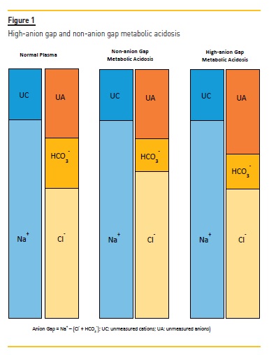
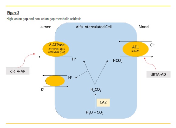
A three-month-old boy was referred to our hospital for the study and management of metabolic acidosis and failure to thrive. The family history was unremarkable, except for his father who had had a possible drug-induced acute kidney injury and uric acid Prenatal and perinatal periods were uneventful and he was born at term (38 weeks of gestational age). His birth weight was 3320 g (50th percentile) and his birth length was 47.4 cm (10th percentile). The neonatal screening for inherited metabolic disorders was normal. The hearing screening at birth showed no response to otoacoustic emissions on the right side. This hearing screening test was repeated one month later and the result was normal. During the first month of life, the patient presented with constant regurgitation and constipation, associated with poor weight gain, leading to downward weight-for-age percentile crossing (from the 50th percentile to the 5th percentile). He was breast-fed for the first month of life and formula-fed thereafter. Infant formula was prepared and administrated correctly. At three months of age he was admitted to his local hospital for failure to thrive. The laboratory workup revealed hemoglobin 10.8 g/dL, leucocytes 13350/uL, sedimentation rate 26 mm/h, urea 47 mg/dL, creatinine 0.44 mg/dL, sodium 137 – 143 mmol/L, potassium 2.4 – 3.3 mmol/L, chloride 120 – 125 mmol/L. Venous blood gases showed pH 7.095 – 7.192, carbon dioxide 19.4 – 26.6 mmHg, bicarbonate 7 – 10 mmol/L, base excess -22 – -18 mmol/L, anion gap 10.4 – 15.1 mmol/L. Urinary pH was ≥6.0, with no glycosuria, proteinuria, or any other alterations in the urine. The sweat chloride test was normal. Transfontanellar ultrasound imaging showed no abnormalities. At our hospital, laboratory assessment also showed non-anion gap metabolic acidosis and hypokalemia. A renal ultrasound was also performed, revealing “increased bilateral diffuse echogenicity of the medullary pyramids suggesting nephrocalcinosis”. IS METABOLIC ACIDOSIS ASSOCIATED WITH FAILURE TO THRIVE? Metabolic acidosis can be caused by three mechanisms: 1) increased acid generation and bicarbonate consumption (bicarbonate acts as a buffer in the presence of increased endogenous or exogenous acid load); 2) loss of bicarbonate, either through the gastrointestinal tract (diarrhoea) or due to a decreased renal tubular capacity to reabsorb the filtered bicarbonate (proximal renal tubular acidosis); 3) diminished renal acid excretion, either due to a reduction in glomerular filtration rate or due to an impaired tubular capacity to acidify the urine (as in distal renal tubular acidosis). Evaluating the anion gap (Na+ – [Cl- + HCO3-]) is critical to understand the etiology of the metabolic acidosis (Figure 1). Metabolic acidosis with increased anion gap is caused by acid accumulation (as occurs in metabolic diseases, intoxications, ketoacidosis, renal insufficiency, etc) with an increase in non-measurable anions concentration; bicarbonate acts as a buffer to this acid load and, therefore, there is a decrease in bicarbonate concentration and chloride concentration remains unchanged1. In metabolic acidosis with normal anion gap (or non-gap metabolic acidosis), there is a decrease in serum bicarbonate concentration either due to renal causes (such as renal tubular acidosis) or due to increased intestinal bicarbonate losses (extra-renal causes). To compensate for the loss of negative charge from decreased serum bicarbonate, the serum chloride concentration increases through renal or intestinal reabsorption, in order to maintain the electroneutrality of the blood. Measurement of the urinary anion gap (AGu = Nau – Clu – Ku) is helpful in the evaluation of patients with hyperchloremic metabolic acidosis (metabolic acidosis with normal anion gap). The normal renal response to metabolic acidosis with acedemia is to increase both the tubular secretion of protons and ammoniagenesis (NH3 synthesis). In the tubular lumen, the secreted NH3 binds to H+ generating ammonium (NH4+). The main defect in dRTA is impaired tubular hydrogen secretion2. As a result, there is a reduced ammonium formation and excretion in the urine. By contrast, ammonium urinary excretion is appropriately increased in other causes of hyperchloremic metabolic acidosis in which distal tubular capacity to acidify the urine is preserved, such as diarrhea and proximal renal tubular acidosis. Therefore, measuring NH4+ in the urine could help differentiate hyperchloremic metabolic acidosis due to a distal defect in urinary acidification from other causes. Direct measurement of urine NH4+ is not readily available in clinical practice. As ammonium is excreted with chloride, urine chloride is a “surrogate marker” for urinary NH4+, enabling the evaluation of ammoniagenesis in response to acedemia. A positive urinary anion gap (+20 or greater) is usually indicative of a normal or low NH4+ urinary excretion, which in patients with non-gap metabolic acidosis suggests dRTA. A negative urinary anion gap (-20 or less) is usually indicative of an appropriately increased NH4+ urinary excretion, as in patients with diarrhoea or proximal tubular renal acidosis. In our patient, the association of failure to thrive and hyperchloremic metabolic acidosis with normal anion gap, in the absence of intestinal losses, points to a renal cause; the presence of hypokalemia, elevated urinary pH and nephrocalcinosis prompted the diagnosis of distal renal tubular acidosis (dRTA). Unfortunately, the measurement of urinary sodium, potassium and chloride was not performed at the time our patient was admitted. In dRTA there is wasting of sodium. This leads to polyuria and a contraction of extracellular volume, which causes secondary hyperaldosteronism and consequent potassium wasting and hypokalemia. Hypokalemia can cause constipation and may also contribute to polyuria3. In situations of chronic metabolic acidosis, the bone will act as a plasma acid buffer, interfering with bone growth and development, thereby affecting weight progression. Furthermore, acidosis inhibits growth hormone and increases protein catabolism4. The acidosis, electrolytic disturbances and chronic dehydration observed in dRTA can therefore interfere with normal weight gain and growth. WHAT IS THE EXPLANATION FOR THE ELEVATED URINARY PH AND NEPHROCALCINOSIS? A urinary pH greater than 6 in the presence of systemic academia denotes a defect in urinary acidification. Despite metabolic acidosis, the urinary pH value does not reach values below 5.5 due to the inability to increase the secretion of hydrogen ions, impairing the elimination of the excessive acid load2,4-6. Hypercalciuria causes calcium crystal deposition, which may lead to nephrocalcinosis and nephrolithiasis7, also promoted by low urinary citrate excretion, which increases the risk of calcium crystal deposition1. Hypercalciuria can also contribute to polyuria2. CLINICAL AND LABORATORY EVOLUTION Due to the presumptive diagnosis of dRTA, the patient was started on oral therapy with Uralyt-U© (citric acid 145 mg/g, potassium citrate 463 mg/g, and sodium citrate 390 mg/g). Each measuring spoon (2.5 g of powder) contains 11 mEq of potassium, 11 mEq of sodium, and 27 mEq of citrate. A dose equivalent to 5.4 mEq/Kg/dia of sodium citrate (2.5 g/day of the powder) was prescribed. An improvement in weight progression, dietary tolerance, and analytical evaluation was observed (Table 1). At 3 years-old, due to a significant language delay, an otorhinolaryngological evaluation was performed, which diagnosed bilateral sensorineural deafness. An auricular prosthesis was placed. IS THERE A LINK BETWEEN SENSORINEURAL DEAFNESS AND THE PATIENT’S RENAL CONDITION? There are primary and secondary causes of dRTA. The primary causes are a group of inherited genetic disorders. The most frequent mutations occur in the SLC4A1 gene, with an autosomal dominant transmission (dRTA-AD) or an autosomal recessive transmission (dRTAAR). Mutations in the ATP6V1B1 and ATP6V0A43,8 genes are associated with autosomal recessive transmission of dRTA (dRTA-AR). In dRTA-AR the first manifestations appear earlier and are more profound: severe hyperchloremic metabolic acidosis associated with significant hypokalemia, nephrocalcinosis, vomiting, dehydration, poor weight progression, possible bone disease (rickets), and sometimes sensorineural hearing loss3,4,8 The secretion of hydrogen ions in the urine is performed by alpha intercalated cells of the collecting ducts, this process being conducted by the V-ATPase transporters (Figure 2). Carbonic anhydrase (CA2) produces hydrogen ions at the same time as bicarbonate reabsorption occurs, promoted by AE1 transmembrane glycoprotein (Cl-/HCO3-), encoded in the SLC4A1 gene. This transporter has a specific isoform in the kidney and erythrocytes. When both isoforms are mutated, it can manifest as dRTA-AD associated with hereditary spherocytic anemia. The vacuolar hydrogen pump (V-ATPase) acts on the intercalated α-cells of the collecting tubule, promoting hydrogen ion secretion. This enzyme consists of 14 subunits. Mutations in the β1 (encoded by the ATP6V1B1 gene) and 4 (encoded by the ATP6V0A4 gene) subunits cause dRTA-AR. As the β1 and α4 subunit are also expressed in the epithelial cells of the cochlea, mutations in this subunits may also cause sensorineural deafness. In the occidental population, 40% of dRTA are caused by mutations in ATP6V0A4 gene, 30% in ATP6V1B1 gene, and 15% in SLC4A1 gene. Recently, mutations responsible for dRTA in FOXI1 and WRD72 genes were associated with deafness and imperfect amelogenesis, respectively. However, in 15% of patients with a clinical diagnosis of dRTA, no mutation was found, which may suggest the existence of other candidate genes9-10. GENETIC TESTING The patient underwent a next-generation sequencing (NGS) fivegene panel for renal tubular acidosis (ATP6V0A4, ATP6V1B1, CA2, SLC4A1, SLC4A4). The variant c.2420G>A (p.Arg807Gln) in the exon 21 of the ATP6V0A4 gene was found to be present in homozygosity. This single-nucleotide variant is known to be associated with dRTA and late-onset sensorineural hearing loss. Genetic characterization of the father may help clarify whether the previous episode of acute kidney injury and nephrolithiasis are truly unrelated or attributable to dRTA (or at least partial dRTA). CASE FOLLOW-UP At 5 years of age, the patient underwent a cochlear implant surgery with good adaptation and evolution. Currently, at the age of 9, the patient remains asymptomatic and has shown a weight and height evolution between the 3rd and the 15th percentiles, under treatment with citrate. The last blood gas results showed normal pH and normal bicarbonate. The urinary calcium-to-creatinine ratio was also normal. As expected, the renal ultrasound still shows nephrocalcinosis, currently grade III/IV. The most relevant clinical outcome regarding bone health response to treatment is the improvement in linear growth. In addition, bone densitometry is not validated in this population for this purpose. The urinary calcium-to-creatinine ratio tends to normalize with citrate/bicarbonate supplementation and salt restriction. Although thiazide diuretics may be used to decrease urinary calcium excretion, this strategy is not routinely used in pediatric patients due to the risk of aggravating hypokalemia and polyuria. Thiazides are reserved for the cases in which urinary calcium excretion does not ameliorate with citrate/bicarbonate supplementation, especially if there is aggravating nephrolithiasis. With this case, the authors intend to highlight the importance of the clinical suspicion of this pathology, given that with the correct therapy and early follow-up, it is possible to recover weight and height, and that an early diagnosis of deafness may have a profound impact on the child’s development. References 1. Greenbaum LA. Composition of body fluids. In Nelson Textbook of Paediatrics. Behrman RE, Kliegman RM, Blum NJ, Shas SS, Geme JW, Tasker RC, Wilson KM [eds]. 21st Edition. Philadelphia. Elsevier. 2020: 389-391 [ Links ] 2. Dixon BP. Renal tubular acidosis. In Nelson Textbook of Paediatrics. Behrman RE, Kliegman RM, Blum NJ, Shas SS, Geme JW, Tasker RC, Wilson KM [eds]. 21st Edition. Philadelphia. Elsevier. 2020:2762-2764 [ Links ] 3. Chan JC, Scheinman JI, Roth KS. Renal tubular acidosis. Pediatr Rev 2001;22(8):277-287 [ Links ] 4. Lopez RS, Román LE. Tubulopatías. In Manual de Diagnóstico y Terapéutica em Pediatria. Fernandez JG, Sánchez AC, Bonis AB, Suso JM, Domínguez JR [eds]. 6ª Edición. Madrid. Panamericana; 2017: 1738-1747 [ Links ] 5. Besouw MTP, Bienias M et al. Clinical and molecular aspects of distal renal tubular acidosis in children. Pediatr Nephrol 2017;32(6):987-996 [ Links ] 6. Matto TK. Etiology and ckinical manifestations of renal tubular acidosis in infants and children. Available: https://www.uptodate.com (Accessed on September 2019) [ Links ] 7. Lum MG. Kidney and urinary tract. In Current Diagnosis and Treatment Pediatrics. Hay WW, Levin MJ, Deterding RR, Abzug MJ [eds]. 22nd Edition. United States of America; 2014: 769-771 [ Links ] 8. Escobar L, Mejía N et al. La acidosis tubular renal distal: una enfermidade hereditária en la que no se pueden eliminar los hidrogeniones. Nefrologia 2013;33(3):289-296 [ Links ] 9. Rodríguez FS. Acidosis tubular renal distal: introducción, epidemiología y genética. In Actualización en Acidosis Tubular Renal Distal. Rodríguez FS, García Nieto VM, Ariceta G [eds]. Madrid, Comunicación Y ediciones Sanitarias, S. L. 2019:1-5 [ Links ] 10. Brum S, Santos AR, Silveira C, Nolasco F, Rueff J, Calado J. An adult patient with hypokaliemia. Port J Nephrol Hypert 2018;32 (1): 89-91. [ Links ] Filipa Valadares, MD Serviço de Pediatria Médica, Área da Mulher, Criança e Adolescente Hospital de Torres Novas, Centro Hospitalar do Médio Tejo E-mail: fvaladaresb@gmail.com Disclosure of potential conflicts of interest: none declared Received for publication: May 26, 2020 Accepted in revised form: Sep 8, 2020


