CLINICAL PRESENTATION
We present a case of a 47-year-old male with past medical history of Wilson disease with neurologic involvement, submitted to liver transplant in 2008. Immunosuppression consisted of tacrolimus, mycophenolate mofetil (MMF) and prednisolone (PDN). He had a history of hypertension and diabetes mellitus under insulin.
Twelve years after transplant he was admitted for liver failure with total serum bilirubin (TB) peaking at 15mg/dL and direct bilirubin (DB) at 10 mg/dL. A chronic allograft rejection was revealed by liver biopsy and attributed to non-compliance. Kidney function was normal. The patient started pulses of methylprednisolone and chronic immunosuppression was increased with addition of everolimus and titration of PDN; MMF was suspended. One month later he was admitted for altered mental status and vomiting with reduced urine output. Noradrenalin and empirical piperacilintazobactam were started for hemodynamic instability and presumed infection, respectively. Lab work showed acute kidney injury (AKI) KDIGO 3 with serum creatinine (sCr) of 7.12mg/dL and urea of 204mg/dL; hyperbilirubinemia of 16.5 mg/dL (DB of 12.3 mg/dL) and elevated liver enzymes). Despite hemodynamic recovery and fluid correction, the patient remained oliguric, starting on hemodialysis 48 hours after admission. One month later, showing no improvement (TB 29mg/dL and sCr 6 mg/dL), a kidney biopsy was performed.
QUESTIONS
1. What is the most likely diagnosis, given the clinical history and presentation?
2. What is the diagnosis considering light microscopy and immunofluorescence studies?
3. What is the differential diagnosis?
4. How should this patient be managed?
ANSWERS
1.What is the most likely diagnosis, given the clinical history and presentation?
AKI in patients with liver disease is common and has several causes. In our patient, prerenal AKI due to gastrointestinal losses, acute tubular necrosis secondary to hemodynamic instability, sepsis on an immunosuppressed host or hepatorenal syndrome in the context of transplant/liver failure were all our main suspects. However, the absence of improvement after correcting or supporting the abovementioned insults led us to perform a renal biopsy.
2. What is the diagnosis considering light microscopy and immunofluorescence studies?
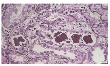
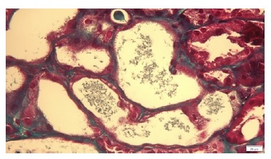
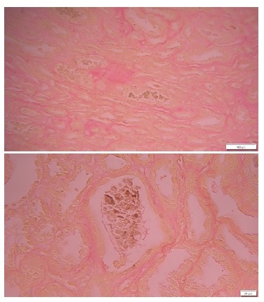
The kidney biopsy specimen consisted of two fragments with medulla, capsule, cortex, ten glomeruli and a medium-sized artery. In light microscopy, one glomerulus showed global and one showed segmental sclerosis; the remaining were unremarkable. Interstitial compartment was also preserved. Figure 1 (PAS) demonstrates the presence of intratubular casts of a brownish pigment. The tubules are dilated with isometric vacuolization of epithelial cells. Figures 2 (TM) shows dilated tubules, with acute tubular necrosis, occupied by granular and red intratubular cast. Figure 3 (Hall stain) confirms the presence of bile within the pigmented granular casts with abundant cellular debri.
The diagnosis is bile cast nephropathy (BCN). BCN is a rare entity characterized by AKI in the context of severe hyperbilirubinemia and elevation of serum bile salts.1,2It can occur as a part of a wide spectrum of cholestasic liver disorders with severe jaundice.3

Figure 1 (PAS) demonstrates the presence of intratubular casts of a brownish pigment. The tubules are dilated with isometric vacuolization of epithelial cells.

Figure 2 (TM) shows dilated tubules, with acute tubular necrosis, occupied by granular and red intratubular cast.

Figure 3 (Hall stain) confirms the presence of bile within the pigmented granular casts with abundant cellular debri.
The exact mechanism of BCN remains unknown; however, various mechanisms may be involved.1,2 The key finding is the tubular obstruction and injury caused by the bilirubin cast formation within the distal tubules.1 Bilirrubin is a low water-soluble pigment filtered by glomerulus and transported by the proximal tubules back into the circulation. When in excess, the tubular transporters became saturated, leading to cast formation and tubular obstruction.1,2
Bilirubin also exerts oxidative damage of the tubular cell membranes, leading to tubular cell direct toxicity and hypertrophy. It also inhibits mitochondrial oxidative phosphorylation, increasing permeability of membranes and resulting in modified electrolyte contente and cell volume. Inhibition of the Na-H, Na-K, Na-Cl pumps by bile salts in proximal tubules and in the loop of Henle results in an acidic environment which may favor bile cast deposition and tubular injury.1,2,3
Lastly, hyperbilirubinemia affects systemic and renal hemodynamics by having negative chronotropic and ionotropic effects, with cardiovascular instability and decreased renal perfusion. Additionally, systemic endotoxemia and nitric oxide-mediated mechanisms result in splanchnic and systemic vasodilatation, decreased renal blood flow and ischemia to the kidney.1,2
Despite clinical suspicion, usually in the setting of AKI associated with TB levels greater than 20 mg/dL, and/or the presence of bilirubin casts on urinalysis, the gold standard for diagnosing BCN is kidney biopsy.2,3,4
The commonest finding is the presence of dilated tubules, with pigmented intraluminal casts predominantly in distal tubules. Acute tubular necrosis is also a prominent feature. Casts are dark green by Hall stain, which uses Fouchet reagent to convert bilirubin to biliverdin.1,3,4,5Note that this stain is insensitive and a negative result does not exclude BCN.1,4,5
Glomeruli are usually spared.2 Immunofluorescence is noncontributory.4 Electron microscopy, although not routinely performed, may show dilated mitochondrial cristae and bile acid accumulation within lysosomes.2 Tubular casts correlate with presence of jaundice.5
3. What is the differential diagnosis?
Cast nephropathy, as defined by the presence of intratubular casts, may emerge as part of a vast number of pathologies. Casts related to cellular debris in acute tubular necrosis are the most common. They are usually granular in its appearance and are negative in Hall stain.4,5
Myoglobin cast nephropathy in rhabdomyolysis and hemolysisassociated hemoglobin casts in hemoglobinuria, are characterized by pigmented granular eosinophilic/reddish casts in distal nephron. Characteristically, they stain positive by myoglobin or by iron immunostain (with Prussian blue), respectively.4,5
Lastly, on light chain cast nephropathy, intratubular casts are typically polychromatic, with sharp edge or fractured appearance and syncytial multinucleated giant cell reaction. Immunofluorescence microscopy with monoclonal light chain staining is diagnostic.4,5
4. How should this patient be managed?
Since it is a rare condition, there are currently no accepted management guidelines for BCN.2
Treatment should focus on prompt correction of hyperbilirubinemia to reverse kidney injury.1,2 After attempting to resolve the cause of cholestasis, removal of excess bilirubin and bile salts could be tried by extracorporeal therapies (ECT)1,2.
ECT can be divided into two major types: biological and non-biological devices.1,6
Biological devices incorporate a component consisting of living liver cells (human or porcine hepatocytes or cancer-derived cell line) in na extracorporeal device and support the whole range of liver functions.1,6
Nonbiological devices are dialysis-derived systems that use artificial membranes and adsorbents for detoxification. They are based on the principle of blood purification by albumin dialysis or by plasma separation and filtration.1,6
Albumin dialysis includes the Molecular Adsorbent Recirculating System (MARS), the Fractional Plasma Separation Adsorption and Dialysis system (Prometheus®) and the single-pass albumin dialysis (SPAD). All three are based on the removal of albumin-bound and water-soluble bilirubin, bile acids, and hepatotoxins.1,6
Plasma separation and filtration includes selective plasma filtration therapy (SEPET) and high-volume plasmapheresis.1,6
Other therapies such as steroids, ursodeoxycholic acid, cholestyramine, or lactulose have shown little or no benefit.1,2


