CASE REPORT
We report a case of a 40-year-old male from Cape Verde, with history of epilepsy and systemic lupus erythematosus (SLE) with cutaneous and ocular involvement, diagnosed in 1995. In 2003, the diagnosis of lupus nephritis class IV (ISN/RPS Society) was made. Despite treatment with cyclophosphamide and corticosteroids he developed end-stage kidney disease and started hemodialysis in 2005. The patient was also on oral anticoagulants after he was diagnosed of antiphospholipid syndrome (APS). Clinical criteria were assumed by multiple vascular access thrombosis as clinical criteria and he had lupus anticoagulant, anticardiolipin antibody and anti-beta 2 glycoprotein 1 as laboratorial criteria.
In August 2021, he underwent a kidney transplant from deceased donor with expanded criteria, 61 years-old and hemorrhagic stroke as cause of death, with two human leukocyte antigen (HLA) mismatches (one HLA class I and one HLA class II), with no donor-specific antibodies (DSAs) and cold ischemia time of 10 hours. He had no history of previous kidney transplant.
Thymoglobulin was administered as the induction immunosuppression, followed by cyclosporine, mycophenolate mofetil (MMF) and prednisolone as maintenance. Cyclosporine was used due to the epilepsy history to prevent tacrolimus neurotoxicity. Importantly to note that, before surgery, the patient received fresh frozen plasma and K vitamin due to warfarin use, restarting anticoagulation after the surgery (first with enoxaparin followed by warfarin), although with infratherapeutic INR levels.
The post-surgical period was complicated by an infected fluid collection adjacent to the graft and delayed graft function. He was submitted to percutaneous drainage that revealed methicillin-susceptible Staphylococcus Aureus and Candida albicans and was subsequently treated with flucloxacillin and fluconazole for 21 days, with dimensional improvement of the fluid collection. Other complications in posttransplant period were transient thrombocytopenia and anemia that were resolved after suspension of thymoglobulin and trimethoprimsulfamethoxazole.
Cyclosporine levels were within acceptable range and cytomegalovirus (CMV) and BK viral charge were undetectable. Despite the resolution of the fluid collection and adequate urine output, there was slow recovery of kidney function with serum creatinine stabilizing in 3-4 mg/dL. Kidney ultrasound and Doppler did not show
alterations apart from the fluid collection previously reported. Thus, a kidney biopsy was performed.
QUESTIONS
1. What is the most likely diagnosis?
2. Considering the kidney biopsy results, what is the final diagnosis?
3. What treatment options can we offer to the patient?
„
ANSWERS
1. What is the most likely diagnosis, considering the clinical history and presentation?
Given the medical history of APS and the presence of a period of anticoagulation below adequate levels, the most likely hypothesis to explain the acute graft dysfunction would be a thrombotic microangiopathy (TMA) due to APS. In cases of recurrence after kidney transplant, it can be caused by withdrawal of warfarin before the surgery which can lead to an increased synthesis of fibrin and thrombin with transient rebound hypercoagulability. This, in addition to endotelial activation caused by surgery, transplant ischemia-reperfusion injury, alloimmunity and exposure to CNI, could culminate in the activation of complement and development of TMA.1 However, he did not have evidence of microangiopathic anemia and thrombocytopenia was resolved after thymoglobulin suspension. After the result of the kidney biopsy, donor specific antibodies were analyzed and an antibody mediated rejection was excluded. Also, antiphospholipid antibodies were evaluated and positive levels of anticardiolipin antibody (870 UQ),
lupus anticoagulant2-5 and anti-beta 2 glycoprotein 1 antibody (2852,3 UQ) were found.
In addition to this hypothesis, it should be considered other potential early acute dysfunction diagnosis, such as2:
• Postischemic acute tubular necrosis is the most common cause of delayed graft function, and common with expanded criteria donors, normally self-limited and diagnosed histologically. Against this hypothesis is the fact that patient was not oliguric and there was no mention of hemodynamic instability during surgery.
• Acute rejection is one of the most common causes of allograft dysfunction in the early post transplantation period. Acute rejection should be suspected among all transplant recipients with early acute dysfunction and requires kidney biopsy to make a definite diagnosis, being acute antibody mediated rejection less probable due to the absence of donor specific antibodies.
• Fluid collections that can occur after transplantation and cause acute kidney allograft dysfunction include lymphoceles, urinary leaks, and perinephric hematomas. These can cause acute dysfunction if there is compression of the graft. In that case it would be expected to resolve after drainage of the fluid collection, which did not happen in our patient.
• Acute interstitial nephritis either due to antibiotic use or to trimethoprim-sulfamethoxazole. It presents with nonspecific signs and symptoms, acute kidney dysfunction and most frequently preserved urine output. The definite diagnosis requires kidney biopsy and treatment is based on corticosteroids and drug discontinuation; both were done in our patient without clinical improvement.
• Vascular thrombosis of the allograft is an uncommon event, normally asymptomatic and presents as a sudden decrease of urine output with either increase in the creatinine level or failure of the creatinine level to decrease after transplant. In this patient it should be considered due to his personal history of APS that is associated with increased risk of vascular thrombosis. It can be diagnosed by Doppler ultrasound which was performed in our patient without evidence of thrombosis.
• Urinary obstruction unlikely due to normal ultrasound and preserved urine output.
• CMV or BK infection and calcineurin inhibitor toxicity are less likely since cytomegalovirus (CMV) and BK virus levels were undetectable and cyclosporine levels were within normal range.
2. What is the diagnosis considering light microscopy and immunofluorescence studies?
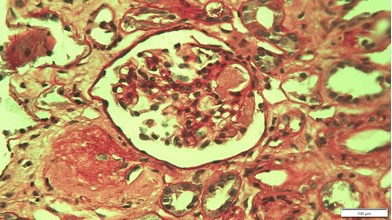
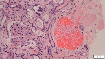
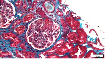
The fragment obtained from kidney biopsy consisted of renal capsule, cortex containing 6 glomeruli, and small sized arteries. In light microscopy, one glomerulus showed an arteriole intraluminal thrombus and other showed glomerular capillary congestion. The remaining four glomeruli did not show alterations. Around 30% of the sample revealed patchy cortical necrosis and interstitial hemorrhage, more visible in subcapsular area. There was an Interstitial mononuclear infiltrate of medium intensity. Moderate acute tubular necrosis. Small arteries had no changes. The immunofluorescence (IMF) study showed 3 glomeruli and was negative for all tested molecules (IgA, IgM, IgG, C3 and C4d).
Therefore, the diagnosis of cortical necrosis due to arteriolar and capillary thrombosis, probably secondary to APS was made. Foci of cortical necrosis have been described and occur due to occlusion of small isolated parenchymatous renal vessels.1,3Although normally asymptomatic, cortical necrosis can be generalized and cause oligúria and severe hypertension which has worst outcomes.
3. What are the treatment options and prognosis?
Prevention is an important part of the management of these patients. This can be achieved by administering perioperative anticoagulation therapy with low molecular weight or unfractionated heparina which has been linked to a lower rate of graft failure in patients with APS. Nevertheless, allograft loss due to vascular thrombosis can occur even with anticoagulation use.1,3-5
Treatment in cases of recurrent APS is also anticoagulation of the patient. In our case, although the patient was under anticoagulation drugs, their effect was neutralized before the surgery by using vitamin K and fresh frozen plasma which despite preventing bleeding complications, predisposes the patient to thrombotic events. This fact associated with INR levels below the goal range, significantly increases the risk of APS recurrence. In refractory cases to anticoagulation, treatment with plasma exchange, rituximab or eculizumab, improved renal outcomes in some case reports of patients with recurrent APS posttransplantation.1,3,6Our patient was treated with anticoagulation intensification and an antiplatelet drug was started. He stopped warfarin due to difficulty to obtain therapeutic levels and started enoxaparin and screening of anti-Xa levels was done to adjust dose. A renal scintigraphy showed small areas of hypofixation on superior and inferior polar regions, with no other important changes. Despite these foci of cortical necrosis, he had a favorable evolution and serum creatinine stabilized at around 2 mg/dL.
Reduced rates of allograft survival are reported in patients with APS secondary do SLE, when compared to patients with SLE alone. Also, increasing evidence has showed a higher risk of early graft loss, especially due to vascular thrombosis or TMA, in APS patients. The presence of lupus anticoagulant in pre-transplant period seems to correlate with development of thrombotic events in the allograft.7
However, more efficient strategies are needed to prevent these events in the perioperative period.

Figure 1 Periodic acid-Schiff (PAS) stain, magnification x400: glomerulus with thrombus in arteriole. Another adjacent glomerulus is sclerotic

Figure 2 Masson’s trichrome stain, magnification x200: glomerulus with evidence of capillary thrombus

Figure 3 Hematoxycilin-eosin (H&E) stain, magnification x200: glomerulus with ghost-like outlines without discernible nuclei


