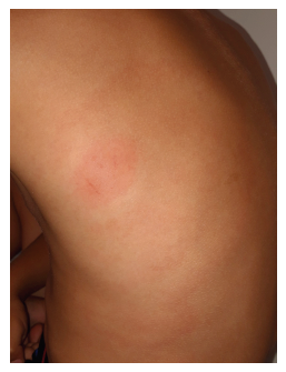
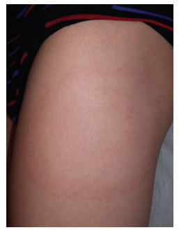
A seven-year-old boy, living in Switzerland, was brought to an Emergency Department in Portugal on July due to appearance of slightly pruritic and erythematous skin lesions on the back, thighs, and buttocks. Lesions had been expanding gradually over the previous week. Fever and other systemic symptoms were denied and parents did not recall a history of insect bite. On physical examination, the boy presented four erythematous annular lesions: the first to appear was on the back, with 20 cm in diameter and a central macula (Figure 1); the remaining were also annular, with around 10 cm in diameter (Figure 2). All lesions were flat and without scale.
Discussion
Lyme disease is a spirochaetal infection caused by Borrelia Burgdorferi sensu lato, which is transmitted by infected ticks of the genus Ixodes. Lyme disease is endemic in areas with certain ecologic conditions, like woodlands with sufficient humidity that enable the development and survival of ticks.1 It is therefore more prevalent in regions with moderate climates, like Scandinavian and Central countries in Europe and some areas in the United States and Asia. However, its geographic distribution is expanding, and cases have been reported throughout Europe.1,2 In Portugal, Lyme disease incidence seems to be rising, particularly in northern and central regions, with a total of 20 cases reported in 2017.3,4
Lyme disease has a broad spectrum of clinical manifestations divided in three stages: early localized disease, early disseminated disease, and late disease. Single erythema migrans lesion is the manifestation of early localized disease and characterized by a rash developing at the site of the tick bite between three to 30 days − typically seven to 14 − after the bite. The lesion gradually expands, reaching between 5 to up to 70 cm. It is most commonly uniformly erythematous but can also have enhanced central erythema or a “bull’s eye” appearance (Figure 1). Erythema migrans is usually asymptomatic but can be pruritic or painful. Systemic symptoms, such as fever, fatigue, headache, neck pain, arthralgia, or myalgia may be reported at this stage. Multiple erythema migrans is the most common manifestation of early disseminated disease and thought to be due to hematogenous dissemination. It consists of multiple annular erythematous lesions similar to the primary lesion but usually smaller (Figure 2), and generally appears three to five weeks after the bite. Together, single and multiple erythema migrans represent about 90% of Lyme disease presentations. Other clinical manifestations of early disseminated Lyme disease include cranial nerve palsy, meningitis, and carditis, which typically presents with prolonged PR interval or rarely with complete heart block. Like in early localized stage, systemic symptoms are also common at this stage. Late Lyme disease generally presents with arthritis and occurs weeks to months after the initial infection.2,5 Another cutaneous manifestation of Lyme disease is Borrelial lymphocytoma which, although rare, is more prevalent in children. Borrelial lymphocytoma is characterized by a nontender bluish-red nodule with one to five centimetres of diameter, which can appear simultaneously or after erythema migrans. The most frequent location in children is the ear lobe and helix.1,4
Lyme disease diagnosis can be established on clinical grounds if the patient presents the typical single or multiple erythema migrans lesions and lives or has recently travelled to an endemic area. In this case, laboratory and serologic testing are not necessary.1,4,6 A history of tick bite is a strong epidemiological link, but is reported in only 30 to 40% of patients with erythema migrans.4 In patients with non-erythema migrans presentations, laboratory confirmation with a two-tier serologic testing is required, in which a sensitive enzyme-linked immunosorbent assay (ELISA) is the first step. If first-tier ELISA test is positive or equivocal, an immunoblot test should be performed. A positive IgG immunoblot test is considered diagnostic of B burgdorferi infection.1,6
In single or multiple erythema migrans, oral antibiotic treatment with doxycycline (for patients ≥ 8 years), amoxicillin, or cefuroxime axetil for 14 to 21 days is recommended to prevent dissemination and development of late Lyme disease. When treated properly, the prognosis is excellent.2,7
As there is no available vaccine, the best way to prevent the disease is to reduce the risk of a tick bite. Some preventive measures are recommended, as avoiding environments potentially infested by ticks, covering bare skin, and using tick repellents. Skin surface, including scalp, should be daily inspected after a possible tick exposure and attached ticks identified should be removed. In Portugal, as in the rest of Europe, prophylaxis after a tick bite is not recommended, as tick infection rates are less than 20%. Clinical observation for the following 30 days is the only recommended measure in case of tick bite.1,4
In this case report, and based on clinical and epidemiologic findings, Lyme disease multiple erythema migrans diagnosis was considered. Basic laboratory testing and electrocardiogram were normal, and the patient was treated with a two-week course of oral amoxicillin, with complete erythema migrans clearance within three days. Diagnosis was later supported by ELISA serology testing positivity and immunoblot testing.
In conclusion, with rising prevalence of Lyme disease in some areas, it is increasingly important to recognize the different stages of the disease, to provide early treatment and prevent hematogenous dissemination and sequels. Lyme disease still has a low prevalence in Portugal, but is endemic in Central Europe, with an estimated incidence of 156 cases per 100.000 people in Switzerland.3,8 Although evidence of typical erythema migrans is sufficient to establish the diagnosis, other manifestations are broad and unspecific, highlighting the importance of the epidemiologic history as a clue to diagnosis.


