SciELO - Scientific Electronic Library Online

 
vol.30 número2Caso clínico dermatológicoOmbros desalinhados índice de autoresíndice de assuntosPesquisa de artigos
Home Pagelista alfabética de periódicos  

Serviços Personalizados

Journal

Artigo

Indicadores

Links relacionados

  • Não possue artigos similaresSimilares em SciELO

Compartilhar


Nascer e Crescer

versão impressa ISSN 0872-0754versão On-line ISSN 2183-9417

Nascer e Crescer vol.30 no.2 Porto jun. 2021  Epub 30-Jun-2021

https://doi.org/10.25753/birthgrowthmj.v30.i2.19222 

Imaging cases

Gynecological clinical case

Caso clínico ginecológico

Daniela Alves Silva¹ 
http://orcid.org/0000-0003-1140-4265

Sofia Dias Costa² 
http://orcid.org/0000-0002-8887-8680

Maria Carlota Cavazza³³ 

Alexandra Luz¹ 
http://orcid.org/0000-0002-9243-6527

1 Department of Pediatrics, Hospital Santo André, Centro Hospitalar de Leiria. 2410-197 Leiria, Portugal. danielafalvess@hotmail.com; alexandra.luz@hotmail.com

2 Hospital Pediátrico de Coimbra, Centro Hospitalar e Universitário de Coimbra. 3000-602 Coimbra, Portugal. sofiacosta89@hotmail.com

3 Department of Gynecology and Obstetrics, Hospital Santo André, Centro Hospitalar de Leiria. 2410-197 Leiria, Portugal. mcarlota.cavazza@gmail.com


Abstract

Herein is reported the case of a child observed in the Emergency Department due to urinary retention, suprapubic pain, and dysuria. Urine culture was performed upon cystitis suspicion, which was positive for Escherichia Coli, and the patient was treated with amoxicillin and clavulanic acid. Three days later, the child was reevaluated for persistent urinary retention, fever, and vaginal bleeding. At gynecological examination, she had an imperforate hymen. Imaging evaluation was performed, revealing an abdominal mass, suggestive of piohematocolpos. The patient was submitted to surgical drainage, with complete condition resolution.

Keywords: diagnosis; hematocolpos; menstrual blood; vesical globe

Resumo

É descrito o caso de uma adolescente observada no Serviço de Urgência por retenção urinária, dor suprapúbica e disúria. Foi realizada urocultura devido a suspeita de cistite, que foi positiva para Escherichia Coli, e a rapariga foi medicada com amoxicilina e ácido clavulânico. Três dias depois, foi reavaliada por persistência da retenção urinária, febre e hemorragia vaginal. Ao exame ginecológico, apresentava um hímen imperforado. Foi realizada avaliação imagiológica, que detetou uma massa abdominal sugestiva de piohematocolpos, tendo sido realizada drenagem cirúrgica com resolução completa do quadro.

Palavras-chave: diagnóstico; globo vesical; hematocolpos; hemorragia vaginal

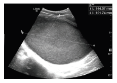
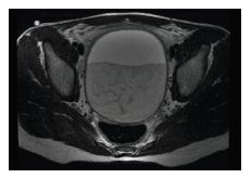
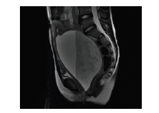
A previously healthy 12-year-old girl presented to the Pediatric Emergency Department complaining of urinary retention, suprapubic pain, and dysuria for two days. She denied fever or gastrointestinal symptoms. Physical examination showed a suprapubic mass compatible with a painful vesical globe and the remaining physical examination was normal. Urine culture was performed to assess a potential cystitis diagnosis, which was positive for multisensitive Escherichia Coli. The girl was discharged medicated with amoxicillin and clavulanic acid. She returned three days later (day 2 of antibiotics) complaining of persistent urinary retention, fever, and vaginal bleeding. She had had her menarche three months before and her last menses occurred during the last fortnight. Upon gynecological examination, diagnosis of microperforated hymen was established. Pelvic ultrasound revealed a hypoechogenic nodular formation with approximately 20 cm in diameter (Figure 1), well defined and homogeneous; uterus and ovaries were not visible, probably pushed by the massive formation. This formation was confirmed by magnetic resonance imaging, which showed a massive lesion occupying space, with well-defined contours and pre-thickened, reaching 16.2 x 9.7 x 8. 9 cm in longitudinal, anteroposterior, and transverse axes, respectively. The lesion had a heterogeneous fluidic content without hypervascularized components in the lesion lumen (Figures 2and3). No changes were detected in the urinary system.

What is your diagnosis?

Figure 1 Pelvic ultrasound 

Figure 2 Pelvic magnetic resonance imaging (transversal view) 

Figure 3 Pelvic magnetic resonance imaging (lateral view) 

Diagnosis

Hematocolpos

Discussion

The girl underwent vaginoscopy and hymenectomy, showing a piohematocolpos which was completely drained. She was revaluated two weeks later, with complete condition resolution.

Hematocolpos is an accumulation of menstrual blood in the vagina, resulting from genital tract obstruction. It is a rare condition that occurs in adolescence, usually secondary to an imperforate or microperforate hymen. This is one of the most common congenital anomalies of the genital tract, and consists of a connective tissue membrane covered by epithelium obstructing the vaginal opening at the introitus level. Hymen is a membrane that usually develops embryologically through fusion of the caudal end of the paramesonephric ducts and the urogenital sinus. The central portion of this membrane perforates through degeneration of epithelial cells. Failure of epithelial cell degeneration and subsequent perforation leads to a hymen that is termed imperforate.1 The differential diagnosis includes transverse vaginal septum, longitudinal vaginal septum, vaginal agenesis, and cervix atresia.2 Symptoms as abdominal pain, dysuria, urinary retention, or amenorrhea can be present, and diagnosis implies a high degree of suspicion.3 Menstrual history and inspection of the external genitalia showing a bluish-colored hymen should be part of the evaluation and aid in diagnosis.4 Surgery is the recommended treatment, consisting of an incision in the vaginal hymen, and should be performed as early as possible to relieve symptoms and prevent complications.5

Lessons from this clinical case

All healthcare professionals attending adolescents should be alert for possible gynecological pathology.

Although hematocolpos is a rare condition, it should be considered in presence of an abdominal mass, for proper guidance.

Rapid recognition allows for timely surgical intervention, preventing complications

References

1. Silva S, Almeida S. Causa rara de dor abdominal numa adolescente. Acta Pediatr Port 2008; 39:216-8. [ Links ]

2. Mwenda AS. Imperforate Hymen - a rare cause of acute abdominal pain and tenesmus: case report and review of the literature. Pan Afr Med J. 2013; 15:28. [ Links ]

3. Sousa H, Fonseca H, Sampaio L. Dor abdominal e retenção urinária aguda em adolescente: apresentação clínica de hematocolpos e revisão da literatura. Nascer e Crescer 2010; 19:152-4. [ Links ]

4. Álvarez CMG, Reymundo MG, Terrón RR, Ahmed FH. Hematocolpos por himen imperforado: causa infrecuente de dolor abdominal y retención aguda de orina. Rev Pediatr Aten Primaria. 2010; 12:621-6. [ Links ]

5. Ercan CM, Karasahin KE, Alanbay I, Ulubay M, Baser I. Imperforate hymen causing hematocolpos and acute urinary retention in an adolescent girl. Taiwanese Journal of Obstetrics and Gynecology 2011; 50:118-20.[/doc] [ Links ]

Received: January 13, 2020; Accepted: August 04, 2020

Correspondence to Daniela Alves Silva Department of Pediatrics Hospital Santo André Centro Hospitalar de Leiria Rua de Santo André 2410-197 Leiria, Portugal. Email: danielafalvess@hotmail.com

Creative Commons License This is an open-access article distributed under the terms of the Creative Commons Attribution License