Introduction
Axillary swelling is a usual cause of concern among children and adolescents and their parents, frequently requiring medical observation. The most common diagnosis is lymph node swelling, caused by lymphadenitis or lymphoma. Rarely, it can also be caused by soft tissue tumors (as rhabdomyosarcoma, lipoma, or fibroma), vascular lesions (as angiodysplasia or aneurysms), or inflammation of the sweat glands,1-5 and by ectopic breast tissue (EBT), an undervalued diagnosis. EBT usually develops as a slowly growing mass and is most commonly identified in young women, with a few cases in the literature reported in adolescents.1,6
Case description
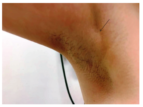
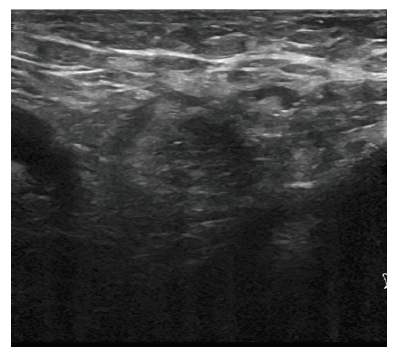
A healthy 14-year-old girl presented in consultation with pain in the right axilla with one year of evolution and swelling for the past two months, gradually increasing in size, especially during menstruation. She had the menarche at the age of ten years and used to razor-shave the axillae. The girl denied other associated symptoms and had no relevant family medical history. On examination, a 4 x 4-cm swelling without inflammatory signs, poorly marginated, freely mobile, and slightly painful on palpation was noted (Figure 1). No changes were identified in both breasts or left axilla. The initial differential diagnoses were hidradenitis and lymphadenitis. Further study through sonography of the region detected the presence of mammary tissue in both axillae (Figure 2) and allowed to establish the diagnosis of ectopic breast tissue (EBT). Given this diagnosis, an abdominal ultrasound was performed to search for associated nephron-urologic anomalies, which revealed a parenchymal septum dividing the two sinusal regions in both kidneys, compatible with duplication of the excretory systems. Due to associated pain and cosmetic reasons, excision of the ectopic tissue of the right axilla was performed, followed by macroscopic and histological confirmation of the diagnosis of EBT. The girl maintains regular clinical surveillance of the left axilla.
Discussion/conclusions
EBT, or accessory breast tissue, is an uncommon entity affecting 2─6% of females and 1─3% of males,1,4, 5,7-10 more frequently diagnosed in Asian people (prevalence of 5% in the Japanese vs. 0.6% in the Caucasian population).3,9,10 It presents bilaterally in two-thirds of cases and in the axillae in 70% (other possible locations are the vulva and groin).1,3-5,7,11,13 The condition usually occurs sporadically, but a hereditary predisposition has been reported.3,5,13,14 Because EBT is under the same hormonal influence as normal breast tissue, it usually appears during periods of hormonal stimulation, as menstruation, pregnancy, and lactation.1-3,5,8,14,15 In the present case, the condition developed in a girl younger than usually reported.
EBT results from an error during embryonic development. During the 6th week, the mammary ridges originate pairs of mammary glands, all of which involute around the third month, except one.1,2,5,8,14,15 When more than one pair remains, the condition is named polymastia (presence of accessory glandular tissue) or polythelia (if supernumerary nipples are present).1,4,5,13-15 In 1915, Kajava devised a classification system of polymastia that is still used nowadays, in which Class I concerns complete breast, with glandular tissue, nipple, and areola; Class II only glandular tissue and nipple, with no areola; Class III only glandular tissue and areola, with no nipple; Class IV only glandular tissue; Class V only nipple and areola, with no glandular tissue; Class VI only the nipple (also designated as polythelia); Class VII only the areola; and Class VIII only a patch of hair.12,14 The present case falls into class IV.
Due to concomitant development of the mammary structure and genitourinary system, the association of EBT and urological anomalies such as supernumerary kidneys, failure of renal development, renal adenocarcinoma, hydronephrosis, and ureteric stenosis has been reported.3-5,8 For this reason, abdominal sonography should be performed as soon as the diagnosis is confirmed. In this case, this exam revealed a duplication of the excretory system in both kidneys.
The clinical presentation is variable. The most common symptoms are swelling of the affected region with associated pain, discomfort, and restriction of movements, which are exacerbated during menstruation and pregnancy.2-4,8,12,13,15 Some women also complain of cosmetic problems, which may lead to psychological disturbances, especially in adolescence.8,9,14 Physical examination should include the contralateral breast since the condition is bilateral in most cases.1,3,7
Sonographic examination is key for diagnosis, allowing the identification of a hypoechoic septate tissue similar to orthotopic mammary tissue. In addition, proper mass characterization is also relevant and should include shape, borders, internal components, vascularity, and relationship with adjacent tissues.5,10 Magnetic resonance imaging can be required in some cases and typically shows an ill-defined subcutaneous mass.2,3,6,8,14
Some authors consider surgical removal as the treatment of choice, either for esthetic reasons or prophylactically to prevent the development of potential complications, like fibroadenoma or carcinoma.1,3-5,8,10,13 However, potential surgical complications, such as contour irregularity, seroma, or recurrence, should also be weighted in the treatment decision.4,12 In the present case, due to pain and cosmetic concerns, the option was to remove the ectopic tissue on the right axilla. In addition, the patient was kept on regular medical surveillance and advised to perform self-examination of the left axilla.
With this case, the authors intend to emphasize the importance of considering EBT in the differential diagnosis of axillary swelling in adolescents. Despite being a rare condition, the pathological and psychological problems associated with the condition require investigation and monitoring.
Take-home messages
The diagnosis of EBT (ectopic breast tissue) should be considered in adolescents and young women presenting with axillary swelling, particularly in those with menstrual cycle changes.
Abdominal ultrasound should be performed at diagnosis due to the acknowledged association between EBT and urologic anomalies.
Similarly to normally located breast tissue, EBT can undergo pathological changes, such as inflammation, fibrosis, fibroadenoma, and carcinoma, requiring continuous surveillance.
Some authors argue in favor of surgical removal for cosmetic and prophylactic reasons.


