


 Inglês (pdf)
 Artigo em XML
 Referências do artigo
 Enviar este artigo por email
 Citado por SciELO
  Similares em
SciELO
Similares em
SciELO 
 Permalink
The authors present two clinical cases of term newborns with congenital nevi. Both pregnancies were uneventful, and no other congenital abnormalities or malformations were identified on physical examination at birth.
The first case concerns a female newborn who presented at birth with multiple small yellow-orange papules in the frontal region, forming a 2 cm diameter plaque near the anterior fontanelle (Figure 1). A transfontanellar ultrasound was performed, which was normal.
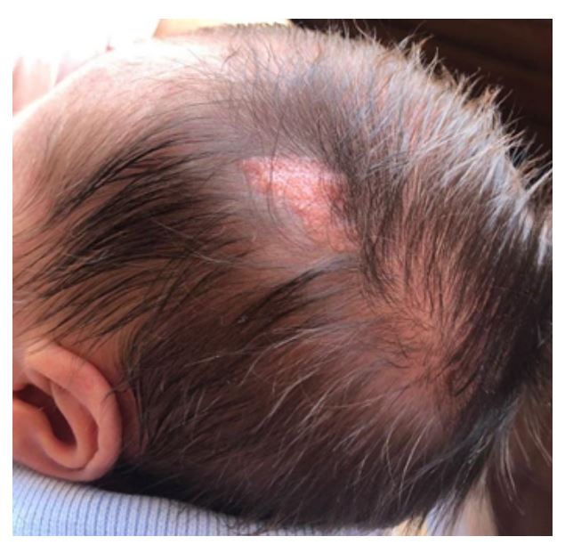
The second case concerns a male newborn who presented at birth with an oval, alopecic, orange-colored lesion approximately 3 cm in diameter in the left temporal region (Figure 2).

Discussion
Two distinct subtypes of epidermal nevi (EN) were identified in these cases. The first case was consistent with a papillomatous EN, while the second case was compatible with a nevus sebaceous (NS). Both patients were evaluated in Dermatology consultations and remained under observation.
Newborns may present with a variety of birthmarks, including congenital EN, which are hamartomas characterized by limited overgrowth of epidermal cell types such as keratinocytes, sebaceous glands, hair follicles, apocrine or eccrine glands, and smooth muscle cells.1),(2
EN is a clinically and genetically heterogeneous condition that encompasses a number of distinct subtypes. These include inflammatory linear verrucous epidermal nevus, epidermolytic epidermal nevus, nevus sebaceous, hair follicle nevus, nevus comedonicus, basaloid follicular hamartoma, eccrine angiomatous hamartoma, porokeratotic adnexal ostial nevus, and Becker nevus.3 The diagnosis is primarily based on the clinical presentation, with a histological examination recommended only for lesions that present diagnostic uncertainty.
EN mostly occur sporadically as an isolated finding and represent a benign condition. They affect approximately 1 to 3 in 1,000 newborns, with nevus simplex (NS) accounting for approximately half of the total cases.1),(4),(5 However, EN can be associated with certain syndromes, collectively referred to as epidermal nevus syndrome, which may involve other organ systems. Notably, NS can be linked to a rare systemic condition known as nevus Sebaceous syndrome or Schimmelpenning syndrome, which is the most well-known type of epidermal nevus syndrome. In this syndrome, the most common extracutaneous manifestation is central nervous system involvement, which can include intellectual disability, seizures, and structural brain abnormalities. Subsequently, involvement of the ophthalmological, musculoskeletal, and, less commonly, the endocrine, cardiovascular, urogenital, and oral systems may occur. Clinicians should remain vigilant for these conditions, especially in the presence of large or extensive nevi associated with additional extracutaneous abnormalities or localized to the head or neck. In fact, individuals with neurological abnormalities are ten times more likely to present with a centrofacial nevus.2),(5
Papillomatous EN typically presents as a single lesion, appearing as an elevated cluster of brown warty-like growths that may extend in a linear distribution along Blaschko lines, suggesting mosaicism due to postzygotic mutations.
NS is a hamartoma of the pilosebaceous follicular unit of unknown cause and is the most common adnexal malformation in pediatric age.4),(6 Most lesions are found on the scalp, often associated with partial or total alopecia, or on the face, but they may also appear on the forehead or neck. The diagnosis of NS is primarily clinical, characterized by a solitary, smooth, yellow, well-circumscribed plaque of oval or linear configuration. During adolescence, the lesion typically becomes warty and thickened.6) Although generally asymptomatic, NS may occasionally be complicated by eczematous reactions.5),(6
In general, children with a small, solitary EN and an otherwise normal physical examination do not require further evaluation beyond dermatologic surveillance. According to recent studies, the risk of malignant transformation is low, typically occurring during or after adolescence, and is slightly higher in cases of NS. The risk of developing basal cell carcinoma in NS is less than 1%; less commonly, it may evolve to sebaceous carcinoma, squamous cell carcinoma, microcystic adnexal carcinoma, or melanoma.2-6
Because of this potential risk, the management of NS remains controversial. Historically, prophylactic excision during adolescence has been widely recommended. However, recent studies have reported low rates of malignant transformation and a higher likelihood of developing secondary benign neoplasms. Despite the low risk of malignancy (approximately 1% develop basal cell carcinoma), surgical excision is still commonly performed, mainly due to concerns about the potential increase in lesion size during puberty and related aesthetic considerations, as well as to provide reassurance to patients and their families. The timing of excision is controversial and should be carefully considered based on factors such as the low inherent risks associated with general anesthesia or the patient’s ability to tolerate the procedure with local anesthesia.1),(2),(7
In conclusion, these case reports highlight the most common presentation of EN as a benign, isolated condition. However, in some cases, EN may be associated with involvement of other organ systems, warranting consideration of the diagnosis of epidermal nevi syndrome. Early identification of EN at birth is critical, as it allows pediatricians to ensure appropriate follow-up, exclude potential syndromic forms, and reassure parents regarding the natural course of these lesions.
Authorship
Cátia Martins - Conceptualization; Bibliographical search; Methodology; Data curation; Writing - original draft; Writing - review & editing; Validation
Laura Correia - Conceptualization; Bibliographical search; Data curation; Writing - review & editing; Validation
Leonor Ramos - Supervision; Visualization; Validation; Writing - review & editing
Rui Castelo - Supervision; Visualization; Validation; Writing - review & editing
Daniela Ramos - Conceptualization; Supervision
Ethical considerations
The authors declare that all procedures were conducted in accordance with the regulations of the Clinical Research and Ethics Committee and the World Medical Association’s Declaration of Helsinki, as updated in 2013.
The authors confirm adherence to their institution’s protocols for publication of patient data.
Written informed consent was obtained in advance.

