


 Inglês (pdf)
 Artigo em XML
 Referências do artigo
 Enviar este artigo por email
 Citado por SciELO
  Similares em
SciELO
Similares em
SciELO 
 Permalink
A previously healthy 11-year-old boy was admitted to the Pediatric Urgency Department with cutaneous lesions in the lower limbs, that caused functional inability for walking.
The lesions first appeared 4 weeks before, when the boy was travelling to Minas Gerais, in Brazil.
Initially, the patient noticed punctiform and erythematous lesions in the lower limbs, pruriginous, apparently due to mosquito bites. The lesions evolved to a pustule with a reddish halo, and later to ulcerations with elevated margins. An adherent hard crust (1 cm of diameter) covered each lesion and yellow exudate appeared.
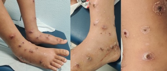
The number of lesions progressively increased in the lower limbs and extended to the upper limbs. The crusts became greyer, thicker and harder, and when removed, showed a shallow punched-out ulceration. The patient noticed swelling in the feet and both ankles, associated with significant pain (figures 1, 2 and 3). At admission, the patient refused walking. No fever or other systemic symptoms were reported.
Discussion
The clinical diagnosis of ecthyma was suspected.
Laboratory analysis revealed leucocytosis (20900/uL), with neutrophilia (76,3%) and elevated C-reactive protein (51,1 mg/L). Blood cultures were negative. Culture of the exudate identified group A beta-hemolytic Streptococcus and methicillin-sensible Staphylococcus aureus.
The patient began treatment with intravenous flucloxacillin (100 mg/kg/day) and clindamycin (30 mg/kg/day). Dermatologist assessment confirmed the diagnosis and recommended additional local procedures, such as daily cleaning of the lesion with chlorhexidine diluted in water, incision and removal of the crusts and application of topic antibiotic (fusidic acid) and colagenase clostridiopeptidase A oinment.
Clinical improvement was noted; therefore, clindamycin was stopped after three days and intravenous flucloxacillin was switched to oral on the fourth day, completing 14 days of treatment.
The patient was discharged after five days, occurring a gradual improvement of the lesions. On six-months follow-up, total resolution of the lesions was observed, while some scars remained on the site of the deeper lesions.
Ecthyma is an ulcerative pyoderma of the skin. Unlike impetigo, ecthyma lesions extend through the epidermis and deep into the dermis, causing scars when healing. It is caused by group A beta-hemolytic Streptococcus, often with a concomitant Staphylococcus aureus infection.1
Patients may be predisposed to develop ecthyma in the presence of immunocompromised states (for example, diabetes mellitus, neutropenia, HIV infection) or preexisting tissue damage, like dermatitis, excoriation or insect bites.
A few factors contribute to the progression of streptococcal skin infections, such as poor hygiene, crowded living conditions and high temperature and humidity (typical of tropical areas). In returning travelers, ecthyma may imitate some zoonotic infections.2,3
Ecthyma lesions usually begin as a vesicle or a pustule; later a hard crust covers an indurated ulcer. It affects more often the lower limbs and can be associated with local adenopathy.
Differential diagnoses include ecthyma gangrenosum, pyoderma gangrenosum, papulonecrotic tuberculids, leishmaniasis and anthrax.
It is essential to provide an early diagnosis and proper treatment to avoid complications related with streptococcal skin infections, as cellulitis, necrotizing fasciitis, lymphadenitis, bacteraemia and, rarely, acute poststreptococcal glomerulonephritis or rheumatic fever.2,4 The most frequent long-term complication is permanent scarring.5
The treatment includes systemic and topical antibiotics and gradual removal of the crusts (as they get soften with topical treatment). Furthermore, it is crucial to improve hygiene in order to control the infection.
Authorship
Ana Foles - Conceptualization; Data Curation; Formal Analysis; Investigation; Writing - original draft
Rita Carvalho - Conceptualization; Data Curation; Investigation; Writing - original draft
Biana Moreira - Conceptualization; Formal Analysis; Writing - review & editing
Joana Cachão - Conceptualization; Formal Analysis; Writing - review & editing

