SciELO - Scientific Electronic Library Online

 
vol.31 número4Doença de Wilson: A História FamiliarDermatomiosite: A Importância do Olho Clínico índice de autoresíndice de assuntosPesquisa de artigos
Home Pagelista alfabética de periódicos  

Serviços Personalizados

Journal

Artigo

Indicadores

Links relacionados

  • Não possue artigos similaresSimilares em SciELO

Compartilhar


Medicina Interna

versão impressa ISSN 0872-671X

Medicina Interna vol.31 no.4 Lisboa dez. 2024  Epub 13-Jan-2025

https://doi.org/10.24950/rspmi.794 

IMAGES IN MEDICINE/ IMAGENS EM MEDICINA

Semiology of Cardiac Tamponade: Jugular Vein Distention

Semiologia de um Tamponamento Cardíaco: Turgescência Venosa Jugular

1Serviço de Medicina Interna, Unidade Local de Saúde Médio Ave, Unidade de Vila Nova de Famalicão, Vila Nova de Famalicão, Portugal

2Serviço de Cardiologia, Unidade Local de Saúde de Braga, Braga, Portugal


Cardiac tamponade is a critical medical condition characterized by dysfunction of cardiac filling due to increased diastolic pressure. This phenomenon may occur due to a pericardial effusion. The increased pressure resulting from this condition may lead to hypotension, muffled heart sounds, and jugular venous distension (JVD), signs that compose the Beck triad.

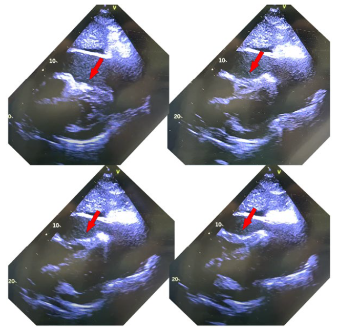
We present a case of a 51-year-old male with advanced lung cancer who sought urgent medical attention for dyspnea that developed for one week and thoracalgia that started on the present day. The patient was obtunded, and hyperhidrotic, with a blood pressure of 89/56 mmHg and a heart rate of 134 bpm. A meticulous examination revealed marked JVD at 45º (Fig. 1), as well as muffled heart sounds, classic signs of this condition.1Swift echocardiography subsequently confirmed the diagnosis of cardiac tamponade (Video 1, Fig. 2) by revealing the presence of pericardial effusion and compressed right cardiac chambers during diastole. The patient was promptly subjected to a pericardiocentesis, which drained 700 cc of serohemorrhagic fluid. During the procedure, the patient improved this hemodynamic profile, normalizing the arterial pressure as well as cardiac frequency.
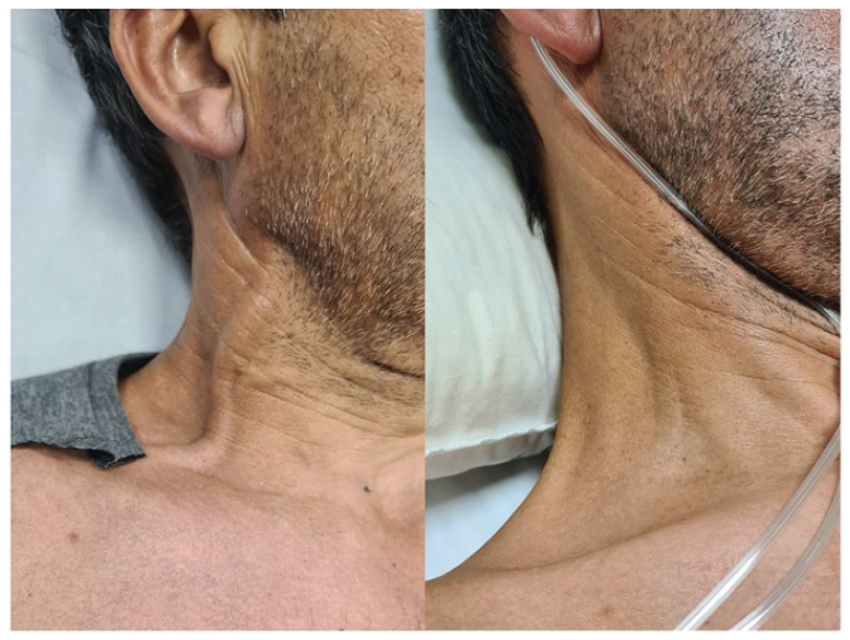
Figure 1: A) Jugular venous distension in a patient with cardiac tamponade; B) Absence of jugular venous distension 24 hours after drainage of the pericardial effusion. 

Video 1: Cardiac tamponade

https://spmi-my.sharepoint.com/:v:/g/personal/ana_silva_spmi_pt/Ea7Df3cBI-A5IhZNzuk8mLXoB-QNWag-68e1nTP26H9PLng?e=GnpPkD

Video 2: Post-pericardiocentesis

https://spmi-my.sharepoint.com/:v:/g/personal/ana_silva_spmi_pt/EW1CPuL-jaOlPrNzh6M0B-pIB76JGFNC70OxTgzj1SS-FmQ?e=Lk1Az0

Figure 2: Echocardiogram showing evidence of a large volume circumferential pericardial effusion with compression of the right ventricle in diastole (cardiac tamponade), shown with red arrow. 

This illustrates the main findings of cardiac tamponade, presenting an example of jugular venous distention with Fig. 1 A, a finding that results from compromised right ventricular filling, stemming from pericardial effusion-induced pressure on the heart chambers.1,2Echocardiography played a crucial role in confirming the diagnosis by presenting both the pericardial effusion and demonstrating the collapsibility of the right ventricle during diastole, confirming the diagnosis of cardiac tamponade. In summary, this case underscores the importance of recognizing clinical findings associated with cardiac tamponade, such as JVD and hypotension, and emphasizes the pivotal role of echocardiography in confirming the diagnosis. Timely pericardiocentesis intervention is indispensable for restoring cardiac function and ultimately enhancing patient outcomes.1

REFERÊNCIAS

1. Brian DH. Cardiac Tamponade. UpToDate [accessed Jan 2024] https://www.uptodate.com/contents/cardiac-tamponadeLinks ]

2. Jensen JK, Poulsen SH, Mølgaard H. Cardiac tamponade: a clinical challenge. E-J Cardiol Pract. 2017;15 [ Links ]

Declaração de Contribuição PFM - Acompanhamento clínico, redação do manuscrito, pesquisa bibliográfica e aprovação final FM - Acompanhamento clínico, pesquisa bibliográfica e revisão crítica PMN - Acompanhamento clínico, pesquisa bibliográfica, revisão crítica e aprovação final Todos os autores aprovaram a versão final a ser publicada. Contributorship Statement PFM - Clinical follow-up, manuscript writing, literature research and final approval FM - Clinical follow-up, bibliographic research and critical review PMN - Clinical follow-up, bibliographic research, critical review and final approval All authors approved the final version of the article.

Conflitos de Interesse: Os autores declaram a inexistência de conflitos de interesse na realização do presente trabalho.

Fontes de Financiamento: Não existiram fontes externas de financiamento para a realização deste artigo.

Confidencialidade dos Dados: Os autores declaram ter seguido os protocolos da sua instituição acerca da publicação dos dados de doentes.

Consentimento: Consentimento do doente para publicação obtido.

Proveniência e Revisão por Pares: Não comissionado; revisão externa por pares.

Conflicts of interest: The authors have no conflicts of interest to declare.

Financing Support: This work has not received any contribution, grant or scholarship.

Confidentiality of Data: The authors declare that they have followed the protocols of their work center on the publication of data from patients.

Patient Consent: Consent for publication was obtained.

Provenance and Peer Review: Not commissioned; externally peer re-viewed.

12© Autor (es) (ou seu (s) empregador (es)) e Revista SPMI 2024. Reutilização permitida de acordo com CC BY-NC 4.0. Nenhuma reutilização comercial. © Author(s) (or their employer(s)) and SPMI Journal 2024. Re-use permitted under CC BY-NC 4.0 commercial re-use.

Received: September 27, 2023; Accepted: February 24, 2024

Correspondence / Correspondência: Pedro Fernandes Moura - pedrofmoura.mi@gmail.com Interno de Formação Específica de Medicina Interna, Unidade Local de Saúde Médio Ave, Unidade de Vila Nova de Famalicão, Vila Nova de Famalicão, Portugal Rua Cupertino de Miranda, s/m, 4761-917 - Vila Nova de Famalicão

Creative Commons License This is an open-access article distributed under the terms of the Creative Commons Attribution License