




 Inglês (pdf)
 Artigo em XML
 Referências do artigo

 Tradução automática
 Enviar este artigo por email
 Citado por SciELO
 Similares em
SciELO

 Permalink


Introduction
Cardiovascular diseases (CVD) remain the leading cause of mortality and disability worldwide,1 with coronary artery disease (CAD) being one of the most prevalent manifestations. CAD imposes a significant burden on healthcare systems, contributing to substantial morbidity, increased hospitalizations, and elevated healthcare costs. The clinical spectrum of CAD ranges from stable angina to acute coronary syndrome (ACS), which can ultimately progress to heart failure (HF) if left untreated or inadequately managed.2 Given this scenario, early diagnosis and risk stratification are fundamental to guiding clinical decision-making and optimizing patient outcomes.
ACS encompasses conditions ranging from unstable angina (UA) to myocardial infarction (MI), with or without ECG changes or troponin elevation. MI is diagnosed based on the fourth universal definition, characterized by cardiomyocyte injury and troponin release, whereas UA involves ischemia without necrosis, presenting as prolonged or worsening angina at rest or minimal exertion.3
Despite continuous advancements in the management of ACS - particularly through improvements in revascularization techniques and pharmacological therapy3 - the incidence of adverse cardiovascular events remains high. Recurrent events, in particular, pose a significant challenge, emphasizing the need for early identification of high-risk patients. Traditional cardiovascular risk factors, such as hypertension, diabetes mellitus, dyslipidaemia, and smoking, are well-established predictors of CAD; however, growing evidence suggests that additional metabolic and inflammatory markers could further enhance risk assessment.
Insulin resistance (IR) is a central feature of metabolic syndrome, which predisposes individuals to a spectrum of cardiovascular complications. In this context, IR has emerged as an important, yet often underrecognized, contributor to atherosclerosis and cardiovascular disease progression.4 IR is strongly associated with metabolic disorders, particularly obesity, type 2 diabetes, and dyslipidaemia,5 and has been shown to accelerate endothelial dysfunction and promote vascular inflammation. Given the complexities involved in measuring IR through gold standard techniques - such as the euglycemic-hyperinsulinemic clamp test, which is labour-intensive and impractical for routine clinical use6 - there has been a growing interest in surrogate markers that can provide a simpler and more accessible assessment of IR. Since 2008, the triglyceride-glucose (TyG) index has been introduced as a reliable and specific marker of IR.7
Studies have demonstrated a strong correlation between the TyG index and IR, with performance comparable to the homeostasis model assessment of IR (HOMA-IR).7 More importantly, emerging evidence supports the clinical relevance of the TyG index in cardiovascular risk assessment.8,9Elevated TyG levels have been consistently associated with increased arterial stiffness and the presence of coronary and carotid atherosclerotic lesions.9 Recent investigations further reinforce its prognostic value, showing that higher TyG levels are predictive of CAD severity, progression, and adverse cardiovascular outcomes.10
Recent evidence suggests that the TyG index is closely associated with various molecular mechanisms contributing to CVD pathogenesis, including metabolic inflexibility, endothelial dysfunction, coagulation disorders, and smooth muscle cell dysfunction.9 These mechanisms collectively impair cardiac function, promote myocardial cell death, facilitate thrombotic and inflammatory processes, and induce structural alterations such as cardiac fibrosis and ventricular stiffness. Consequently, the TyG index serves as a valuable tool in predicting cardiovascular risk and understanding the intricate metabolic pathways linking IR to CVD.
At the molecular level, IR induces metabolic disturbances such as increased lipid oxidation and impaired glucose metabolism, reducing metabolic flexibility and cardiac efficiency.11,12Endothelial dysfunction, characterized by diminished nitric oxide bioavailability and increased mitochondrial reactive oxygen species, exacerbates myocardial cell death and angiogenesis impairment.13,14Additionally, heightened platelet hyperactivity, increased adhesion molecules, and reduced prostaglandin I2bioavailability contribute to coagulation abnormalities, promoting thrombosis and inflammation.15 Smooth muscle cell dysfunction, driven by excessive glycosylation, smooth muscle proliferation, and collagen deposition, results in structural cardiac remodelling, leading to fibrosis and ventricular stiffness.16 Together, these interconnected mechanisms underscore the role of IR in cardiovascular pathology.
Given the increasing recognition of metabolic dysfunction as a key driver of cardiovascular risk, the role of the TyG index in ACS patients warrants further exploration. While conventional risk stratification models primarily rely on traditional risk factors and established scoring systems,17 integrating novel biomarkers such as the TyG index may provide additional prognostic value. The ability to refine risk prediction could lead to earlier therapeutic interventions and personalized treatment strategies, ultimately improving patient outcomes.
The GRACE score is a well-established tool for risk stratification in ACS, predicting major adverse cardiovascular and cerebrovascular events (MACCE). Whether integrating TyG index with the GRACE score enhances prognostic accuracy remains unclear.18
Therefore, this study aims to assess the impact of base-line TyG levels on cardiovascular outcomes in patients admitted with ACS who underwent percutaneous coronary intervention (PCI). Furthermore, we seek to determine whether the combination of the TyG index with current conventional cardiovascular prognostic scores enhances risk stratification and improves prognostic accuracy in this population. By addressing these questions, our findings could contribute to the refinement of risk assessment strategies in contemporary ACS management and potentially support the integration of metabolic markers into clinical practice.
Material and Methods
We performed a retrospective, observational, longitudinal study on adult patients hospitalized with a diagnosis of ACS in an Intermediate Care Unit of a secondary hospital, admitted between January of 2019 and December of 2023. The study population included patients with ST-segment elevation myocardial infarction (STEMI), non-STEMI (NSTEMI), and unstable angina (UA). Patients with suspected or confirmed familial hypertriglyceridemia were excluded, given its distinct metabolic profile. Data were collected through a detailed review of electronic medical records. Demographic data (age, gender, autonomy as assessed by the Clinical Frailty Scale, and body mass index), comorbid diseases (hypertension, diabetes mellitus, dyslipidaemia, smoking, heart failure, atrial fibrillation, chronic kidney disease, previous acute myocardial infarction, previous stroke, previous coronary intervention), and diagnostic information were obtained at admission. Routine laboratory tests, including glycemia, creatinine, low-density lipoprotein cholesterol (LDL-C), high-density lipoprotein cholesterol (HDL-C), total cholesterol (TC), and triglyceride (TG), were performed using fasting blood samples. Echocardiographic findings were also reviewed - particularly left ventricular ejection fraction (LVEF). The angiographic data, including the number of diseased vessels, location of target lesions, number of stents, and number of treated vessels, were also collected. Prescription patterns pre-admission and at hospital discharge were assessed, focusing on beta-blockers, renin-angiotensin-aldo-sterone system (RAAS) inhibitors, mineralocorticoid receptor antagonists, and sodium-glucose co-transporter 2 inhibitors (SGLT2i). Follow-up data were collected, through the outpatient or readmission charts, for up to 12 months post-event, capturing clinical evolution, medication adherence, repeat echocardiography findings, and laboratory results.
All data were collected retrospectively using a standardized data collection form.
The incidence of major adverse cardiovascular and cerebrovascular events (MACCE) was documented, defined as a composite of myocardial infarction (AMI), stroke, cardiovascular and all-cause death, and revascularization.
The TyG index was calculated as ln [(fasting triglyceride (mg/dL)×fasting glucose (mg/dL))/2]. Calculation of the GRACE Score was based on the clinical data obtained at admission (age, heart rate, systolic blood pressure, Killip class, creatinine level, ST-segment deviation, elevated cardiac enzymes, and cardiac arrest).
Both TyG index and GRACE score used cut-offs derived from our study population. Specifically, the thresholds (TyG = 8.80; GRACE = 109) correspond to the median values in our cohort, thereby dividing patients into two equally sized groups. This approach ensured balanced comparison and avoided overfitting to externally defined cut-offs.
All participants were separated into low-risk and high-risk according to the TyG index levels: Low-TyGI (n=267, TyG-I <8.80), High-TyGI (n=267, TyGI ≥8.80); and the GRACE Scores levels: Low-GS (n=267, GS <109), High-GS (n=267, GS≥109).
Statistical analysis was conducted using GraphPad Prism v.10.4.1. Categorical variables were expressed as absolute frequencies (n) and percentages (%), with comparisons performed using the Chi-square test or Fisher’s exact test. Continuous variables were described as means ± standard deviation (SD). Kaplan-Meier curves were used to assess event-free survival, with comparisons between groups made using the log-rank test. A p-value < 0.05 was considered statistically significant.
The study was conducted in accordance with ethical standards and received approval from the local Ethics Committee.
Results
During the selected period, a total of 534 patients diagnosed with ACS were included in the study. Among them, 35,58% (n=190) presented with STEMI, 61.24% (n=327) with NSTEMI, and 3.18% (n=17) with UA. The population consisted of 401 men and 133 women, aged between 31 to 99 years (mean 63.95±12.39). Most patients (89.51%) were previously autonomous, classified as Clinical Frailty Scale (CFS) 1-3 (n=478). A history of coronary artery disease was identified in 20.22% (n=108) of patients, while a family history of cardiovascular dis-ease was reported in 15.17% (n=81).
Only 4 patients had no known or stratified VRF. Among the remaining population, 7.68% had one VRF, 31.65% had two, 37.27% had three, and 22.66% had four or more. Dyslipidaemia (DL) was the most prevalent VRF, affecting 85.21% (n=455) of patients, followed by arterial hypertension (HT) in 65.73% (n=351). Diabetes mellitus (DM) was present in 34.08% (n=182). Smoking was identified as a risk factor in 64.61% (n=345), and obesity was document-ed in 26.97% (n=144).
Patients were stratified into low or high-risk both for TyG Index. The baseline characteristics of the study population are shown in Table 1.
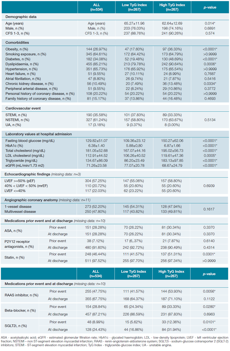
Patients in the High TyG Index group were significantly younger than those in the Low TyG Index group (62.64±12.69 vs 65.27±11.96 years, p=0.014). The prevalence of obesity (36.33% vs 17.60%, p<0.0001), diabetes (48.69% vs 19.48%, p<0.0001), dyslipidaemia (90.64% vs 79.78%, p=0.0006), and chronic kidney disease (13.48% vs 7.49%, p=0.0334) was significantly higher in the High TyG Index group. No significant differences were observed for sex distribution, smoking exposure, hypertension, heart failure, atrial fibrillation, peripheral arterial disease, personal history of coronary disease, or family history of coronary disease between the two groups.
No significant differences were observed in the distribution of STEMI, NSTEMI, or UA between groups (p=0.5134). No significant differences were found in echocardiographic parameters (p=0.6939) or coronary anatomy (p=0.1617). Laboratory values at admission also showed marked differences, with the High TyG Index group presenting significantly higher fasting blood glucose, HbA1c, total cholesterol, LDL cholesterol, and triglyceride - the observed differences in metabolic parameters between the High TyG Index and Low TyG Index groups align with the fundamental components of the TyG Index, which is derived from fasting triglyceride and glucose levels.
At baseline, the overall use of cardioprotective therapies was suboptimal, particularly for statins and RAAS inhibitors, although patients with higher TyG index were more frequently on these drugs as well as on SGLT2 inhibitors, reflecting recognition of their higher metabolic risk.
At discharge, aspirin was prescribed to the majority of patients (96.18% overall), with no significant difference between the Low (95.40%) and High (96.96%) TyG Index groups (p=0.3726). Similarly, the use of P2Y12 receptor antagonists (91.60%), statins (97.52%), RAAS inhibitors (67.75%), and beta-blockers (87.21%) did not significantly differ between groups (p>0.05 for all). However, a significant difference was observed in the prescription of SGLT2 inhibitors, which were more frequently prescribed in the High TyG Index group (31.94%) compared to the Low TyG Index group (16.86%) (p<0.0001).
Fig. 1 illustrates the distribution of vascular risk factors (VRF) among patients stratified by TyG index levels. In the low TyG Index group, the majority of patients had 2 or 3 VRFs (99 patients each, 37.08%), while fewer had 4+ VRFs (33 patients, 12.40%), 1 VRF (32 patients, 11.99%), or no VRFs (4 patients, 1.50%). Conversely, in the High TyG Index group, there was a notable shift toward a higher burden of VRFs, with 100 patients (37.45%) having 3 VRFs and 88 patients (32.96%) having 4+ VRFs, while only 9 patients (3.37%) had 1 VRF and none had 0 VRFs.
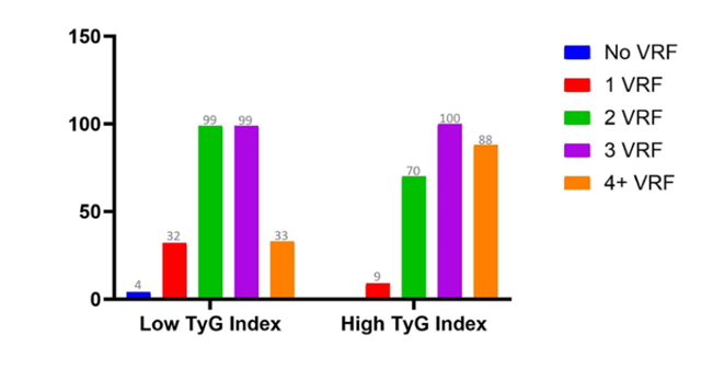
Figure 1: Distribution of the number of traditional vascular risk factors (VRF) among patients stratified by TyG Index category.
During the 12-month follow-up period, 68 patients (12.73% of the total population) experienced primary endpoint events, which consisted of 34 (6.37%) all-cause deaths, out of which 16 (3.00%) cardiovascular-related; 21 (3.93%) myocardial infarctions, 6 (1.12%) ischemic strokes, and 19 (3.56%) ischemia-driven coronary revascularization. The incidence of MACCEs and the individual events showed no significant difference among the groups. The occurrence of MACCEs and individual events per group are summarized in Fig. 2.
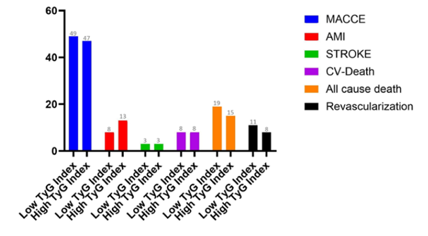
Figure 2: Incidence of major adverse cardiovascular and cerebrovascular events (MACCE) and individual cardiovascular outcomes (acute myocardial infarction (AMI), stroke, cardiovascular death (CV-Death), all-cause death, and revascularization in patients) stratified by TyG Index category.
To address the benefit of TyG Index along GRACE Score, as a prognostic tool, patients were also divided by Low/High GRACE Score, which were then combined, allowing for a division in four groups: (1) Low GS/Low TyGI (n=119), (2) Low GS/High TyGI (n=148), (3) High GS/Low TyGI (n=148), and (4) High GS/High TyGI (n=119).
MACCE incidence was highest in Group 3 (n=40), followed by Group 4 (n=33), Group 2 (n=14) and Group 1 (n=9). Kaplan-Meier survival analysis using only GS showed a significant decline in MACCE-free survival with higher scores (p<0.0001). Using TyGI alone to assess the same risk stratification did not show a relevant difference between groups. By combining TyGI with GS, we found a statistically significant distinction among all groups (p<0.0001), with a trend towards lower MACCE-free survival in Group 4 compared to Group 3. When addressing AMI-free survival, however, TyGI alone, unlike GS, showed a significant decline with higher scores (p=0.046 vs p=0.068).
When combining both scores, Group 4 (High GRACE / High TyG) and Group 2 (Low GRACE / High TyG) showed significantly lower event-free survival compared with their respective low-TyG counterparts (Groups 3 and 1). Overall, event-free survival differed significantly across all four groups (log-rank p = 0.030). Figs. 3 and 4 depict these findings.
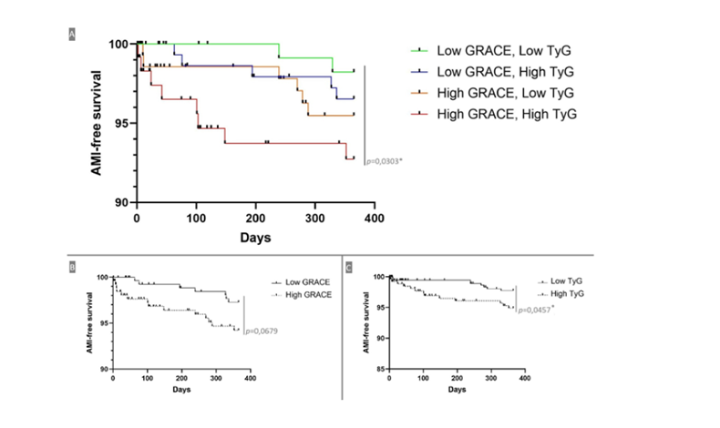
Figure 3: Acute myocardial infarction (AMI)-free survival curves stratified by TyG Index and GRACE score. (A) Kaplan-Meier survival analysis showing recurring AMI-free survival based on the combination of TyG Index and GRACE Score categories. (B) AMI-free survival comparison between Low and High GRACE score groups. (C) AMI-free survival comparison between Low and High TyG Index groups. Significant differences were observed among the groups (p-values indicated in the graphs).
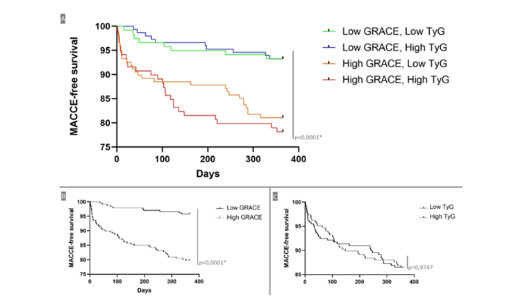
Figure 4: MACCE-free survival curves stratified by TyG Index and GRACE score. (A) Kaplan-Meier survival analysis showing major adverse cardiovascular and cerebrovascular event (MACCE)-free survival based on the combination of TyG Index and GRACE score categories. (B) MACCE-free survival comparison between low and High GRACE score groups. (C) MACCE-free survival comparison between low and High TyG Index groups. Significant differences were observed among the groups (p-values indicated in the graphs).
Discussion
The present study sought to evaluate the prognostic significance of the TyG Index in patients with ACS. Our findings provide valuable insights into the interplay between IR, metabolic dysfunction, and cardiovascular risk, particularly when integrating the TyG index with traditional stratification tools such as the GRACE score. By analysing the association between the TyG index and clinical, biochemical, and prognostic outcomes, we aimed to assess whether this marker could refine existing risk models and guide more personalized therapeutic strategies.
Our results revealed that patients with a high TyG index were significantly younger and had a higher prevalence of obesity, diabetes, dyslipidemia, and chronic kidney disease compared to those in the low TyG index group. This observation aligns with previous studies indicating that an elevated TyG index is strongly associated with metabolic dysfunction and IR, which are well-established risk factors for cardiovascular disease.9 IR fosters a pro-inflammatory, pro-thrombotic environment that accelerates endothelial dysfunction, oxidative stress, and plaque instability - key mechanisms in ACS pathophysiology. The clustering of metabolic comorbidities in patients with elevated TyG index values underscores the need for comprehensive risk modification strategies targeting glucose metabolism, lipid homeostasis, and renal function to mitigate cardiovascular risk.
In analysing the burden of vascular risk factors across TyG index subgroups, we observed that patients with a high TyG index exhibited a markedly greater prevalence of multiple VRFs, particularly in those with three or more risk factors (188 vs 132 in the low TyG group). This pattern again suggests a strong association between IR and the clustering of vascular risk factors. Patients with multiple VRFs face a compounded risk of adverse cardiovascular events, due to the synergistic effects of dysglycemia, dyslipidaemia, and vascular dysfunction, necessitating a multifaceted approach to risk reduction. These findings highlight the need for targeted interventions in high-risk metabolic phenotypes.
Prescription patterns findings suggest that high TyG patients already represent a metabolically high-risk phenotype, often targeted with more intensive therapy even before ACS. Nevertheless, residual risk persists despite treatment, as evidenced by their adverse outcomes. The greater prescription of SGLT2 inhibitors at discharge in this group may reflect clinicians’ awareness of their cardiometabolic burden, supporting the potential role of TyG in guiding earlier and more aggressive preventive strategies.
However, it is key to note that neither TyG index values nor IR do necessarily capture all traditional cardiovascular risk factors, and, therefore, it should not be considered a standalone risk predictor but rather an adjunct to established clinical models to refine stratification.
Laboratory findings demonstrated that the High TyG index group had significantly higher fasting glucose, HbA1c, total cholesterol, LDL cholesterol, and triglyceride levels. These differences are expected, as the TyG index is derived from fasting glucose and triglyceride levels. Nevertheless, the marked disparities in metabolic parameters reinforce the utility of the TyG index as a surrogate marker for IR and cardiometabolic risk.
Despite the metabolic differences between groups, no significant variations were found in the distribution of STEMI, NSTEMI, or UA between the high and low TyG index groups. Likewise, no notable differences were observed in echocardiographic parameters or coronary anatomy. This suggests that while the TyG index reflects an underlying metabolic risk, it may not directly correlate with the acute presentation or anatomical burden of coronary artery disease. This raises the possibility that the TyG index reflects a functional rather than a purely structural risk. Patients with a High TyG index may have more vulnerable plaque phenotypes or a greater propensity for microvascular dysfunction, which is not always captured by conventional imaging modalities. Future research utilizing advanced imaging techniques, such as coronary computed tomography angiography (CCTA),19 optical coherence tomography (OCT)20 or intravascular ultrasound (IVUS) in PCI,21 could help elucidate these potential differences in plaque characteristics between TyG index subgroups.
The increased prescription of sodium-glucose co-transporter 2 (SGLT2) inhibitors in the High TyG index group suggests that clinicians may already recognize the metabolic burden in these patients. The significantly higher prevalence of DM and CKD likely influenced this prescribing pattern on this subset of the population. Given the emerging evidence supporting the cardioprotective benefits of SGLT2 inhibitors22 and GLP-1 receptor agonists,23 it is plausible that integrating the TyG index into routine cardiovascular risk assessment could facilitate early initiation of such therapies. These novel therapeutic agents exert pleiotropic effects beyond glycaemic control, including reductions in inflammation, oxidative stress, and endothelial dysfunction24,25- mechanistic pathways that align closely with the metabolic derangements captured by the TyG index. Thus, identifying High-risk patients via the TyG index could potentially enable more targeted metabolic interventions in the secondary prevention of ACS.
During the 12-month follow-up, 12.73% of the total study population experienced MACCE, including all-cause death, cardiovascular death, myocardial infarction, ischemic stroke, and ischemia-driven coronary revascularization. Notably, no significant differences in MACCE incidence were observed between the high and low TyG index groups when analysed independently. One possible explanation for this observation is that the TyG index predominantly reflects long-term metabolic dysfunction rather than the acute determinants of cardiovascular events. While IR and dyslipidaemia contribute to atherosclerosis and endothelial dysfunction, these processes develop gradually and may not immediately translate into increased short-term event rates, although a trend could already be seen on recurring AMI rates.
The GRACE score remains a dominant predictor of adverse outcomes, likely due to its incorporation of key clinical parameters such as age, heart rate, blood pressure, and renal function,17 which are well-established determinants of short-term prognosis in ACS patients. When integrating the TyG index with the GRACE score, a more nuanced risk stratification approach emerged. Notably, MACCE incidence was highest in patients with a High GRACE score, regardless of TyG category, whereas differences between TyG strata were smaller. This pattern suggests that GRACE remains the predominant determinant of adverse outcomes in our cohort, with TyG contributing additional-but more modest-risk stratification. However, an intriguing finding emerged when AMI-free survival was analysed: the TyG index alone demonstrated significant prognostic value, whereas the GRACE score did not. This raises the hypothesis that while the GRACE score effectively captures acute cardiovascular risk, it may not adequately account for the chronic metabolic and inflammatory milieu that predisposes patients to recurrent myocardial infarction. IR reflected by an elevated TyG index, is closely linked to chronic vascular inflammation, endothelial dysfunction, and atherothrombosis, all of which contribute to plaque instability and recurrent ischemic events. Thus, the TyG index may serve as an independent marker of long-term susceptibility to atherosclerotic progression, even among patients with initially lower GRACE scores.
Kaplan-Meier survival analyses further reinforced these findings by demonstrating that combining the TyG index with the GRACE score improved risk stratification. Patients with both High GRACE scores
and High TyG index values exhibited lower MACCE-free and AMI-free survival rates compared to other groups. This suggests a synergistic effect, where traditional clinical risk factors and metabolic dysfunction collectively drive adverse outcomes. From a clinical standpoint, these findings underscore the potential value of incorporating metabolic markers into contemporary risk assessment models for ACS. As such, the TyG index may enhance prognostic accuracy, particularly in identifying patients at elevated risk for recurrent myocardial infarction who might otherwise be overlooked by traditional scoring systems.
Several limitations should be acknowledged. This was a retrospective, observational study conducted in a single centre, which may limit the generalizability of our findings. The follow-up period was limited to 12 months, and longer-term outcomes were not assessed, which may hinder the detection of long-term trends and underpower statistical analyses.
Future prospective studies with larger sample sizes and extended follow-up periods are needed to validate our findings. Additionally, exploring the relationship between the TyG index and novel biomarkers of inflammation, oxidative stress, and endothelial dysfunction could provide further insights into its pathophysiological significance. Given the growing interest in precision medicine, integrating the TyG index into machine learning models alongside conventional risk scores and genetic risk factors may improve cardiovascular risk prediction. Such an approach could help identify high-risk subgroups that may benefit from intensified preventive measures or novel therapeutic strategies.
Conclusion
This study highlights the potential value of incorporating metabolic markers, such as the triglyceride-glucose (TyG) index, into contemporary cardiovascular risk assessment models. The TyG index is an easily accessible and cost-effective tool, requiring only fasting triglyceride and glucose levels - parameters routinely measured in ACS patients - making it a practical addition to existing risk stratification models. While traditional tools like the GRACE score effectively predict adverse cardiovascular events, it does not account for IR or metabolic dysfunction, which play a pivotal role in atherosclerosis progression. The integration of the TyG index into established risk models may enhance risk stratification, offering a more nuanced understanding of long-term cardiovascular risk in patients with ACS.

