Serviços Personalizados
Journal
Artigo
Indicadores
-
 Citado por SciELO
 -
 Acessos
Links relacionados
-
 Similares em
SciELO
Compartilhar
Jornal Português de Gastrenterologia
versão impressa ISSN 0872-8178
J Port Gastrenterol. vol.19 no.2 Lisboa fev. 2012
Gastric suture line ulcer: An unexpected cause of gastrointestinal bleeding
Úlcera gástrica por fi o de sutura: uma causa inesperada de hemorragia digestiva
Rosa Ferreira,a Pedro Amaro,b Manuela Ferreira,b José Manuel Romãozinhob
aGastroenterology Department, Hospitalar Center of Coimbra, Coimbra, Portugal
bGastroenterology Intensive Care Unit, University Hospital of Coimbra, Portugal
*Corresponding author
Abstract
Gastrointestinal bleeding due to suture line ulceration has been attributed to use of non‑absorbable silk sutures. We report a case of a 63‑year‑old male with gastrointestinal bleeding due to gastric suture line ulceration after splenectomy performed 11 years ago. Emergent upper endoscopy revealed a small gastric ulcer with a visible vessel and raised the suspicion of gastric varices. An endoscopic ultrasound was performed, showing a gastric hyperechogenic area in the mucosa layer with an acoustic shadow. Upper endoscopy with a transparent cap was repeated and a mobile foreign body in the ulcer´s bed was found. It was successfully removed by forceps. After the procedure a silk suture line of 3.5 cm was identified.
KEYWORDS Gastric ulcer; Silk suture; Upper gastrointestinal bleeding; Endoscopic cap; Endoscopic ultrasound
Resumo
A hemorragia digestiva por ulceração da linha de sutura tem sido atribuída ao uso de fios de sutura de seda, não absorvíveis. Os autores relatam um caso de um homem de 63 anos com hemorragia digestiva devido a úlcera gástrica por fio de sutura após esplenectomia realizada 11 anos antes. A endoscopia digestiva urgente revelou uma pequena úlcera gástrica com vaso visível e levantou a suspeita de varizes gástricas. A eco‑endoscopia mostrou uma área hiperecogénica na camada mucosa do estômago, esboçando cone de sombra. Repetiu‑se a endoscopia digestiva com um cap, visualizando‑se um corpo estranho móvel no leito da úlcera, que foi removido com uma pinça e identificado como um fio de sutura de seda de 3,5 cm.
PALAVRAS-CHAVE Úlcera gástrica; Fio de sutura; Hemorragia digestiva alta; Cap endoscópico; Eco‑endoscopia
Introduction
Non‑absorbable silk sutures used in surgery of the gastrointestinal tract have been associated with several complications, such as migration toward the luminal surface, marginal ulcers and suture line ulceration1‑4. Upper gastrointestinal bleeding without mucosal ulceration due to the use of silk suture has also been reported after partial gastrectomy5. The use of non‑absorbable silk sutures in gastric and bowel surgery was therefore discontinued.
We herein report a case of a 63‑year‑old male with massive gastrointestinal bleeding due to gastric suture line ulceration after splenectomy performed 11 years ago.
Case report
A 63‑year‑old man presented to the emergency department with hematemesis. He denied abdominal pain or altered bowel habits. His past medical history was notable for a splenectomy 11 years ago due to an abdominal trauma, diabetes, hypertension and hypercholesterolemia. He was on medication with insulin, statin and angiotensin receptor antagonist with hydrochorothiazid. He denied the use of antiplatelet and nonsteroidal anti‑inflammatory drugs. His alcohol intake was 120 g/day for more than 30 years.
On physical examination he was pale, taquicardic (110 bpm) and hypotensive (50/30 mmHg). His abdomen was soft, painless with no apparent hepatomegaly or fluid wave. The digital rectal exam showed melena.
Laboratory data revealed anemia of 10.1 g/dL (normal value, 12‑16), thrombocytopenia of 114x103/mL (normal value, 150‑400), prothrombin time of 64% (normal value, 70‑120), elevation of alanine aminotransferase of 70 U/L (normal value, 10‑40), aspartate aminotransferase of 57 U/L (normal value, 10‑42) and γ‑glutamyltransferase of 312 U/L (normal value, <30). Bilirrubin and albumin were normal.
After volume resuscitation, an emergent upper Endoscopy was performed and revealed thickening of the folds of the greater curvature of the stomach´s body, raising the suspicion of varices. However, deeply seated and hidden by convergent folds, a small ulcer with a visible vessel was found with great difficulty due to lack of distensibility of this area. Endoscopic hemostasis with adrenaline was done and was apparently effective.
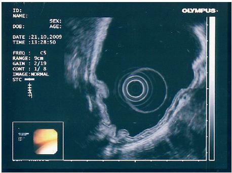
In order to characterize these findings, an endoscopic ultrasound was made. It ruled out the hypothesis of gastric varices, but showed thickening of gastric folds with a hyperechogenic area in the mucosa layer with an acoustic shadow (Fig. 1).

Figure 1 Upper endoscopic ultrasound showing a hyperechogenic area in the mucosa layer with an acoustic shadow in the greater curvature of the gastric body.
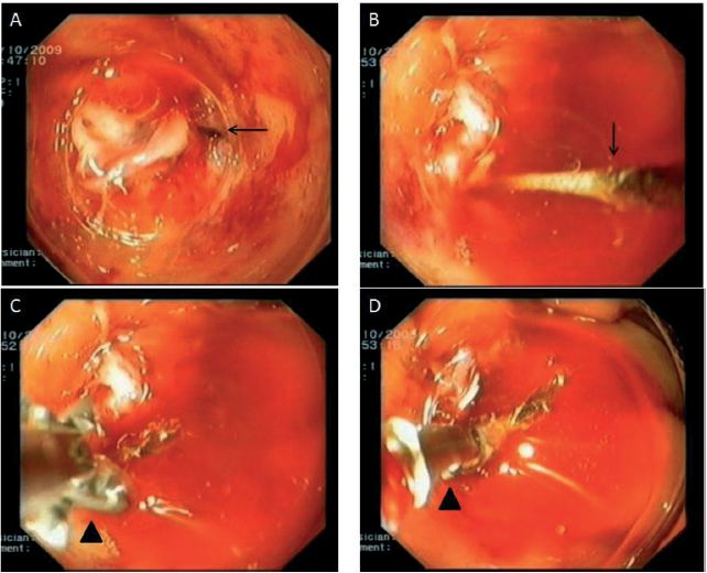
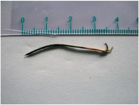
Upper endoscopy was repeated with an auxiliary transparent cap to allow a carefully exploration of the ulcer´s bed that eventually revealed a mobile, tapered, dark‑brown foreign body penetrating into the mucosa (Fig. 2). It was carefully removed with a coagrasper® fórceps without complication (Fig. 2). After this procedure, a silk suture line of 3.5 cm was identified (Fig. 3).

Figure 2 Upper endoscopy showing: A‑ dark brown foreign body (arrow) in ulcer´s bed penetrating into the mucosa; B‑ foreign body (arrow) after been pushed by forceps; C and D‑ foreign body extraction using coagrasper® forceps (arrowhead).

Figure 3 A silk suture line of 3.5 cm in length.
The patient was discharged at day 6 on proton pump inhibitors and iron supplementation. Follow‑up Endoscopy one month later showed no mucosal lesions.
Discussion
To our knowledge this is the first case reported of upper gastrointestinal bleeding due to gastric suture line ulcer after splenectomy attributed to the use of non‑absorbable suture. Nevertheless, suture line ulceration after gastric surgery caused by non‑absorbable suture materials was first described by Paterson in 19096.
Symptoms related to non‑absorbable sutures include abdominal pain, nausea, vomiting, hematemesis and melena2,5. However, massive upper gastrointestinal hemorrhage is rare7.
Mucosal ulceration at the site of the suture is not always present at endoscopy5.
Histologically, it is characterized by a foreign body reaction, with giant cell infiltration, fibrosis and dense capillary proliferation4,8. It has been demonstrated that silk sutures lead to a persistent inflammatory reaction that can promote a dense network of vessels. This vessels may be disrupted by mechanical forces produced by luminal oriented sutures and thereby can cause hemorrhage5.
Like in our case, this complication may present many years after the surgery5.
The suture material can be removed either endoscopically or surgical2,4,5,9. In our patient the non‑absorbable suture line was successfully removed using endoscopic forceps. The use of an auxiliary cap in upper endoscopy proved to be helpful since it improved the visualization of the ulcer´s bed and allowed a detailed exploration oh this area. Surgical removed is indicated when endoscopic therapy fails4.
This case illustrates that gastric suture line ulceration should be considered in the differential diagnosis of upper gastrointestinal bleeding, particularly in those patients who underwent previous abdominal surgery. On the other hand, endoscopic suture removal is feasible, safe and effective in managing suture line ulcers.
References
1. Ueda E, Kitayama J, Seto Y, et al. Postoperative complications after local resection of the stomach. Surg Today. 2002;32:305‑9. [ Links ]
2. Yu S, Jastrow K, Clapp B, et al. Foreign material erosion after laparoscopic Roux‑en‑Y gastric bypass: findings and treatment. Surg Endosc. 2007;21:1216‑20. [ Links ]
3. Sacks BC, Mattar SG, Qureshi FG, et al. Incidence of marginal ulcers and the use of absorbable anastomotic sutures in laparoscopic Roux‑en‑Y gastric bypass. Surg Obes Relat Dis. 2006;2:11‑6. [ Links ]
4. Frezza EE, Herbert H, Ford R, et al. Endoscopic suture removal at gastrojejunal anastomosis after Roux‑en‑Y gastric bypass to prevent marginal ulceration. Surg Obes Relat Dis. 2007;3:619‑22. [ Links ]
5. Pecha R, Prindiville T, Kotfila R, et al. Gastrointestinal hemorrhage consequent to foreign body reaction to silk sutures: case series and review. Gastrointest Endosc. 1998;48:299‑301. [ Links ]
6. Paterson H. Jejunal and gastrojejunal ulcer following gastrojejunostomy. Proc Roy Soc Med. 1909;2:238‑300. [ Links ]
7. Jacob H, Brandt L, Berkowitz D, et al. Massive hemorrhage from suture line ulceration: documentation by endoscopy. Gastrointest Endosc. 1982;28:181‑2. [ Links ]
8. Posthlewait R, Willigan D, Ulin A. Human tissue reaction to sutures. Ann Surg. 1975;181:144‑50. [ Links ]
9. Lee JK, Van Dam J, Morton JM, et al. Endoscopy is accurate, safe, and effective in the assessment and management of complications following gastric bypass surgery. Am J Gastroenterol. 2009;104:572‑82. [ Links ]
*Corresponding author
E-mail address: rosa.l.ferreira@gmail.com
Received January 10, 2011; accepted April 3, 2011


