Serviços Personalizados
Journal
Artigo
Indicadores
-
 Citado por SciELO
 -
 Acessos
Links relacionados
-
 Similares em
SciELO
Compartilhar
Jornal Português de Gastrenterologia
versão impressa ISSN 0872-8178
J Port Gastrenterol. vol.19 no.6 Lisboa nov. 2012
https://doi.org/10.1016/j.jpg.2012.04.025
Giant pseudopolyposis in ulcerative colitis: Case report and review of literature
Pseudopolipose gigante na colite ulcerosa: caso clínico e revisão da literatura
Inês Marquesa,∗, Ana Lagosa, Jorge Reisa, Filomena Medeirosb, António Pintoa, Beatriz Nevesa
a Serviço de Gastrenterologia, Hospital Pulido Valente - Centro Hospitalar Lisboa Norte, Lisboa, Portugal
b Serviço de Anatomia Patológica, Hospital Pulido Valente - Centro Hospitalar Lisboa Norte, Lisboa, Portugal
*Corresponding author.
Abstract
Localized giant pseudopolyposis is an extremely rare feature of pseudopolyposis complicating inflammatory bowel disease. It represents a localized exuberant collection of pseudopolyps, which may simulate neoplasms such as polypoid cancer.
We report a case of a 23-year-old man with the diagnosis of ulcerative colitis, who presented with symptoms of subacute large bowel obstruction. At colonoscopy a large polypoid lesion was found in the transverse colon, causing almost complete luminal obstruction. The patient underwent an extended right hemicolectomy and the surgical specimen examination showed multiple pseudopolyps corresponding to exuberant glandular hyperplasia with mucosal dense mixed cell transmural infiltrate with no dysplasia or malignancy. The diagnosis of giant inflammatory polyposis associated with inflammatory bowel disease was therefore made.
KEYWORDS Giant pseudopolyposis; Inflammatory bowel disease; Ulcerative colitis; Intestinal obstruction
Resumo
A pseudopolipose gigante do cólon é uma complicação rara, habitualmente associada a doença inflamatória intestinal. Corresponde a uma exuberante coleção de pseudopólipos que podem confundir-se com neoplasia.
Descreve-se o caso de um doente de 23 anos com colite ulcerosa que iniciou um quadro de suboclusão intestinal. Realizou colonoscopia que demonstrou no cólon transverso uma massa polipóide que ocupava a quase totalidade do lúmen intestinal. Foi submetido a hemicolectomia direita alargada e a peça cirúrgica em seção demonstrava a presença de múltiplos pseudopólipos de grandes dimensões que microscopicamente correspondiam a exuberante hiperplasia glandular, com denso infiltrado inflamatório transmural, sem evidência de displasia ou neoplasia, permitindo assim o diagnóstico de pseudopolipose gigante do cólon associada a doença inflamatória intestinal.
PALAVRAS-CHAVE Pseudopolipose gigante; Doença inflamatória intestinal; Colite ulcerosa; Obstrução intestinal
Introduction
Inflammatory polyposis of the colon is a recognized common complication of ulcerative colitis, reportedly occurring in 12-18% of the cases.1 However, giant inflammatory pseudopolyposis (GIPs) is a rare feature of pseudopolyposis complicating ulcerative colitis or Crohns disease.2 The lesion represents a localized exuberant collection of pseudopolyps (diameter > 1.5 cm) giving rise to a large intraluminal colonic mass, which may simulate neoplasms such as villous adenoma or polypoid cancer. The pathogenesis is deemed to be abnormal healing in the form of exuberante post inflammatory regeneration.2,3
GIPs have been found in both quiescent and active diseases and clinically it may present in many different ways, including crampy abdominal pain, anemia, weight loss, passage of blood per rectum, obstruction, hypoproteinemia4 and palpable abdominal mass. It rarely regresses with medical management alone and surgical resection is often required.
The greatest challenge is to recognize this entity on small colonoscopic biopsy, as finding just inflammation appears inconsistent with the clinical picture of suspected malignancy.
Case report
A 22-year-old man presented to our department with a twoyear history of ulcerative pancolitis. On first episode, he was hospitalized in another hospital and medical treatment with oral steroids was needed to induce clinical remission. Thereafter, he was put on maintenance therapy with oral mesalamine and remained symptom free.
He was admitted for cramping abdominal pain, nausea, vomiting and scant rectal bleeding. He had no diarrhea. These symptoms had been present for several weeks, and he was put on oral prednisone (20 mg/day) but complaints become worse in the three days preceding presentation. On physical examination the patient was febrile (38.5 ◦C), his abdomen was slightly tender in the right quadrant, but there were no gross peritoneal signs or palpable masses. Bowel sounds were normal. Digital rectal examination was unremarkable. Laboratory findings revealed anemia (Hb = 10.8 g/dL), white blood cell count was 15.30×10⁹/L with neutrophilia (98%) and C-reactive protein was 14.7 mg/dL.
Upright abdominal X-ray demonstrated dilation of the small and large bowel and some air fluid levels. An abdominal computed tomography scan showed a large obstructive mass extending from the ascending to the transverse colon, with marked intestinal distension upstream. There was no lymph node enlargement, ascites or other lesions.
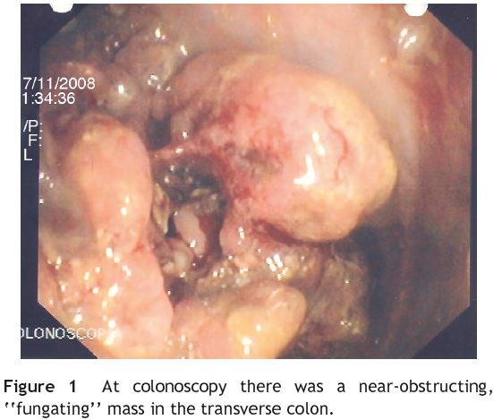
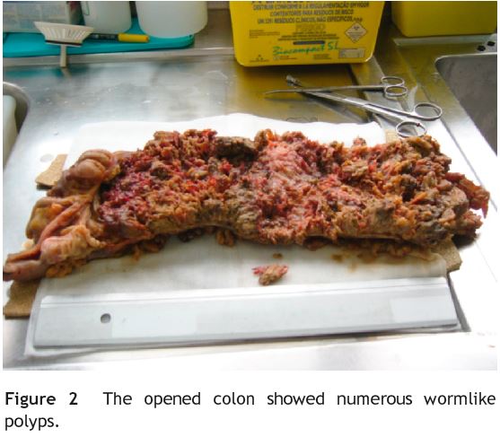
Colonoscopic examination revealed a near-obstructing, fungating mass in the transverse colon, preventing further passage of the endoscope (Fig. 1). The remaining colon and rectum had endoscopic normal-appearing mucosa. Colon carcinoma was suspected and several biopsies were obtained. Histological examination revealed marked acute inflammation, but there was no evidence of dysplasia or malignancy. Because malignancy was suspected and to relieve the obstructive symptoms, the patient underwent an extended right hemicolectomy with primary anastomosis. The surgical specimen was 45 cm in length and interested the right colon, 8 cm of the terminal ileum and the appendix (Fig. 2). In the colon, multiple filliform polypoid lesions (2-3 cm high) were observed over a length of 30 cm, initiating at 4 cm from the ileocecal valve and occupied the whole lumen perimeter. There was no single dominant mass.


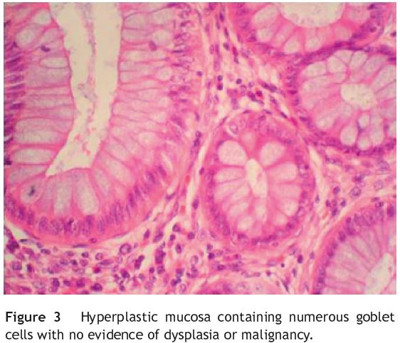
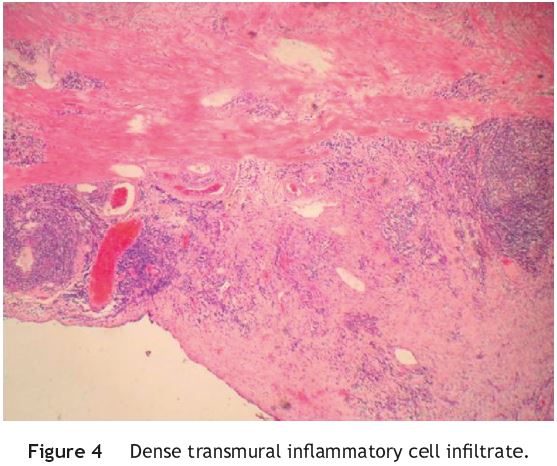
Microscopically, the polypoid projections corresponded to glandular hyperplasia associated with goblet cell hyperplasia (Fig. 3) and intense acute and chronic inflammation, without dysplasia. There were areas of erosion and ulceration of the mucosa and crypt abscesses. The transmural infiltrate included numerous eosinophils, plasma cells and lymphocytes and involved the serosa (Fig. 4). No granulomas were found. In the terminal ileum, mucosa showed similar changes but less exuberant. Microorganisms were observed only into the luminal contents (rare gram−and gram + bacteria). No microorganisms were identified with the Gram, Ziehl Neelsen, PAS, mucicarmine and Warthin Starry stains. There was marked lymphoid hyperplasia of the ileum and of the resected lymph nodes. These changes were interpreted as active inflammatory bowel disease associated with giant pseudopolyposis.


The patients postoperative recovery was uneventful. He was discharged home after nine days tolerating a regular diet and producing normal bowel movements. He continued on maintenance therapy with oral mesalamine and follow-up colonoscopy six months later showed no residual lesion.
Discussion
First described in 1965,5,6 giant inflammatory polyposis represents an extreme variant of inflammatory polyps and is usually associated with inflammatory bowel disease, although it may occur in patients with no prior history of IBD.7 It occurs most commonly in females with pancolitis and there is a predilection for the left colon.3,7
In this case report, the patient had ulcerative pancolitis for two years and was on oral therapy with steroids for what seemed to be a flare-up. Since colonoscopy showed normal-appearing mucosa in the remaining colon, patients symptoms were probably related with the exuberant mass of pseudopolyps, rather than with inflammatory bowel disease per se. As described in other case reports, GIP formation could be related to exuberant postinflammatory regeneration of the surviving colonic mucosa between areas of ulceration7 and may be found in quiescent disease.
Patients may present with a variety of symptoms including anemia, weight loss, abdominal pain and rectal bleeding, but like in our case, most go undiagnosed until they develop signs and symptoms of obstruction.1
Although carcinoma was unlikely in this 23-year-old man with a 2-year history of colon disease, endoscopic findings were strongly suggestive of malignancy and so extended right hemicolectomy was performed. Giant inflammatory polyposis is broadly considered a benign entity.5 In our literature review, we found only one reported case of an occult carcinoma8 and another with dysplasia9 arising in localized GIP.
As most patients present with obstructive symptoms, surgery is usually the first approach. However, nonsurgical management may be an option. There is a case report of a rectal GIP successfully treated with budesonide.10 Initial proper diagnosis and familiarization with this entity may allow medical treatment with steroids.11 We truly recognize that further studies on medical treatment are required.
Six months after surgery, our patient had no colon lesions and was symptom free. Although some authors advocate that residual disease may predict potential recurrence,7 we suggest an individualized approach on long-term follow-up.
References
1. Okayama N, Itoh M, Yokoyama Y, Tsuchida K, Takenchi T, Kobayashi K, et al. Total obliteration of colonic lumen by localized giant inflammatory polyposis in ulcerative colitis: report of a Japanese case. Intern Med. 1996;35:24-9. [ Links ]
2. Ooi BS, Tjandra J, Pedersen S, Bhathal P. Giant pseudopolyposis in inflammatory bowel disease. Aust N Z J Surg. 2000;70: 389-93. [ Links ]
3. Zeki S, Catnach S, King A, Hallan R. An obstructing mass in a young ulcerative colitis patient. World J Gastroenterol. 2009;15:877-8. [ Links ]
4. Bryan L, Newman M, Alexander J. Giant inflammatory polyposis in ulcerative colitis presenting with protein losing enteropathy. J Clin Pathol. 1990;43:346-7. [ Links ]
5. Goldgraber M. Pseudopolyps in ulcerative colitis. Dis Colon Rectum. 1965;8:355-63. [ Links ]
6. Bernstein J, Ghahremani G, Paige M, Rosenberg J. Localized giant pseudopolyposis of the colon in ulcerative and granulomatous colitis. Gastrointest Radiol. 1978;3:431-5. [ Links ]
7. Sheikholeslami M, Schaefer R, Mukunya P. Diffuse giant inflammatory polyposis. Arch Pathol Lab Med. 2004;128:1286-8. [ Links ]
8. Kusunoki M, Nishigami T, Yanagi H, Okamoto T, Shoji Y, Sakamoue Y, et al. Occult cancer in localized giant pseudopolyposis. Am J Gastroenterol. 1992;87:379-81. [ Links ]
9. Pilichos C, Preza A, Demonakou M, Kapatsoris D, Bouras C. Topical budesonide for treating giant rectal pseudopolyposis. Anticancer Res. 2005;25:2961-4. [ Links ]
10. Koinuma K, Togashi K, Konishi F, Kirii Y, Horie H, Okada M. Localized giant inflammatory polyposis of the cecum associated with distal ulcerative colitis. J Gastroenterol. 2003;38:880-3. [ Links ]
11. Wyse J, Lamoureux E, Gordon P, Bitton A. Occult dysplasia in a localized giant pseudopolyp in Crohns colitis: a case report. Can J Gastroenterol. 2009;23:477-8. [ Links ]
Conflicts of interest
The authors have no conflicts of interest to declare.
*Corresponding author.
E-mail address: inesnmarques@zonmail.pt (I. Marques).
Received 9 October 2010; accepted 15 December 2010


