Serviços Personalizados
Journal
Artigo
Indicadores
-
 Citado por SciELO
 -
 Acessos
Links relacionados
-
 Similares em
SciELO
Compartilhar
Jornal Português de Gastrenterologia
versão impressa ISSN 0872-8178
J Port Gastrenterol. vol.21 no.2 Lisboa abr. 2014
https://doi.org/10.1016/j.jpg.2014.02.001
CLINICAL CASE
Pancreatic metastasis from renal cell carcinoma: A different cause for recurrent duodenal bleeding
Metástase intrapancreática de tumor de células renais: causa rara de hemorragia digestiva alta recorrente
Tito Miguel Correiaa,∗, Nuno Miguel Almeidaa, Ana Isabel Torres-Oliveiraa, Ana Velezb, Pedro Oliveirab, Maria Augusta Ciprianob, Nuno Diasc, Carlos Manuel Rico-Sofiaa
a Gastroenterology Department - Centro Hospitalar e Universitário de Coimbra, Portugal
b Surgery Department - Centro Hospitalar e Universitário de Coimbra, Portugal
c Pathology Department - Centro Hospitalar e Universitário de Coimbra, Portugal
*Corresponding author
ABSTRACT
A 64-year-old man with a renal cell carcinoma (RCC) underwent right nephrectomy. There was no identifiable recurrence during follow-up. Six years after surgery he presented with melena and ferropenic anaemia. Endoscopic evaluation demonstrated a vascular lesion in duodenum and haemostasis was performed. However, the bleeding recurred and further endoscopies revealed an enlarged multilobulated infiltrative and ulcerative lesion over the duodenum bulb. Histological and imagiological investigations were, even so, suggestive of a vascular lesion adjacent to the duodenal wall. Given the uncertain diagnosis and recurrent bleeding a surgical resection was deemed unavoidable. A cephalic duodenopancreatectomy was performed and histologic evaluation revealed an intrapancreatic RCC metastasis with duodenal infiltration. No evidence of recurrence after 12 months was observed.
In conclusion, RCC metastasis should be considered in patients with a pancreatic mass as it gives the past history of RCC. Awareness of this entity and a high index of suspicion would help in proper diagnosis and treatment.
Keywords: Renal cell carcinoma; Intrapancreatic metastasis; Duodenal bleeding
RESUMO
Doente do sexo masculino de 64 anos de idade, submetido a nefrectomia direita há 6 anos, sem recidiva identificável durante o follow-up, apresentou-se com melena e anemia ferropriva. A avaliação endoscópica mostrou lesão vascular no duodeno tendo sido submetido a hemóstase com sucesso. No entanto, após 3 meses, ocorreu recidiva hemorrágica, observando-se lesão infiltrativa e ulcerada multilobulada no bulbo duodenal. O estudo histológico e imagiológico eram sugestivos de uma lesão vascular adjacente a parede do duodenal. Dado o diagnóstico incerto e hemorragia recorrente, optou-se por realização de ressecção cirúrgica. A gastroduodenopancreatectomia cefálica revelou a presença de metástase de carcinoma de células renais intrapancreática com infiltração duodenal. Não foi observada evidência de recidiva após 12 meses.
Em conclusão, as metástases do carcinoma de células renais devem ser consideradas em pacientes com massa pancreática e história de RCC. A consciência desta entidade e um alto índice de suspeita é necessário para o diagnóstico e tratamento adequado.
Palavras-Chave: Carcinoma de células renais; Metástase intrapancreática; Hemorragia duodenal
Background
Renal cell carcinoma (RCC) has the potential to metastasize to almost any site. Metastatic disease may be present in up to 25% of patients at the time of diagnosis while another 50% develop metastasis during follow-up.1,2 Its tendency to vascular spreading as well as expansible ostetolytic skeletal metastization is well known. However, it is less expected that RCC presents as an unpredictable tumour which can manifest with very late metastases at unexpected sites, even a long period after successful resection of early stage lesion.3,4 Common metastatic sites for RCC are lungs, bone, liver, adrenal glands and brain, being rarely detected in thyroid gland, gallbladder, pancreas and orbit.5
In this paper, we report a case of a 64-years-old man with solitary pancreatic metastasis with duodenal infiltration manifested as recurrent upper gastrointestinal bleeding 6 years after nephrectomy.
Case report
A 64-year-old man presented in the emergency room with melena. He also referred fatigue and generalized weakness for the previous 3 days. He had no associated symptoms and denied hematemesis, fresh rectal bleeding, abdominal pain or weight loss. There was no history of recent use of non-steroidal, anti-inflammatory, anticoagulant and antiagregant drugs. His past medical history was significant for hypertension and radical right nephrectomy, 6 years before, for pseudocapsulated renal cell carcinoma involving the central part of the kidney. Microscopically, the tumour was classified as clear-cell with eosinophilic and granular cells, grade II/III in the Furhmans nuclear grading system, with no calyx, capsular or vascular involvement and the ureter and hilar lymph nodes were also free of tumour. Abdominal contrast enhanced computed tomography (CT) revealed no metastization and the patient was staged as T1N0M0. No adjuvant chemotherapy was administered in view of a favourable tumour histopathology. He was placed on regular oncology follow-up and had been disease free up to his last visit.
On clinical examination he was hemodynamically stable and appeared pale. Abdominal examination was unremarkable (except for a surgical scar of right nephrectomy). Laboratory investigation on admission was significant for normocytic anaemia with haemoglobin 8.1 g/dl and leucocytosis (14.6×10⁹/l). The upper gastrointestinal Endoscopy (UGIE) showed an oozing haemorrhage from a solitary vascular lesion, without ulceration, in the duodenum bulb. It was injected with diluted (1:10,000) epinephrine and three endoclips (EZ clip, HX-610-090 l, Olympus, Pennsylvania, EUA) were applied, with proper haemostasis at the end of the procedure. After endoscopic review he was discharged on day 4, with proton-pump inhibitor (pantoprazole 40 mg id). As an outcome patient, he did the Helicobacter pylori urea breath test, which was negative.
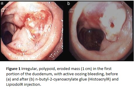
Three months later, the patient suffered from another episode of melena, without haemodynamic repercussion, but with mild anaemia (10.5 g/dl). Upper GI Endoscopy revealed an active oozing bleeding originating from na irregular, polypoid, eroded mass (1 cm) in the first portion of the duodenum (Fig. 1a). The lesion showed violet prominente structures, consistent with vascular nature. We chose to inject n-butyl-2-cyanoacrylate glue (HistoacrylR) and LipiodolR (0.5 ml + 0.5 ml) in the central region of the lesion, with haemostatic success (Fig. 1b). The patient was discharged on the 5th day of hospitalization after endoscopic revision, which identified different characteristics of the lesion, that was now very irregular, with central ulcerated pigmented prominences (Fig. 2a). Biopsies were taken and histological examination revealed morphological findings compatible with an angiomatous lesion.
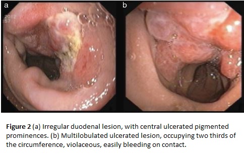
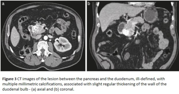
He was referred for detailed imaging and laboratory investigation, including abdominal angio-computerized tomography (CT) and endoscopic ultrasonography (EUS). The CT scan revealed a lesion between the pancreas and the duodenum with 42mm×30mm, but ill defined, with no obvious mass effect, with multiple millimetric calcifications. This lesion was associated with slight regular thickening of the wall of the duodenal bulb, which could correspond to angiomatous lesion (Fig. 3a and b). No other alterations were identified, including tumour recurrence at the nephrectomy site. In the duodenal bulb, EUS revealed a multilobulated ulcerated lesion, occupying two thirds of the circumference, violaceous, easily bleeding on contact (Fig. 2b), which was reflected in ultrasound as heterogeneous wall thickness (12 mm). Hemogram (including MCV) and biochemical tumour markers (CEA and CA 19.9) were normal.
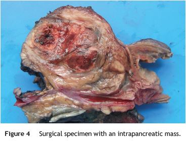
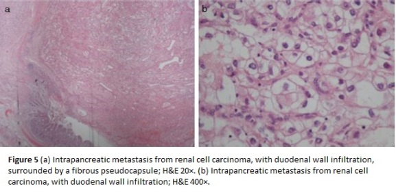
After the third upper gastrointestinal bleeding (with visualization of a multilobulated, ulcerated and violaceous bleeding lesion on the duodenum) and based on clinical history, the patient underwent elective laparotomy. Intraoperatively, a 4 cm lesion was identified in the pancreatic head with infiltration of the duodenum Wall and endoluminal growth, which was resected by classic pancreaticoduodenectomy - Whipples procedure (Fig. 4). Histology and immunohistochemistry studies revealed na intrapancreatic metastasis from renal cell carcinoma, with duodenal wall infiltration, surrounded by a fibrous pseudocapsule (Fig. 5a and b). The surgical margins were free of tumour and no metastases were found in the regional lymph odes.
The follow-up was uneventful with no evidence of recurrence at 12 months.
Discussion
Pancreatic metastasis is a rarity and seen in only 3-12% of patients with disseminated malignancy at autopsy. Majority forms are metastasis from primary sites such as lungs, breast, renal cell carcinoma, colon and melanoma.6 The incidence of metastasis from primary renal cell carcinoma to pancreas ranges from 0.5 to 3% of all metastatic RCC.7-9 However, when these rarities converge, it forms a unique association in which RCC is the most common primary tumour in 30% of all patients with pancreatic metastasis.6,10 These are usually detected many years after nephrectomy, ranging from 6 to 8 years.10-12 The metastization from RCC to pancreas may occur by haematogenous or lymphatic dissemination, the direct spread to the pancreas being unusual.13 In 2006, Sellner et al.14 identified 236 cases of isolated pancreatic metastasis of RCC, either asymptomatic in 35% of the cases, or presenting with abdominal pain (20%), GI bleeding (20%), obstructive jaundice (9%), weight loss (9%), pancreatitis and diabetes (3% each). Symptoms were tumour diameter-dependent, more frequent in those with more than 45mm.9 In the case presented, imagiological evaluation demonstrated a typical well defined lesion, with characteristic hypervascularity manifested by contrast enhancement, that is usually seen in over 75% patients.3,11
It is also difficult to diagnose primary or metastatic RCC in small biopsies because of the wide variety of histologic appearance,15 which may explain why there was no histologic confirmation and definitive diagnosis until surgical exploration. The natural history of duodenal ulcers is benign, and there is no association with carcinoma.16 It is difficult to differentiate malignant from benign ulcers in initial endoscopic evaluation. However, the possibility of malignancy of the duodenal ulcer should be considered if the ulcer does not heal after 8 weeks of medical treatment or if there are polypoid or submucosal masses with elevation and ulceration at the apex or multiple nodules of varying sizes with tip ulceration on repeated endoscopic examination. This kind of lesion may need aggressive biopsy techniques using Jumbo forceps or surgical biopsy to obtain suficiente tissue for diagnosis.17
Surgical resection of primary renal cell carcinoma and metastatic deposits remains the most effective treatment since chemotherapy, radiotherapy and hormonal therapy have proven ineffective.7,10 A metachronous solitary resectable metastasis of an RCC or multiple metastasis confined to one organ (like pancreas) are suitable surgical candidates.7,18 Surgical procedures vary according to the site and the extent of the lesion, including distal pancreatectomy, pancreatoduodenectomy or tumour enucleation. A relatively good prognosis has been observed in the absence of lymphatic spread and in local recurrence.7,9 The outcome of isolated pancreatic RCC metastasis is clearly more favourable with a mean 5 years survival ranging from 43 to 88%, even better than that of primary adenocarcinoma of pancreas.7,8 In a systematic review of more than 400 patients with RCC pancreatic metastasis, Tanis et al.18 found that long disease-free interval (preferably more than 2 years) after resection of primary tumour, a single, asymptomatic metastatic deposit with central necrosis and complete excision of the secondary lesion (with negative margins) are associated with good prognosis. In addition, other authors found that synchronous had worse prognosis when compared to metachronous solitary metastasis.19 The prognosis is also poorer when the metachronous metastasis develop before one year after nephrectomy.20 Recently, immunotherapy has shown encouraging results in advanced RCC (e.g. Bevacizumab, Sunitinib, and Sorafenib). It has been used in cases with aggressive growth pattern, extrarenal disease at exploration or as neoadjuvant therapy in locally unresectable disease.18 Though in isolated lesions, liable to excision, surgery is still the first line therapy.
We report this case to highlight the importance of investigating patients with history of RCC presenting with any gastro-intestinal tract manifestations looking for metastasis. In our patient, three episodes of GI bleeding from na intrapancreatic metastasis presented after a long diseasefree interval of 6 years. The two endoscopies performed in the context of the two episodes of upper gastrointestinal bleeding described in the case report were considered relatively innocent. The first episode was described as probable simple duodenal vascular injury - duodenal Dieulafoy. The second episode, despite identification of a polypoid eroded structure, the endoscopic appearance (confirmed by histology) suggested a vascular lesion (angiomatous structure). However, endoscopic revision evidenced a sudden and significant change in the lesion characteristics and growth. It was then described as an infiltrating and ulcerated mass. CT did not allow a precise etiological characterization of the lesion. Unfortunately, the radial EUS was suboptimal in terms of quality due to technical constraints, limiting the identification of lesions to the most superficial layers of the wall, which was not coincident with the CT images. Surgical decision was based not only on the endoscopic appearance of the lesion and risk of bleeding, but also taking into account the neoplastic background. We opted not to perform EUSFNA before surgery because negative results do not change the previously established surgical strategy due to low sensitivity of this technique.
In conclusion, we think that RCC metastasis should be considered if any patient with a pancreatic tumour gives past history of surgery for RCC. On the other hand post-nephrectomy patients with RCC suffering from gastrointestinal bleeding must have a complete evaluation, especially endoscopic examination, because late recurrent renal cell carcinoma metastasis to the GI tract should be kept in mind, although rare. Awareness of this entity and a high index of suspicion by the physician and pathologist would help in proper diagnosis and treatment.
References
1. Rini BI, Campbell SC, Escudier B. Renal cell carcinoma. Lancet. 2009;373:1119-32. [ Links ]
2. Matveev VB, Gurari˘ı LL, Began-Bogatski˘ı KM. Surgical treatment of late metastasis of kidney cancer. Urol Nefrol. 1999;2:51-2. [ Links ]
3. Kassabian A, Stein J, Jabbour N, Parsa K, Skinner D, Parekh D, et al. Renal cell carcinoma metastatic to the pancreas: a single-institution series and review of the literature. Urology. 2000;56:211-5. [ Links ]
4. Thyavihally YB, Mahantshetty U, Chamarajanagar RS, Raibhattanavar SG, Tongaonkar HB. Management of renal cell carcinoma with solitary metastasis. World J Surg Oncol. 2005;3:48. [ Links ]
5. Figlin RA. Renal cell carcinoma: management of advanced disease. J Urol. 1999;161:381-6. [ Links ]
6. Scatarige JC, Horton KM, Sheth S, Fishman EK. Pancreatic parenchymal metastasis: observations on helical CT. Am J Roentgenol. 2001;176:695-9. [ Links ]
7. Zerbi A, Oatolano E, Balzano G, Borri A, Beneduce AA, Di Carlo V. Pancreatic metastasis from renal cell carcinoma: which patients benefit from surgical resection? Ann Surg Oncol. 2008;15:1161-8. [ Links ]
8. Sellner F, Tykalsky N, De Santes M, Pont J, Klimpfinger M. Solitary and multiple isolated metastasis of clear cell renal cell carcinoma to the pancreas: an indication for pancreatic surgery. Ann Surg Oncol. 2006;13:75-85. [ Links ]
9. Bassi C, Butturini G, Falconi M, Sargenti M, Mantovani W, Pederzoli P. High recurrence rate after atypical resection for pancreatic metastasis from renal cell carcinoma. Br J Surg. 2003;90:555-9. [ Links ]
10. David AW, Samuel R, Eapen A, Vyas F, Joseph P, Sitaram V. Pancreatic metastasis from renal cell carcinoma 16 years after nephrectomy: a case report and review of literature. Trop Gastroenterol. 2006;27:175-6. [ Links ]
11. Akatsu T, Shimazu M, Aiura K, Ito Y, Shinoda M. Clinicopathological features and surgical outcome of isolated metastasis of renal cell carcinoma. Hepato-gastroenterol. 2007;54: 1836-40. [ Links ]
12. Moussa A, Mitry E, Hammel P, Sauvanet A, Nassif T, Palazzo L. Pancreatic metastasis: a multicentric study of 22 patients. Gastroenterol Clin Biol. 2004;2:872-6. [ Links ]
13. Law CH, Wei AC, Hanna SS, Al-Zahrani M, Taylor BR, Greig PD, et al. Pancreatic resection for metastatic renal cell carcinoma: presentation, treatment and outcome. Ann Surg Oncol. 2003;10:922-6. [ Links ]
14. Sellner F, Tykalsky N, De Santis M, Pont J, Klimpfinger M. Solitary and multiple isolated metastasis of clear cell renal cell carcinoma to the pancreas: an indication for pancreatic surgery. Ann Surg Oncol. 2006;13:75-85. [ Links ]
15. Chang WT, Chai CY, Lee KT. Unusual upper gastrointestinal bleeding due to late metastasis from renal cell carcinoma: a case report. Kaohsiung J Med Sci. 2004;20:137-41. [ Links ]
16. Konar A, Das AS, De PK, Roy A, Mazumder DN. Natural history of severe duodenal ulcer disease. Indian J Gastroenterol. 1998;17:48-50. [ Links ]
17. Nabi G, Gandhi G, Dogra PN. Diagnosis and management of duodenal obstruction due to renal cell carcinoma. Trop Gastroenterol. 2001;22:47-9. [ Links ]
18. Tanis PJ, van der Gaag NA, Busch OR, van Gulik TM, Gouma DJ. Systematic review of pancreatic surgery for metastatic renal cell carcinoma. Br J Surg. 2009;96:579-92. [ Links ]
19. Motzer RJ, Bander NH, Nanus DM. Renal-cell carcinoma. N Engl J Med. 1996;335:865-75. [ Links ]
20. Tongaonkar HB, Kulkarni JN, Kamat MR. Solitary metastasis from renal cell carcinoma: a review. J Surg Oncol. 1992;49: 45-8. [ Links ]
*Corresponding author
E-mail address: Titocorreia@gmail.com (T.M. Correia).
Ethical disclosures
Protection of human and animal subjects. The authors declare that no experiments were performed on humans or animals for this investigation.
Confidentiality of data. The authors declare that they have followed the protocols of their work centre on the publication of patient data and that all the patients included in the study have received sufficient information and have given their informed consent in writing to participate in that study.
Right to privacy and informed consent. The authors have obtained the informed consent of the patients and/or subjects mentioned in the article. The author for correspondence is in possession of this document.
Conflicts of interest
The authors have no conflicts of interest to declare.
Received 14 January 2014; accepted 11 February 2014


