SciELO - Scientific Electronic Library Online

 
 número14Editorial Millenium 14Relevância da informação no regresso a casa do idoso após cirurgia da catarata: perspetiva dos enfermeiros índice de autoresíndice de assuntosPesquisa de artigos
Home Pagelista alfabética de periódicos  

Serviços Personalizados

Journal

Artigo

Indicadores

Links relacionados

  • Não possue artigos similaresSimilares em SciELO

Compartilhar


Millenium - Journal of Education, Technologies, and Health

versão impressa ISSN 0873-3015versão On-line ISSN 1647-662X

Mill  no.14 Viseu abr. 2021  Epub 30-Abr-2021

https://doi.org/10.29352/mill0214.20456 

Life and health sciences

Infrared thermography for risk reduction of nosocomial cross infections during the COVID-19 pandemic: potential application in healthcare facilities

Uso da termografia infravermelha para redução do risco de infecções nosocomiais cruzadas durante a pandemia de COVID-19: aplicação potencial em instituições de cuidado à saúde

Termografía infrarroja para la reducción del riesgo de infecciones cruzadas nosocomiales durante la pandemia de COVID-19: posible aplicación en centros de salud

Renata Rodrigues Teixeira Castro1  2  3 
http://orcid.org/0000-0001-5560-693X

João Giffoni da Silveira Neto1  3 
http://orcid.org/0000-0002-8678-5501

Leonardo de Souza Moreira Alves4 
http://orcid.org/0000-0001-8296-2745

Adalgiza Mafra Moreno1  3 
http://orcid.org/0000-0003-3681-7314

Marco Orsini1 
http://orcid.org/0000-0002-8526-6937

Roberta Rodrigues Teixeira Castro1  3 
http://orcid.org/0000-0001-6096-9727

1 Universidade Iguaçu, Faculdade de Medicina, Nova Iguaçu, Brasil

2 Hospital Naval Marcílio Dias, Clínica de Cardiologia, Rio de Janeiro, Brasil

3 Cardiologia do Esporte, Rio de Janeiro, Brasil

4 Universidade Federal Fluminense, Pós-graduação em Ciências Cardiovasculares, Niterói, Brasil


Abstract

Introduction:

Among millions of people who may be infected with COVID-19, patients with cardiovascular and oncologic diseases exhibit the highest risks of having worse outcomes. These patients are subject to chronic disease decompensation and may be subjected to cross-infection while visiting health facilities that are taking care of COVID-19 infected patients.

Objective:

To present scientific evidence and propose the use of infrared thermography for the reduction of viral cross-infection in healthcare facilities.

Methods:

Theoretical essay

Results:

Previous experience in pandemic show favorable results with the use of infrared thermography identifying infected patients and reducing the possibility of cross infections.

Conclusion:

Infrared thermography is a radiation-free, relatively inexpensive, noncontact, and noninvasive technology that could be used for mass-screening of patients and visitors with fever, especially in services where patients with cardiovascular disease seek for medical care, reducing the risk of cross-infection.

Keywords: infrared thermography; Covid-19; pandemic; cardiovascular disease; cross-infection

Resumo

Introdução:

Entre milhões de pessoas que podem estar infetadas pelo COVID-19, os pacientes com doenças cardiovasculares e oncológicas apresentam os maiores riscos de ter piores resultados. Esses pacientes estão sujeitos à descompensação crônica da doença e à infeção cruzada enquanto visitam unidades de saúde que cuidam de pacientes infetados com COVID-19.

Objetivo:

Apresentar as evidências científicas e propor o uso de termografia infravermelha para redução de infeções virais cruzadas em instituições de saúde.

Métodos:

Ensaio teórico

Resultados:

Experiências em pandemias prévias sugerem resultados favoráveis com o uso de termografia infravermelha na identificação de pacientes infetados e redução da possibilidade de infeção cruzada.

Conclusão:

A termografia por infravermelho é uma tecnologia sem radiação, relativamente barata, sem contato e não invasiva que pode ser usada para triagem em massa de pacientes e visitantes com febre, especialmente em serviços onde pacientes com doença cardiovascular buscam atendimento médico, reduzindo o risco de infeção cruzada.

Palavras-chave: termografia infravermelha; Covid-19; pandemia; doença cardiovascular; infecção cruzada

Resumen

Introducción:

Entre millones de personas que pueden estar infectadas con COVID-19, los pacientes con enfermedad cardiovascular y oncologica exhiben los mayores riesgos de tener peores resultados. Estos pacientes están sujetos a descompensación de enfermedades crónicas y pueden estar sujetos a infección cruzada mientras visitan los centros de salud que atienden a pacientes infectados con COVID-19.

Objetivo:

Presentar evidencia científica y proponer el uso de la termografía infrarroja para la reducción de infecciones virales cruzadas en instalaciones de salud.

Métodos:

Ensaio teórico

Resultados:

La experiencia previa en pandemia muestra resultados favorables con el uso de termografía infrarroja que identifica a pacientes infectados y reduce la posibilidad de infecciones cruzadas.

Conclusión:

La termografía infrarroja es una tecnología libre de radiación, relativamente barata, sin contacto y no invasiva que podría usarse para el cribado masivo de pacientes y visitantes con fiebre, especialmente en servicios donde los pacientes con enfermedades cardiovasculares buscan atención médica, reduciendo el riesgo de infección cruzada.

Palabras Clave: Termografía infrarroja; Covid-19; Pandemia; Enfermedad cardiovascular; Infección cruzada

Introduction

Knowledge about COVID-19 increases every day, while the novel coronavirus spreads across the globe. Among millions of people who may be affected, patients with cardiovascular and oncologic diseases exhibit the highest risks of having worse outcomes (Liu et al., 2020; Yang et al., 2020; Zhou et al., 2020).

The American Heart Association, in conjunction with the United States´ National Institutes of Health, has just published the annual report annually on the most up-to-date statistics related to heart disease, stroke, and cardiovascular risk factors(Virani et al., 2020). This report shows that according to the 2017 National Health Interview Survey, the age-adjusted prevalence of all types of heart disease was 10.6% in the United States(Virani et al., 2020). The aging and growth of populations all over the world contribute to cardiovascular disease prevalence. In fact, almost a third of all deaths globally were due to cardiovascular disease in 2017 (Collaborators, 2018). This is not different in Brazil, where cardiovascular diseases are the major cause of death, accounting for nearly 20% of all deaths in adults(Mansur & Favarato, 2012).

Most cardiovascular diseases are chronic and need continuous care with frequent visits to medical facilities. In fact, cardiovascular ambulatory care has a pivotal role in reducing cardiovascular deaths(Tu et al., 2017). Aristizábal et al(Aristizábal et al., 2015), found a 40% reduction in emergency room visits and rehospitalizations related to new cardiovascular and coronary events in patients with a previous acute coronary event who received care under a comprehensive ambulatory care model. In Brazil, not only ambulatory services, but also the family medicine program(Silva et al., 2019) reduced hospitalizations due to cardiovascular disease(Lentsck & Mathias, 2015).

When the World Health Organization upgraded COVID-19 to pandemic status, most countries urged to promote social distancing, which means shutting down schools, prohibiting group gatherings and public events, working from home (as much as possible) and staying away from each other as much as possible. Social distancing instructions also closed many health facilities considered non-essential. Although there is no universal definition for “essential health care”(Monekosso, 1984), social distancing policies and the shortage of health staff and medical supplies resulted in closing or reducing operating hours of most public and private ambulatory general practice services.

With this scenario, we can anticipate that soon, patients with COVID-19 infection symptoms and patients with acute decompensation of cardiovascular diseases will be sharing seats in emergency waiting rooms. Thus, screening measures that can identify and isolate potential COVID-19 cases, could prevent disease transmission in health facilities.

Jiang et al. (Jiang et al., 2020) published a brief review summarizing published studies as of late February 2020 on the clinical features, symptoms, complications, and treatments of COVID-19 and found that the main clinical manifestations are fever (90% or more), cough (around 75%), and dyspnea (up to 50%). As fever is the most prevalent symptom, it seems reasonable that this vital sign should be screened as soon as patients arrive to health facilities. Most health facilities use probe electronic thermometers, which must be in contact with patient´s skin or mucosa for some minutes before temperature can be taken. After that, they should be properly disinfected before the next use. This would limit screening speed.

Objective

The aim of this paper is to discuss the potential use of infrared thermography for early detection of patients infected with COVID-19 in health facilities, proposing its use for risk reduction of cross infection.

Methods

We have performed an integrative review searching in Pubmed about the following MESH terms: infrared thermography, viral, pandemic. This review was used to the proposition of this theoretical essay.

Literature review

Infrared thermography

Infrared thermal imaging, or thermography, is a noncontact and noninvasive approach that has been used in medicine since the early 1960s(Ghassemi et al., 2018). Infrared thermal imaging does not require irradiation as it uses infrared radiation emitted from biological tissues to calculate temperature distributions(Usamentiaga et al., 2014). The last five decades witnessed the increase in the utility of thermal imaging cameras to correlate skin temperature and thermal physiology(Jiang et al., 2005; Merla & Romani, 2006; Ring & Ammer, 2012; Ring et al., 2010) and they are currently being used in oncologic imaging(Arora et al., 2008; Lee et al., 2010; Wishart et al., 2010), ischemic monitoring(Bagavathiappan et al., 2009; Peleki & da Silva, 2016), sports medicine(Moreira et al., 2017) and also fever screening(Childs, 2018; Dagdanpurev et al., 2018; Ghassemi et al., 2018) (figure 1).

Figure 1 Infrared thermography image of a patient with fever. 

All objects or bodies with temperature above absolute zero emit electromagnetic radiation, which is known as infrared radiation or thermal radiation(Jones, 1998). The infrared energy emitted from an object or person is directly proportional to its temperature. Infrared cameras create an image by converting radiant heat energy into a signal that can be displayed on a monitor. Therefore, temperatures are accurately measured by the infrared camera, where pixels are the data acquisition points for thermal temperature.

Infrared thermography for fever screening in individuals

Comparison of infrared temperature readings and oral temperatures has shown high sensitivity and specificity in different studies (Chan et al., 2004; Ng et al., 2004; Nguyen et al., 2010). Chamberlain et al.(Chamberlain et al., 1995) evaluated 2447 subjects aged 12 to 103 years who denied recent potentially febrile illness and ingestion of medications that affect normal body temperature. The mean ear infrared emission temperature was 36.51 ± 0.46oC. The reproducibility was better than that of electronic thermometer at the oral and axillary sites. Based on this study, the 99th percentile was 37,6oC, which the authors(Chamberlain et al., 1995) considered the appropriate cutoff for fever screening using infrared ear thermometers.

International organizations, as the International Eletrotechnical Commission(IEC/ISO, 2017) and the European Association of Thermology(Mercer & Ring, 2009) conducted clinical studies that proved the accuracy of infrared thermometers for fever screening, as far as appropriate procedures are applied. Noteworthy, there are standard and technical reports recommending best practices for thermographic fever screening(Ghassemi et al., 2018; IEC/ISO, 2017; ISO, 2009).

Since ambient temperature influences body temperature, the deployment of these systems could be more challenging in hot climates, either in tropical or subtropical regions or temperate countries during summer months. Nevertheless, Tay et al(Tay et al., 2015) found high sensitivity and specificity for fever detection using and infrared thermal detection system in a tropical healthcare setting. Also, Suzuki et al.(Suzuki et al., 2010), found that measurement of body temperature with infrared thermometer was effective for mass body temperature screening even in warm environment. Most studies analyzing feasibility of medical infrared imaging took thermal pictures in climatized rooms. Nevertheless, a practical and feasible protocol for its use in emergency rooms, where temperature is not easily controlled, is available(Coats et al., 2018).

Infrared thermography for mass detection of fever in travelers

The first studies describing infrared thermography use for mass fever screening in airports were published in 2004 (Chan et al., 2004; Ng et al., 2004). Infrared thermal cameras and noncontact infrared thermometers are the only viable temperature measurement approaches for mass screening of infectious disease pandemic(Ghassemi et al., 2018) like the current COVID-19 outbreak.

Diverse national efforts implemented the use of infrared thermography for mass detection of fever at boarders and quarantine stations. After WHO´s global alert for H1N1 pandemic in 2009 many national health agencies start to screen travelers to delay local transmission. Cowling et al (Cowling et al., 2010). reviewed these screening policies and found that they could delay local transmissions for 1-2 weeks. This period could be used to better planning and preparation for local epidemics.

Cho & Yoon(Cho & Yoon, 2014) retrieved data from arrivals’ health declaration forms and questionnaires for febrile arrivals at an international airport collected by a Korean quarantine station during 2012 and found that thermal camera temperature and tympanic (or ear) temperature was not statistically significant. Despite low fever prevalence, this manuscript suggests that self-reported questionnaires and thermal camera scanning may serve as effective tool for mass detection of fever.

During the Ebola virus disease outbreak (2014 to 2016) in Sierra Leone all people (n=166,242) passing through their International airport underwent screening with fixed infrared thermal scanners and five individuals were denied air travel from Sierra Leone(Wickramage, 2019). Since 2006, the Taiwan Notifiable Diseases Surveillance System for dengue fever has been using remote-sensing infrared thermography and quarantine stations of all harbors and international airports to detect febrile passengers(McKerr et al., 2015). This simple and robust system enabled timely and accurate reporting of dengue fever cases.

Studies that evaluated the implementation of mass fever detection in airports faced the same limitation: there were very few patients with fever (table 1) (Cho & Yoon, 2014; Priest et al., 2011; St John et al., 2005; Wickramage, 2019). Thus, it is difficult to justify the implementation of a screening system that uses new technology and personnel efforts to detect a rare condition (fever in airports).

Table 1 Summary of studies that analyzed infrared thermography for mass detection of fever in airports 

Infrared thermography for detection of fever in health facilities

As prevalence of fever is expected to be higher in medical facilities, mass screening of individuals entering hospitals should be detect more people with fever in hospitals than in airports. A prospective observational study(Holm et al., 2018) included 198 medical patients admitted to the Emergency Department. Researchers took standardized thermal picture and temperatures of the inner canthus (central temperature) and three peripheral temperatures (earlobe, nose tip, and tip of the third finger). Gradients between central and peripheral temperatures showed a significant association with 30-day mortality, suggesting prognostic value.

During the severe acute respiratory syndrome (SARS) epidemics in 2003, Taiwan implemented a protocol where all patients and visitors should be screened for fever at the entrance of every hospital building, aiming to reduce the risk of nosocomial cross infections. The infrared thermal imaging system screened 72,327 outpatients and visitors at the Taipei Medical University-Wan Fang Hospital and detected 305 febrile patients(Chiu et al., 2005), three of them with SARS.

Reducing cross-infection of patients with cardiovascular disease in medical facilities

During pandemics medical services are overwhelmed and patient triage is of paramount importance. Unfortunately, healthcare resources are limited, and patients are facing long waiting times in medical facilities. It is important to identify and prioritize patients who requires urgent attention and intervention (Tam et al., 2018). In pandemic times, proper and fast patient triage can also guide right-hospital allocation accordingly to possible infective status. If fast triages strategies are not implemented, patients with cardiac decompensation and with COVID-19 infection will be sharing the same waiting rooms, increasing the risk of cross-infection. Noteworthy, symptoms-focused triage evaluation may not be effective in identifying infected patients, as patients with cardiovascular decompensation and the ones infected with COVID-19 share symptoms (breathlessness, fatigue and chest pain).

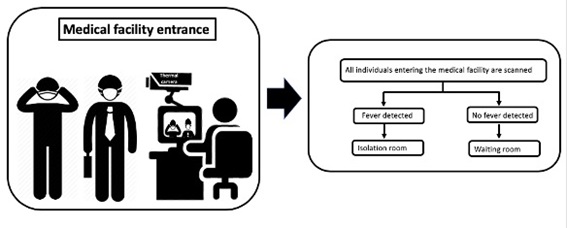
As temperature readings obtained by infrared thermal imaging system could be used to screen patients and visitors in medical facilities(Ataş Berksoy et al., 2018; Chiang et al., 2008) we propose a flowchart (Figure 2) to guide the thermal screening of everyone entering medical facilities. Each person with high temperature should be rapidly isolated and evaluated. This relatively simple flowchart could potentially reduce cross-infection risk at waiting rooms.

Figure 2 Suggested flowchart for thermal scanning of people entering medical facilities 

Limitations of infrared thermography implementation in medical facilities

Despite the promising results of infrared thermography use in emergency rooms, there are some limitations that must be considered. Asymptomatic patients may transmit the virus (Wei et al., 2020). While thermography is useful to identify patients with elevated central temperature, it would not be able to identify those asymptomatic patients with temperature values within the normal range. Nevertheless, asymptomatic patients would seldom, if ever, seek medical assistance during pandemics. Noteworthy, taking of an antifebrile drug results in body temperature reduction, which can affect thermography efficacy (Chiang et al., 2008; Nishiura & Kamiya, 2011).

Currently available infrared cameras show high sensitivity, excellent time resolution and should be calibrated according to heat emissivity, room temperature, humidity, and distance to the object of interest. Most published studies lack complete and detailed descriptions of how the camera and/or the software were calibrated and what settings have been used, limiting their reproducibility (KJ et al., 2020; Shterenshis, 2017).

Finally, normative range of surface skin temperature is still not established(Shterenshis, 2017) and accuracy of fever detection by infrared thermography is determined by the selected fever temperature cutoff. Noteworthy, the use of different cutoff values would impact in method´s sensitivity and specificity. In fact, maximizing accuracy by choosing highest specificity may not be desirable in a real-world pandemic setting, where secondary evaluation is available. By the other hand, setting thermo scanners to high sensitivity would increase the demand to secondary evaluation.

Conclusion

Patients with comorbidities and chronic disease, especially the ones with cardiologic or oncologic diseases, are at increased risk of bad prognosis when infected by COVID-19(Liu et al., 2020; Yang et al., 2020; Zhou et al., 2020). Despite social isolation policies, these patients must keep continuous healthcare to reduce disease decompensation, emergency room visits and rehospitalizations.

Limiting contact between infected and non-infected patients is pivotal to reduce nosocomial cross infections during the COVID-19 pandemic. Previous experiences show that infrared thermography is a radiation-free, relatively inexpensive, noncontact, and noninvasive technology that could be used for mass-screening of patients and visitors in health care facilities. Implementation of thermography use in healthcare facilities entrance can detect and rapidly isolate people with fever, providing them appropriate care and reducing nosocomial cross-infection.

References

Aristizábal, D., Gallo, J., Valencia, Á., Jaime, J., Correa, M., Aristizábal, A., Montoya, M., & Abad, J. (2015). Effect of a comprehensive ambulatory care model on outcomes of patients with acute coronary syndrome in Colombia. Rev Panam Salud Publica, 38(5), 362-369. [ Links ]

Arora, N., Martins, D., Ruggerio, D., Tousimis, E., Swistel, A. J., Osborne, M. P., & Simmons, R. M. (2008). Effectiveness of a noninvasive digital infrared thermal imaging system in the detection of breast cancer. Am J Surg, 196(4), 523-526. https://doi.org/10.1016/j.amjsurg.2008.06.015 [ Links ]

Atas Berksoy, E., Bag, Ö., Yazici, S., & Çelik, T. (2018). Use of noncontact infrared thermography to measure temperature in children in a triage room. Medicine (Baltimore), 97(5), e9737. https://doi.org/10.1097/MD.0000000000009737 [ Links ]

Bagavathiappan, S., Saravanan, T., Philip, J., Jayakumar, T., Raj, B., Karunanithi, R., Panicker, T. M., Korath, M. P., & Jagadeesan, K. (2009). Infrared thermal imaging for detection of peripheral vascular disorders. J Med Phys, 34(1), 43-47. https://doi.org/10.4103/0971-6203.48720 [ Links ]

Chamberlain, J. M., Terndrup, T. E., Alexander, D. T., Silverstone, F. A., Wolf-Klein, G., O'Donnell, R., & Grandner, J. (1995). Determination of normal ear temperature with an infrared emission detection thermometer. Ann Emerg Med, 25(1), 15-20. https://doi.org/10.1016/s0196-0644(95)70349-7 [ Links ]

Chan, L. S., Cheung, G. T., Lauder, I. J., & Kumana, C. R. (2004). Screening for fever by remote-sensing infrared thermographic camera. J Travel Med, 11(5), 273-279. https://doi.org/10.2310/7060.2004.19102 [ Links ]

Chiang, M. F., Lin, P. W., Lin, L. F., Chiou, H. Y., Chien, C. W., Chu, S. F., & Chiu, W. T. (2008). Mass screening of suspected febrile patients with remote-sensing infrared thermography: alarm temperature and optimal distance. J Formos Med Assoc, 107(12), 937-944. https://doi.org/10.1016/S0929-6646(09)60017-6 [ Links ]

Childs, C. (2018). Body temperature and clinical thermometry. Handb Clin Neurol, 157, 467-482. https://doi.org/10.1016/B978-0-444-64074-1.00029-X [ Links ]

Chiu, W. T., Lin, P. W., Chiou, H. Y., Lee, W. S., Lee, C. N., Yang, Y. Y., Lee, H. M., Hsieh, M. S., Hu, C. J., Ho, Y. S., Deng, W. P., & Hsu, C. Y. (2005). Infrared thermography to mass-screen suspected SARS patients with fever. Asia Pac J Public Health, 17(1), 26-28. https://doi.org/10.1177/101053950501700107 [ Links ]

Cho, K. S., & Yoon, J. (2014). Fever Screening and Detection of Febrile Arrivals at an International Airport in Korea: Association among Self-reported Fever, Infrared Thermal Camera Scanning, and Tympanic Temperature. Epidemiol Health, 36, e2014004. https://doi.org/10.4178/epih/e2014004 [ Links ]

Coats, T. J., Morsy, M., Naseer, S., Keresztes, K., Hussain, S., Dexter, K., & Sims, M. R. (2018). A pilot study of the Leicester ED medical infrared imaging protocol in fever and sepsis. PLoS One, 13(7), e0201562. https://doi.org/10.1371/journal.pone.0201562 [ Links ]

Collaborators, G. C. o. D. (2018). Global, regional, and national age-sex-specific mortality for 282 causes of death in 195 countries and territories, 1980-2017: a systematic analysis for the Global Burden of Disease Study 2017. Lancet, 392(10159), 1736-1788. https://doi.org/10.1016/S0140-6736(18)32203-7 [ Links ]

Cowling, B. J., Lau, L. L., Wu, P., Wong, H. W., Fang, V. J., Riley, S., & Nishiura, H. (2010). Entry screening to delay local transmission of 2009 pandemic influenza A (H1N1). BMC Infect Dis, 10, 82. https://doi.org/10.1186/1471-2334-10-82 [ Links ]

Dagdanpurev, S., Sun, G., Choimaa, L., Abe, S., & Matsui, T. (2018). Clinical Application of Multiple Vital Signs-Based Infection Screening System in a Mongolian Hospital: Optimization of Facial Temperature Measurement by Thermography at Various Ambient Temperature Conditions Using Linear Regression Analysis. Conf Proc IEEE Eng Med Biol Soc, 2018, 5313-5316. https://doi.org/10.1109/EMBC.2018.8513513 [ Links ]

Ghassemi, P., Pfefer, T. J., Casamento, J. P., Simpson, R., & Wang, Q. (2018). Best practices for standardized performance testing of infrared thermographs intended for fever screening. PLoS One, 13(9), e0203302. https://doi.org/10.1371/journal.pone.0203302 [ Links ]

Holm, J. K., Kellett, J. G., Jensen, N. H., Hansen, S. N., Jensen, K., & Brabrand, M. (2018). Prognostic value of infrared thermography in an emergency department. Eur J Emerg Med, 25(3), 204-208. https://doi.org/10.1097/MEJ.0000000000000441 [ Links ]

Jiang, F., Deng, L., Zhang, L., Cai, Y., Cheung, C. W., & Xia, Z. (2020). Review of the Clinical Characteristics of Coronavirus Disease 2019 (COVID-19). J Gen Intern Med. https://doi.org/10.1007/s11606-020-05762-w [ Links ]

Jiang, L. J., Ng, E. Y., Yeo, A. C., Wu, S., Pan, F., Yau, W. Y., Chen, J. H., & Yang, Y. (2005). A perspective on medical infrared imaging. J Med Eng Technol, 29(6), 257-267. https://doi.org/10.1080/03091900512331333158 [ Links ]

Jones, B. F. (1998). A reappraisal of the use of infrared thermal image analysis in medicine. IEEE Trans Med Imaging, 17(6), 1019-1027. https://doi.org/10.1109/42.746635 [ Links ]

KJ, H., JB, M., & RE, S. (2020). Infrared thermography for mass fever screening: repeating the mistakes of the past? Thermology international, 30(1), 2. [ Links ]

Lee, C. H., Dershaw, D. D., Kopans, D., Evans, P., Monsees, B., Monticciolo, D., Brenner, R. J., Bassett, L., Berg, W., Feig, S., Hendrick, E., Mendelson, E., D'Orsi, C., Sickles, E., & Burhenne, L. W. (2010). Breast cancer screening with imaging: recommendations from the Society of Breast Imaging and the ACR on the use of mammography, breast MRI, breast ultrasound, and other technologies for the detection of clinically occult breast cancer. J Am Coll Radiol, 7(1), 18-27. https://doi.org/10.1016/j.jacr.2009.09.022 [ Links ]

Lentsck, M., & Mathias, A. (2015). Internações por doenças cardiovasculares e a cobertura da estratégia saúde da família. Rev. Latino-Am. Enfermagem, 23(4), 9. [ Links ]

Liu, K., Chen, Y., Lin, R., & Han, K. (2020). Clinical features of COVID-19 in elderly patients: A comparison with young and middle-aged patients. J Infect. https://doi.org/10.1016/j.jinf.2020.03.005 [ Links ]

Mansur, A. e. P., & Favarato, D. (2012). Mortality due to cardiovascular diseases in Brazil and in the metropolitan region of São Paulo: a 2011 update. Arq Bras Cardiol, 99(2), 755-761. https://doi.org/10.1590/s0066-782x2012005000061 [ Links ]

McKerr, C., Lo, Y. C., Edeghere, O., & Bracebridge, S. (2015). Evaluation of the national Notifiable Diseases Surveillance System for dengue fever in Taiwan, 2010-2012. PLoS Negl Trop Dis, 9(3), e0003639. https://doi.org/10.1371/journal.pntd.0003639 [ Links ]

Mercer, J., & Ring, E. (2009). Fever screening and infrared thermal imaging: concerns and guidelines. Thermology International, 19(3), 3. [ Links ]

Merla, A., & Romani, G. L. (2006). Functional infrared imaging in medicine: a quantitative diagnostic approach. Conf Proc IEEE Eng Med Biol Soc, 2006, 224-227. https://doi.org/10.1109/IEMBS.2006.260267 [ Links ]

Monekosso, G. L. (1984). Essential health care: a framework for its definition and implementation in health districts. Trop Doct, 14(4), 146-150. https://doi.org/10.1177/004947558401400402 [ Links ]

Moreira, D. G., Costello, J. T., Brito, C. J., Adamczyk, J. G., Ammer, K., Bach, A. J. E., Costa, C. M. A., Eglin, C., Fernandes, A. A., Fernández-Cuevas, I., Ferreira, J. J. A., Formenti, D., Fournet, D., Havenith, G., Howell, K., Jung, A., Kenny, G. P., Kolosovas-Machuca, E. S., Maley, M. J., Merla, A., Pascoe, D. D., Priego Quesada, J. I., Schwartz, R. G., Seixas, A. R. D., Selfe, J., Vainer, B. G., & Sillero-Quintana, M. (2017). Thermographic imaging in sports and exercise medicine: A Delphi study and consensus statement on the measurement of human skin temperature. J Therm Biol, 69, 155-162. https://doi.org/10.1016/j.jtherbio.2017.07.006 [ Links ]

Ng, E. Y., Kaw, G. J., & Chang, W. M. (2004). Analysis of IR thermal imager for mass blind fever screening. Microvasc Res, 68(2), 104-109. https://doi.org/10.1016/j.mvr.2004.05.003 [ Links ]

Nguyen, A. V., Cohen, N. J., Lipman, H., Brown, C. M., Molinari, N. A., Jackson, W. L., Kirking, H., Szymanowski, P., Wilson, T. W., Salhi, B. A., Roberts, R. R., Stryker, D. W., & Fishbein, D. B. (2010). Comparison of 3 infrared thermal detection systems and self-report for mass fever screening. Emerg Infect Dis, 16(11), 1710-1717. https://doi.org/10.3201/eid1611.100703 [ Links ]

Nishiura, H., & Kamiya, K. (2011). Fever screening during the influenza (H1N1-2009) pandemic at Narita International Airport, Japan. BMC Infect Dis, 11, 111. https://doi.org/10.1186/1471-2334-11-111 [ Links ]

Peleki, A., & da Silva, A. (2016). Novel Use of Smartphone-based Infrared Imaging in the Detection of Acute Limb Ischaemia. EJVES Short Rep, 32, 1-3. https://doi.org/10.1016/j.ejvssr.2016.04.004 [ Links ]

Priest, P. C., Duncan, A. R., Jennings, L. C., & Baker, M. G. (2011). Thermal image scanning for influenza border screening: results of an airport screening study. PLoS One, 6(1), e14490. https://doi.org/10.1371/journal.pone.0014490 [ Links ]

Ring, E. F., & Ammer, K. (2012). Infrared thermal imaging in medicine. Physiol Meas, 33(3), R33-46. https://doi.org/10.1088/0967-3334/33/3/R33 [ Links ]

Ring, E. F., McEvoy, H., Jung, A., Zuber, J., & Machin, G. (2010). New standards for devices used for the measurement of human body temperature. J Med Eng Technol, 34(4), 249-253. https://doi.org/10.3109/03091901003663836 [ Links ]

Shterenshis, M. (2017). Challenges to Global Implementation of Infrared Thermography Technology: Current Perspective. Cent Asian J Glob Health, 6(1), 289. https://doi.org/10.5195/cajgh.2017.289 [ Links ]

Silva, M. V. M. D., Oliveira, V. D. S., Pinto, P. M. A., Razia, P. F. S., Caixeta, A. C. L., Aquino, É., & Morais Neto, O. L. (2019). Trends of hospitalizations for ambulatory care-sensitive cardiovascular conditions in the municipality of Senador Canedo, Goiás, Brazil, 2001-2016. Epidemiol Serv Saude, 28(1), e2018110. https://doi.org/10.5123/S1679-49742019000100018 [ Links ]

St John, R. K., King, A., de Jong, D., Bodie-Collins, M., Squires, S. G., & Tam, T. W. (2005). Border screening for SARS. Emerg Infect Dis, 11(1), 6-10. https://doi.org/10.3201/eid1101.040835 [ Links ]

Suzuki, T., Wada, K., Wada, Y., Kagitani, H., Arioka, T., Maeda, K., & Kida, K. (2010). The validity of mass body temperature screening with ear thermometers in a warm thermal environment. Tohoku J Exp Med, 222(2), 89-95. https://doi.org/10.1620/tjem.222.89 [ Links ]

Tam, H. L., Chung, S. F., & Lou, C. K. (2018). A review of triage accuracy and future direction. BMC Emerg Med, 18(1), 58. https://doi.org/10.1186/s12873-018-0215-0 [ Links ]

Tay, M. R., Low, Y. L., Zhao, X., Cook, A. R., & Lee, V. J. (2015). Comparison of Infrared Thermal Detection Systems for mass fever screening in a tropical healthcare setting. Public Health, 129(11), 1471-1478. https://doi.org/10.1016/j.puhe.2015.07.023 [ Links ]

Tu, J. V., Maclagan, L. C., Ko, D. T., Atzema, C. L., Booth, G. L., Johnston, S., Tu, K., Lee, D. S., Bierman, A., Hall, R., Bhatia, R. S., Gershon, A. S., Tobe, S. W., Sanmartin, C., Liu, P., Chu, A., & Panel, C. P. P. I. E. (2017). The Cardiovascular Health in Ambulatory Care Research Team performance indicators for the primary prevention of cardiovascular disease: a modified Delphi panel study. CMAJ Open, 5(2), E315-E321. https://doi.org/10.9778/cmajo.20160139 [ Links ]

Usamentiaga, R., Venegas, P., Guerediaga, J., Vega, L., Molleda, J., & Bulnes, F. G. (2014). Infrared thermography for temperature measurement and non-destructive testing. Sensors (Basel), 14(7), 12305-12348. https://doi.org/10.3390/s140712305 [ Links ]

Virani, S. S., Alonso, A., Benjamin, E. J., Bittencourt, M. S., Callaway, C. W., Carson, A. P., Chamberlain, A. M., Chang, A. R., Cheng, S., Delling, F. N., Djousse, L., Elkind, M. S. V., Ferguson, J. F., Fornage, M., Khan, S. S., Kissela, B. M., Knutson, K. L., Kwan, T. W., Lackland, D. T., Lewis, T. T., Lichtman, J. H., Longenecker, C. T., Loop, M. S., Lutsey, P. L., Martin, S. S., Matsushita, K., Moran, A. E., Mussolino, M. E., Perak, A. M., Rosamond, W. D., Roth, G. A., Sampson, U. K. A., Satou, G. M., Schroeder, E. B., Shah, S. H., Shay, C. M., Spartano, N. L., Stokes, A., Tirschwell, D. L., VanWagner, L. B., & Tsao, C. W. (2020). Heart Disease and Stroke Statistics-2020 Update: A Report From the American Heart Association. Circulation, 141(9), e139-e596. https://doi.org/10.1161/cir.0000000000000757 [ Links ]

Wei, W. E., Li, Z., Chiew, C. J., Yong, S. E., Toh, M. P., & Lee, V. J. (2020). Presymptomatic Transmission of SARS-CoV-2 - Singapore, January 23-March 16, 2020. MMWR Morb Mortal Wkly Rep, 69(14), 411-415. https://doi.org/10.15585/mmwr.mm6914e1 [ Links ]

Wickramage, K. (2019). Airport Entry and Exit Screening during the Ebola Virus Disease Outbreak in Sierra Leone, 2014 to 2016. Biomed Res Int, 2019, 3832790. https://doi.org/10.1155/2019/3832790 [ Links ]

Wishart, G. C., Campisi, M., Boswell, M., Chapman, D., Shackleton, V., Iddles, S., Hallett, A., & Britton, P. D. (2010). The accuracy of digital infrared imaging for breast cancer detection in women undergoing breast biopsy. Eur J Surg Oncol, 36(6), 535-540. https://doi.org/10.1016/j.ejso.2010.04.003 [ Links ]

Yang, J., Zheng, Y., Gou, X., Pu, K., Chen, Z., Guo, Q., Ji, R., Wang, H., Wang, Y., & Zhou, Y. (2020). Prevalence of comorbidities in the novel Wuhan coronavirus (COVID-19) infection: a systematic review and meta-analysis. Int J Infect Dis. https://doi.org/10.1016/j.ijid.2020.03.017 [ Links ]

Zhou, M., Zhang, X., & Qu, J. (2020). Coronavirus disease 2019 (COVID-19): a clinical update. Front Med. https://doi.org/10.1007/s11684-020-0767-8 [ Links ]

Received: June 26, 2020; Accepted: November 08, 2020

Corresponding Author Renata Castro Rua Franz Weissman, 410/1110 22775-051 Rio de Janeiro - Brasil castrorrt@gmail.com

Creative Commons License This is an open-access article distributed under the terms of the Creative Commons Attribution License