Serviços Personalizados
Journal
Artigo
Indicadores
-
 Citado por SciELO
 -
 Acessos
Links relacionados
-
 Similares em
SciELO
Compartilhar
Motricidade
versão impressa ISSN 1646-107X
Motri. vol.14 no.4 Ribeira de Pena dez. 2018
https://doi.org/10.6063/motricidade.14094
ARTIGOS ORIGINAIS
Neuromuscular efficiency of the knee joint muscles in the early- phase of resistance training: effects of antagonist’s muscles pre- activation
Euler Alves Cardoso1[*], Frederico Ribeiro Neto1, Wagner Rodrigues Martins2, Martim Bottaro1, Rodrigo Luiz Carregaro2
1College of Physical Education, Universidade de Brasília, UnB, Brasília/DF, Brazil.
2School of Physical Therapy, Campus UnB Ceilândia, Universidade de Brasília, UnB, Brasília/DF, Brazil.
ABSTRACT
It was our objective to compare the neuromuscular efficiency (NME) adaptations between resistance exercise methods (with and without pre-activation of the antagonist’s muscles) after six-weeks training. This randomized controlled trial assigned forty-nine men (mean age 20.9 ± 2.2 years; height 1.80 ± 0.1 m; body mass 75.0 ± 8.2 kg) into two groups: 1) Reciprocal Training group (RT, concentric knee flexion immediately followed by concentric knee extension at 60°.s-1); and Conventional Training (CT, concentric knee extension exercise). Both training adopted three sets, 10 repetitions at 60°.s-1, 2 days/week for 6 weeks. NME of knee extension and flexion were assessed pre and post-training. The groups were similar at baseline, for all variables. We found significant effects on NME only for the rectus femoris muscle in the RT group (ES = 0.31; 95%CI [0.30-0,92]; p<0.01). There were no significant differences at NME pre- and post-training in CT and Total Work did not differ between groups. Reciprocal training provided better neuromuscular efficiency, but effects were limited to the rectus femoris muscle. The small effect sizes suggest caution in the results.
Keywords: athletic performance, electromyography, muscle strength, strength training.
Introduction
Resistance training programs have long been recognized as one of the most useful intervention strategies in the context of rehabilitation (Kannus, 1994), for improving health-related outcomes (Garber et al., 2011) and sports performance (Brill, Macera, Davis, Blair, & Gordon, 2000). Moreover, there is high-quality evidence demonstrating positive strength training effects on balance (Costill, Wilmore, & Kenney, 2012), increased muscle strength (Deschenes et al., 2002; Harvey, Lin, Glinsky, & De Wolf, 2009) and functionality (Serra-Añó et al., 2012). In this context, previous studies (Beck et al., 2007; Coburn et al., 2006; Holtermann, Roeleveld, Vereijken, & Ettema, 2005) have shown that the adoption of short-term training generates acute effects on muscle strength which, consequently, represent an interesting alternative if the aim is to improve the muscle function in a short period of time. Likewise (Coburn et al.,2006; Holtermann et al., 2005), early phase strength training showed improvements in the muscle strength and rate of force development (Brown & Whitehurst, 2003). An interesting strategy to potentiate the functional performance in the acute phases is the adoption of antagonist muscle’s pre-activation (Cunha et al., 2013).
The adoption of the pre-activation of antagonist’s muscles has been recognized as an interesting strategy to improve the muscle function and performance (Baker & Newton, 2005; Jeon, Trimble, Brunt, & Robinson, 2001). One exercise modality that uses the pre- activation of the antagonist’s muscles is referred to as reciprocal training (RT) and consists of a pre-activation immediately prior to the agonist muscle contraction. Previous studies demonstrated larger increments of muscle work and force generation (Carregaro, Gentil, Brown, Pinto, & Bottaro, 2011; Robbins, Young, Behm, & Payne, 2010) influenced by pre-activation exercises when compared to conditions without the pre-activation. Additional studies using surface electromyography (sEMG) during pre- activation of antagonist muscles also demonstrated neural responses after the resistance training, such as an increased EMG activity of the agonist muscle after the activation of the antagonists (Jeon et al., 2001; Maynard & Ebben, 2003; Robbins et al., 2010). It is worth mentioning that the EMG signal amplitude is related to motor units activation by neural firing rate and, consequently, could be associated with the values of maximum torque (David, Mora, & Perot, 2008; Hassani et al., 2006). _For instance, Baker and Newton (2005) demonstrated increases of 4.7% of the capacity to generate power after bouts of eight repetitions using the pre-activation of upper limbs antagonist muscles. According to the authors, the results were explained by an increased firing rate of the agonist muscles, influenced by a neural stimulation of the prior antagonistic contraction.
Neuromuscular Efficiency (NME) is defined as the individual ability to generate force in relation to its muscle activation level, and improvements in NME are obtained when muscle contractions against high loads are sustained by a lower neural recruitment (Aragão et al., 2015; David et al., 2008; Deschenes et al., 2002). Thus, NME is an index that evaluates a muscle group functional status (David et al., 2008), and it is related to muscle responsiveness and neural excitability (Deschenes et al., 2002). Hence, this index is an important screening tool that can allow athletic trainers and health professionals to evaluate the muscle responsiveness and functional performance arising from specific sports training programs and/or specific rehabilitation processes (Aragão et al., 2015; Deschenes et al., 2002). For instance, Deschenes et al. (2002) adopted the NME to verify if strength reductions resulting from 14-day muscle immobilization occurred due to decreases in the neural input. Their results demonstrated a decrease in total work and EMG activity of the knee extensors and flexors, though with no changes in the NME. These findings suggested that decreases of muscle work and EMG were primarily due to neural effects, not to contractile or morphological factors. These findings demonstrated that the NME could be more sensitive to the neural aspects and might be useful to the assessment of early-phase resistance training. Notwithstanding, despite the usefulness of this index, to the best of our knowledge there is a lack of studies addressing the NME across different modalities of resistance training exercises. Therefore, this study aimed to compare the NME adaptations between two resistance exercise methods (with and without pre- activation of the antagonist’s muscles) after a six- week training program. Our hypothesis is that Reciprocal Training will lead to greater improvements in NME than Traditional Training, without pre-activation.
Method
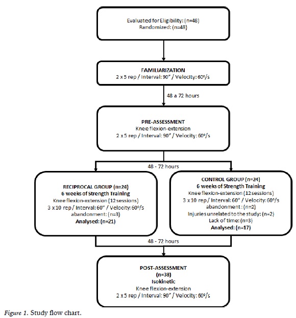
This is a randomized controlled trial reported in accordance with the Consort Statement (Schulz, Altman, & Moher, 2010). The study flowchart is depicted in Figure 1. The participants underwent a resistance-training program two times a week for six weeks.
Participants
Forty-eight healthy young males were consecutively enrolled in the study (mean age 20.9 ± 2.2 years; height 1.80 ± 0.1 m; body mass 75.0 ± 8.2 kg). All participants were recruited through posters and verbal contacts in the University Campus. All volunteers were notified of the research procedures and were invited to participate by signing informed consent. The Institutional Research Ethics Committee approved the study (Protocol n. 112/12).
Participants were interviewed with a questionnaire containing personal and demographic information; and clinical history. The inclusion criteria were: 1) age between 18 to 30 years; 2) no physical training in the past six months prior to the study. Participants were excluded if presented any cardiorespiratory impairment, metabolic diseases, ligament ankle and/or knee injury, diseases or neurological signal and/or proprioceptive deficits.
Participants were randomly divided into two groups: Conventional Training - with no pre-activation (CT) and Reciprocal Training group - with pre-activation (RT). For the randomization process, a table with random numbers was generated in the website “www.random.org”. Sequentially sealed opaque envelopes were used, containing the intervention groups´ name. A researcher, blinded to the purposes of the research, was responsible for this procedure.
Familiarization and evaluation procedure
Participants attended the laboratory on three different occasions. On the first visit, all participants performed a familiarization procedure, composed by two sets of five sub- maximum concentric isokinetic repetitions of knee flexion-extension with using the speed of 60°.s-1 and an interval of 90 seconds between sets. On the second visit, and after an interval of at least 24 hours, the pre-training (baseline) assessment was performed. Participants performed the same protocol as the familiarization visit; except during the pre- and post-training evaluations, in which maximum effort was adopted, in two sets of five repetitions. For the pre-training assessment, the electromyographic activity of the vastus medialis, rectus femoris and biceps femoris was acquired. On the third visit (post-training), the assessment was performed with the same procedures of the previous evaluations (with a time lag of 72hs from the last training session). During testing and intervention, participants were instructed to position their arms against their chest. Participants were instructed to perform maximum strength throughout all range of motion. For all participants, visual feedback on the computer screen and verbal encouragement were given, in an attempt to reach the maximum effort level. The testing sessions were performed in the morning period, for all participants.
Isokinetic Dynamometry
For the present study, an isokinetic dynamometer was used (Biodex System 4, Biodex Medical, Shirley, NY). Calibration and positioning procedures followed the manufacturer’s specifications. The hip position was standardized to 100º flexion. A range of motion of 80º was used (excursion from 90° flexion to 10°, using knee extension parameter as 0°) to prevent knee hyperextension. During testing, participants were asked to keep their arms crossed over their thoracic region. Verbal encouragement and visual feedback were provided to encourage participants to reach their maximal exertion level. The participants were stabilized in the equipment using restraints on their thighs, pelvis, and trunk to prevent unwanted movement. The same investigator performed all tests for all participants and was not blind to group allocation.
Surface Electromyography
Portable 4-channel surface electromyography (Miotec Biomedical Equipment LTDA, Brazil) was used. The equipment has a resolution of 14 bits, noise level < 2 LBS and common mode rejection of 110 dB. The simple differential active electrodes (input impedance of 1010 ohms) have polyethylene foam with hypoallergenic medical adhesive, adherent solid gel to contact bipolar Ag/AgCl (silver/silver chloride) with a distance of 20 mm between poles.
Electrodes Placement was based on SENIAM guidelines (Hermens, Freriks, Disselhorst-Klug, & Rau, 2000). The reference electrode was connected to the bony prominence of the seventh cervical vertebra (C7) to measure the EMG signal. A specific seat was positioned on dynamometer chair so that the electrodes would not remain in contact with the seat surface to measure the EMG signal from biceps femoris. Electrodes were positioned over the muscle belly of the vastus medialis, rectus femoris, and biceps femoris, in parallel to the muscle fibers. Before placing electrodes, the area was shaved and lightly abraded with 70% alcohol.
Signal Processing
Total work and peak torque were processed by the Biodex Advantage software and used to calculate the NME. The EMG signal amplitude was calculated using the RMS (root mean square) during the repetition in which the peak torque occurred. Electromyography analysis was performed in the Miograph Software (Miotec- Biomedical equipment®). The signal was filtered with a band-pass frequency of 20 Hz and 450 Hz (4th order Butterworth filter). The EMG and dynamometer signals were synchronized in time. Neuromuscular Efficiency was calculated according to the following equation (Deschenes et al., 2002):
In which PT is the Peak torque in N.m and RMS is the Root mean square in μV.
Intervention protocol
Participants underwent an exercise program twice a week over six weeks (at a minimum interval of at least 48 hours between each session). Training sessions consisted of three sets of ten maximal concentric isokinetic repetitions at 60°.s-1 with 1-minute rest interval (Parcell, Sawyer, Tricoli, & Chinevere, 2002). One group performed the reciprocal exercise sessions (RT), characterized by agonist and antagonist knee extensors and flexors muscles contractions at 60º.s-1 (knee flexion immediately followed by knee extension, at each repetition). The Conventional Training (CT) performed sessions characterized by a concentric knee extension at 60º.s-1, while the knee flexion was performed passively (without pre-activation of the antagonist’s muscles). For both groups, there was a warm-up on the isokinetic dynamometer characterized by two sets of five concentric isokinetic repetitions (50% of the maximum strength of knee flexion-extension at 60°.s-1, with 45 seconds interval between sets). The training was performed bilaterally, however, as there were no significant differences between the dominant vs non-dominant limb before and after the resistance training for both groups, we chose to present the data of the dominant limb only (leg used to kick a ball). The same researcher trained and evaluated all participants. During the study period, participants were advised not to do any additional resistance exercises. The training sessions were performed in the morning or afternoon period, according the participant’s availability. However, the weekly sessions remained at the same period (2 morning sessions or 2 afternoon sessions per week) from the beginning until the end of the training program.
Statistical analysis
The sample size calculation was based on a pilot study. We used the G*Power software (version 3.1.9). The statistical power was stablished at 80% (1-β), α value of 5%, and an effect size of 0.25, considering differences on neuromotor variables between groups (RT and CT). The calculation demonstrated a required sample size of 34 participants.
Data normality assumptions were confirmed by the Shapiro-Wilk test. The independent variable was the training group (RT and CT), and the dependent variables were: NME, total work (in Joules) and RMS (root mean square). A mixed model ANOVA for repeated measures (group [RT and CT] x time [pre and post]) was adopted to verify differences between groups, with the Bonferroni post hoc test. The effect size was calculated, in order to determine the magnitude of the intervention, based on the Cohen´s d and classified according to Rhea (2004) as: trivial (<0.35); small (>0.35 and <0.80); moderate (>0.81 and <1.50) and large (>1.50). The confidence interval of 95% was determined, and the statistical significance was set at 5% (P<0.05, two-tailed).
Results
Throughout the study, ten participants dropped out (n=3 due to injury in daily activities; n=7 not able to participate in all assessments). Thus, the final sample was composed by 38 participants (17 participants allocated in the CT group and 21 participants in the RT group) (Figure 1).
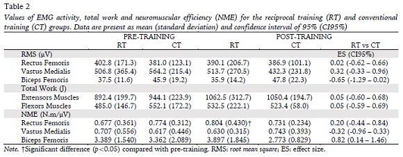
There were no significant differences between groups for age, height, and body mass index (Table 1). Also, groups presented no significant differences in pre-training assessment for all variables (Table 2).
For the RT group, the NME of the rectus femoris presented a significant increase after six weeks of training, with a small effect size (ES = 0.31; 95%CI [-0.30; 0.92]; F = 4,92) p<0.05).
However, there were no significant differences in the NME of the vastus medialis and biceps femoris muscles (ES = - 0.17, 95% CI [0.78;0.44]; ES = 0.30, 95%CI [0.31-0.91], respectively, p>0.05).
The CT presented no significant differences after the training program for neither of the muscles groups, and the effect sizes were trivial for the rectus femoris (ES = 0.16; 95%CI [0.83; 0.52]; p=0.08), vastus medialis (ES = 0.30; 95%CI [0.38; 0.98]; p=0.10) and biceps femoris (ES = 0.37; 95%CI [1.05; 0.37]; p=0.13) (Table 2).
No significant differences were found for the Total Work (between and within groups), considering the knee extensor and knee flexor muscles (Table 2).
Discussion
The present study hypothesized that the Reciprocal Training would lead to greater improvements in NME than conventional training without pre-activation. The comparison between the modalities showed that the exercise performed with reciprocal contractions presented a higher NME for the rectus femoris muscle after six-weeks of training, though with small effect size. The total work did not differ between groups or after six weeks of training.
The present findings demonstrated that the rectus femoris presented a higher NME after six- weeks of training. Hence, it could be assumed that smaller neural recruitment was necessary to achieve an equal or higher amount of muscle strength. These results may be explained by the influence of varied resistance and concentric antagonist speed contractions on subsequent concentric agonist efforts (Burke, Pelham, & Holt, 1999), in which the contraction of the flexor muscles activates Golgi tendon organs, depolarizing Ib axon and firing nerve impulses that are propagated to the spinal cord (Purves et al., 2001). The consequence of this mechanism could be a strength reduction of the flexor muscles associated with the extensor muscle spindles depolarization, thus leading to a more efficient contraction of the rectus femoris. Although the classical findings of Moritani and deVries (1979) showed a prevalence of neural factors responsible for strength gains up to four weeks of training, recent studies (Bickel et al., 2005; Seynnes, de Boer, & Narici, 2007) have reported earlier involvements of hypertrophic components. Accordingly, muscle stimuli arising from the training with duration of six weeks may have provided a morphological adaptation in the skeletal muscle (Bickel et al., 2005; Seynnes et al., 2007). The authors assumed that an increased cross-sectional area, the increment of muscle fibers contractile capacity and molecular adaptations could have led to neuromuscular performance improvement. Hence, our rectus femoris NME findings may also be explained by a hypertrophy mechanism, which can be related to an increased strength gain associated with a stabilization of the electromyography signal at the post-training. The study of Seynnes et al. (2007) demonstrated that physically active male participants submitted to leg extension exercises during 20 days of training, significantly increased their rectus femoris cross-sectional area, concomitant to small or no changes in the electromyographic activity, and could also corroborate our NME findings. Additionally, it is possible to assume that the performance of the rectus femoris muscle was augmented due to post activation potentiation enhancements and by increases in the rate of force development, leading to a better contractile capacity (Sale, 2002). The use of a pre-activation strategy (knee flexors movements prior to the knee extension) may have been able to induce this post activation potentiation, corroborating the study of Batista et al. (2007), in which intermittent contractions were able to improve the neuromuscular performance during knee extension exercises.
A surprising finding was that the biceps femoris NME did not present a significant improvement for the RT group after six weeks of training. Apparently, the flexor muscles need a higher training volume or greater intervention period compared to the extensor muscles. However, no results were found in the literature to support this hypothesis, and further studies are necessary to elucidate the influence of different training volume on flexor muscles early phase-adaptations. Even though our study did not measure morphological variables, the mechanisms underlying our findings seem to be different for different muscle groups. EMG augmentation is attributed to synchronization of motor units (De Luca, 1984; Milner-Brown, Mellenthin, & Miller, 1986) and conduction velocity increases (De Luca, 1984). For the rectus femoris, it is possible to assume that hypertrophy and cross-bridges increases were more prevalent compared to neural adaptations (Moritani & deVries, 1979), as there was an increased NME. It is suggested that a specific assessment of cross- sectional muscle area could contribute to better comprehension and are warranted in future research. Moreover, the NME of the flexor muscles did not change compared to the pre- training, though it was higher in RT compared to the CT at the post-training. Thus, it is assumed that the NME was reduced in the CT group by a probable increase of the muscle fibers recruitment despite an equal muscle strength production, leading to decreased muscle efficiency at the post-training. Also, the characteristic of the exercise program adopted in the present study (three sets of ten repetitions), may have been insufficient to generate adequate strength gains. Accordingly, Robbins et al. (2010) compared three groups with different training volumes of lower limbs exercises after six weeks. Their findings demonstrated that the group with eight sets had a significantly greater strength gain compared to groups that adopted one and four sets. Also, the group with four sets was not significantly different from the one set group. Thus, it is hypothesized that if we had adopted a biceps femoris training periodization, perhaps this would enhance the training volume and would have led to significant results of the NME for the RT group. In addition, we recommend that future studies investigate the effects of the reciprocal training on the knee flexors muscles adaptations and compare the effects of this training between knee’s flexor and extensors muscles.
Neuromuscular efficiency is an interesting muscle responsiveness measure toward neural excitation (Deschenes et al., 2002). Typically, NME is used in isometric contractions (Arabadzhiev, Dimitrov, Dimitrova, & Dimitrov, 2010; Aragão et al., 2015; Milner-Brown et al., 1986; Schimidt, Machado, Vaz, & Carpes, 2014) for muscle fatigue evaluation (Remaud, Cornu, & Guevel, 2005) for different cross-sectional studies outcomes (David et al., 2008; Schimidt et al., 2014) or even to compare isokinetic exercises and dynamic protocols (Remaud et al., 2005). However, we did not find in the literature studies that have used NME to compare training methods in an experimental design. Thus, it is suggested that future resistance training studies address the NME as a dependent variable.
Limitations
Our study presented some limitations. Firstly, the lack of a proper periodization (characterized by an increase in training volume throughout the training) might have influenced our findings. Secondly, the outcome assessor was not blind to group allocation; therefore, estimates of intervention effects might have been influenced by some bias (Chess & Gagnier, 2013).
Practical applications
Our findings demonstrated that if the aim is to improve the neuromuscular efficiency of the knee muscles during a short-term training period, the adoption of reciprocal contractions could be recommended.
CONCLUSION
Our study demonstrated that the use of reciprocal muscle contractions training presented a better rectus femoris neuromuscular efficiency after six weeks of resistance training of young and healthy men. However, we recommended caution considering the small effect sizes.
REFERENCES
Arabadzhiev, T. I., Dimitrov, V. G., Dimitrova, N. A., & Dimitrov, G. V. (2010). Interpretation of EMG integra or RMS and estimates of "neuromuscular efficiency" can be misleading in fatiguing contraction. Journal of electromyography and kinesiology, 20(2), 223-232. DOI: 10.1016/j.jelekin.2009.01.008. [ Links ]
Aragão, F. A., Schäfer, G. S., Albuquerque, C. E. d., Vituri, R. F., Mícolis de Azevedo, F., & Bertolini,G. R. F. (2015). Eficiência neuromuscular dos músculos vasto lateral e bíceps femoral em indivíduos com lesão de ligamento cruzado anterior. Revista Brasileira de Ortopedia, 50(2), 180-185. DOI: https://doi.org/10.1016/j.rbo.2014.03.004.
Baker, D., & Newton, R. U. (2005). Acute effect on power output of alternating an agonist and antagonist muscle exercise during complex training. Journal of Strength and Conditioning Research, 19(1), 202-205. DOI: 10.1519/1533- 4287(2005)19<202:Aeopoo>2.0.Co;2. [ Links ]
Batista, M. A., Ugrinowitsch, C., Roschel, H., Lotufo, R., Ricard, M. D., & Tricoli, V. A. (2007). Intermittent exercise as a conditioning activity to induce postactivation potentiation. Journal of Strength and Conditioning Research, 21(3), 837-840. DOI: 10.1519/r-20586.1. [ Links ]
Beck, T. W., Housh, T. J., Johnson, G. O., Weir, J. P., Cramer, J. T., Coburn, J. W., . . . Mielke, M. (2007). Effects of two days of isokinetic training on strength and electromyographic amplitude in the agonist and antagonist muscles. Journal of Strength and Conditioning Research, 21(3), 757-762. DOI: 10.1519/r-20536.1. [ Links ]
Bickel, C. S., Slade, J., Mahoney, E., Haddad, F., Dudley, G. A., & Adams, G. R. (2005). Time course of molecular responses of human skeletal muscle to acute bouts of resistance exercise. Journal of applied physiology, 98(2), 482-488. DOI: 10.1152/japplphysiol.00895.2004. [ Links ]
Brill, P., Macera, C., Davis, D., Blair, S., & Gordon, N. (2000). Muscular strength and physical function. Medicine and Science in Sports and Exercise, 32, 412- 416. [ Links ]
Brown, L., & Whitehurst, M. (2003). The effect of short-term isokinetic training on force and rate of velocity development. Journal of Strength and Conditioning Research, 17(1), 88-94. [ Links ]
Burke, D. G., Pelham, T. W., & Holt, L. E. (1999). The influence of varied resistance and speed of concentric antagonistic contractions on subsequent concentric agonistic efforts. Journal of Strength and Conditioning Research, 13(3), 193-197. [ Links ]
Carregaro, R. L., Gentil, P., Brown, L. E., Pinto, R. S., & Bottaro, M. (2011). Effects of antagonist pre-load on knee extensor isokinetic muscle performance. Journal of sports sciences, 29(3), 271- 278. DOI: 10.1080/02640414.2010.529455. [ Links ]
Chess, L. E., & Gagnier, J. (2013). Risk of bias of randomized controlled trials published in orthopaedic journals. BMC Medical Research Methodology, 13, 76. DOI: 10.1186/1471-2288-13-76. [ Links ]
Coburn, J. W., Housh, T. J., Malek, M. H., Weir, J. P., Cramer, J. T., Beck, T. W., & Johnson, G. O. (2006). Neuromuscular responses to three days of velocity-specific isokinetic training. Journal of Strength and Conditioning Research, 20(4), 892-898. DOI: 10.1519/r-18745.1. [ Links ]
Costill, D., Wilmore, D., & Kenney, W. (2012). Physiology of sport and exercise (6th ed.): Human Kinetics.
Cunha, R., Carregaro, R. L., Martorelli, A., Vieira, A., Oliveira, A. B., & Bottaro, M. (2013). Effects of short-term isokinetic training with reciprocal knee extensors agonist and antagonist muscle actions: a controlled and randomized trial. Brazilian Journal of physical therapy, 17(2), 137- 145. DOI: 10.1590/s1413-35552012005000077. [ Links ]
David, P., Mora, I., & Perot, C. (2008). Neuromuscular efficiency of the rectus abdominis differs with gender and sport practice. Journal of Strength and Conditioning Research, 22(6), 1855-1861. DOI: 10.1519/JSC.0b013e31817bd529. [ Links ]
De Luca, C. J. (1984). Myoelectrical manifestations of localized muscular fatigue in humans. Critical reviews in biomedical engineering, 11(4), 251-279. [ Links ]
Deschenes, M., Giles, J., McCoy, R., Volek, J., Gomez, A., & Kraemer, W. (2002). Neural factors account for strength decrements observed after short-term muscle unloading American Journal of Physiology. Regulatory, Integrative and Comparative Physiology, 282, R578-R583. [ Links ]
Garber, C., Blissmer, B., Deschenes, M., Franklin, B., Lamonte, M., Lee, I., . . . Swain, D. (2011). Quantity and quality of exercise for developing and maintaining cardiorespiratory, musculoskeletal, and neuromotor fitness in apparently healthy adults: Guidance for prescribing exercise. Medicine and Science in Sports and Exercise, 43(1), 334-1359. [ Links ]
Harvey, L., Lin, C., Glinsky, J. V., & De Wolf, A. (2009). The effectiveness of physical interventions for people with spinal cord injuries: A systematic review. Spinal Cord, 47, 184-195. [ Links ]
Hassani, A., Patikas, D., Bassa, E., Hatzikotoulas, K., Kellis, E., & Kotzamanidis, C. (2006). Agonist and antagonist muscle activation during maximal and submaximal isokinetic fatigue tests of the knee extensors. Journal of electromyography and kinesiology, 16(6), 661-668. DOI: 10.1016/j.jelekin.2005.11.006. [ Links ]
Hermens, H. J., Freriks, B., Disselhorst-Klug, C., & Rau, G. (2000). Development of recommendations for SEMG sensors and sensor placement procedures. Journal of electromyography and kinesiology, 10(5), 361-374. [ Links ]
Holtermann, A., Roeleveld, K., Vereijken, B., & Ettema, G. (2005). Changes in agonist EMG activation level during MVC cannot explain early strength improvement. European Journal of Applied Physiology, 94, 593-601. [ Links ]
Jeon, H. S., Trimble, M. H., Brunt, D., & Robinson, M. E. (2001). Facilitation of quadriceps activation following a concentrically controlled knee flexion movement: the influence of transition rate. The Journal of orthopaedic and sports physical therapy, 31(3), 122-129. DOI: 10.2519/jospt.2001.31.3.122. [ Links ]
Kannus, P. (1994). Isokinetic Evaluation of Muscular Performance. International Journal of Sports Medicine, 15, S11-S18. [ Links ]
Maynard, J., & Ebben, W. P. (2003). The effects of antagonist prefatigue on agonist torque and electromyography. Journal of Strength and Conditioning Research, 17(3), 469-474. [ Links ]
Milner-Brown, H. S., Mellenthin, M., & Miller, R. G. (1986). Quantifying human muscle strength, endurance and fatigue. Archives of physical medicine and rehabilitation, 67(8), 530-535. [ Links ]
Moritani, T., & deVries, H. A. (1979). Neural factors versus hypertrophy in the time course of muscle strength gain. American journal of physical medicine, 58(3), 115-130. [ Links ]
Parcell, A. C., Sawyer, R. D., Tricoli, V. A., & Chinevere, T. D. (2002). Minimum rest period for strength recovery during a common isokinetic testing protocol. Medicine and Science in Sports and Exercise, 34(6), 1018-1022. [ Links ]
Purves, D., Augustine, G., Itzpatrick, F., Hall, W., LaMantia, A., McNamara, J., & Williams, S. (2001). Neuroscience (4th ed.). Sunderland (MA): Sinauer Associates.
Remaud, A., Cornu, C., & Guevel, A. (2005). A methodologic approach for the comparison between dynamic contractions: Influences on the neuromuscular system. Journal of Athletic Training, 40(4), 281-287. [ Links ]
Rhea, M. R. (2004). Determining the magnitude of treatment effects in strength training research through the use of the effect size. Journal of Strength and Conditioning Research, 18(4), 918-920. DOI: 10.1519/14403.1. [ Links ]
Robbins, D. W., Young, W. B., Behm, D. G., & Payne, W. R. (2010). The effect of a complex agonist and antagonist resistance training protocol on volume load, power output, electromyographic responses, and efficiency. Journal of Strength and Conditioning Research, 24(7), 1782-1789. DOI: 10.1519/JSC.0b013e3181dc3a53. [ Links ]
Sale, D. G. (2002). Postactivation potentiation: role in human performance. Exerc Sport Sci Rev, 30(3), 138-143. [ Links ]
Schimidt, H. L., Machado, Á. S., Vaz, M. A., & Carpes, F. P. (2014). Isometric muscle force, rate of force development and knee extensor neuromuscular efficiency asymmetries at different age groups. Revista Brasileira de Cineantropometria & Desempenho Humano, 16, 307-315. [ Links ]
Schulz, K. F., Altman, D. G., & Moher, D. (2010). CONSORT 2010 statement: updated guidelines for reporting parallel group randomised trials. BMJ, 340, c332. DOI: 10.1136/bmj.c332. [ Links ]
Serra-Añó, P., Pellicer-Chenoll, M., García-Massó, X., Morales, J., Giner-Pascual, M., & González, L. (2012). Effects of resistance training on strength, pain and shoulder functionality in paraplegics. Spinal Cord, 50, 827-831. [ Links ]
Seynnes, O. R., de Boer, M., & Narici, M. V. (2007). Early skeletal muscle hypertrophy and architectural changes in response to high- intensity resistance training. Journal of applied, 102(1), 368-373. DOI: 10.1152/japplphysiol.00789.2006. [ Links ]
Acknowledgments: Nothing to declare.
Conflict of interests: Nothing to declare.
Funding: Nothing to declare.
Manuscript received at March 20th 2018; Accepted at December 10th 2018
[*]Corresponding author: Email: prof.euleralves@gmail.com


