Introduction
The arrangement of the front teeth in prosthetic replacement remains a challenge for most clinicians, and reliable anatomical parameters are essential to plan the face contour reconstruction.1,2In the case of teeth absence, the maxillary alveolar ridge undergoes intense resorption, with bone remodeling taking place in the palatine and apical directions.2 Dental absence and bone support resorption in the maxillary anterior region result in lip support loss; consequently, aesthetic and phonetic changes are inevitable.1,2
Soft‑tissue‑based landmarks have been extensively described in the literature, and the incisive papilla (IP) has been pointed out as an important anatomical reference.3-7 However, despite having a constant position, the IP is commonly found buccally relocated and/or with a rounded shape in atrophic alveolar ridges due to teeth loss,8,9thus compromising its reliable use as a reference for determining the position of the artificial incisor prosthesis.(3,4,8,10,11)
On the other hand, the incisive foramen (IF), an opening in the bone of the hard palate located immediately under the IP and behind the incisors, is stable even in the context of ridges with extensive bone resorption, regardless of teeth presence or absence.12‑14 This anatomical stability has justified its use in studies involving processes such as morphological evaluations of palatine sutures as anatomical references in anesthesia innervation coming from the palatal foramen, assessments of facial growth, and examinations of the spatial orientation of upper incisor implant replacement.15,16,17,18 Prior biometric and linear measurements have shown that this anatomical structure is reliable.12,18‑20
The premaxilla is the first palate portion to fuse in the seventh week of uterine life.21 It is a stable area in adults, with regular growth occurring during its development.21,22Since the IF is clearly identifiable in computer tomography, this anatomical landmark could be a pertinent reference point for proper maxillary incisor arrangement.
The objective of this study was to obtain data supporting the establishment of bony anatomical parameters to guide the proper arrangement of anterior artificial teeth in the maxilla.
The following hypothesis was tested: reliable measurements of the maxilla can be obtained using the IF in fully dentate dry skulls with an Angle Class I molar relationship and suitable overjet and overbite.
Material and methods
This research received institutional review board approval. The authors used a dry skull collection of the Department of Orthodontics at the Federal University of Bahia for the purposes of this ex‑vivo study. The initial study sample included 62 skulls. Inclusion criteria were an identified cause of death, presence of all permanent anterior teeth, Angle Class I molar relationship, overjet and overbite of up to 3 mm, alignment between the maxillary central incisors, and absence of any hard tissue pathology in the maxillary anterior region. After a sample exam, two skulls were excluded due to not meeting all inclusion criteria.
Study measurements were performed by two previously calibrated independent observers, directly on the dry skulls, using the same digital caliper (Mitutoyo, Kawasaki, Japan). All steps were performed with an accuracy of 0.01 mm.
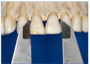
The measurements were performed according to the following anatomical landmarks: 1) Measurement A: The distance from the gingival third of the labial surface of the maxillary central incisors (LS‑MCI) to the IF posterior wall (Figure 1); 2) Measurement B: The distance from the IF posterior wall and the distal portion of the posterior nasal spine (PNE) (Figure 2); 3) Measurement C: The width of the maxillary central incisors (W‑MCI) (Figure 3).
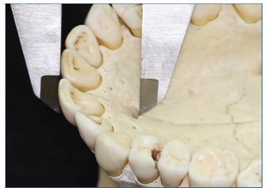
Figure 1 Measurement A: distance from the labial surfasse of the maxillary central incisors to the IF posterior wall
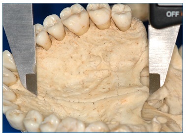
Figure 2 Measurement B: distance from the IF posterior wall to the distal portion of the posterior nasal spine
The first observer measured all 60 skulls. To evaluate intraexaminer reliability, the measurements were repeated in 20% of the sample, randomly selected, with a two‑day interval.
The second observer, who was not present during the first stage, measured, with the same caliper, 20 skulls (33.33% of the first stage). A paired t‑test revealed no significant differences between the intra‑and interexaminer measurements.
Normal distribution corroboration using the Lilliefors test verified the normality of all sample groups. A paired t‑test evaluated the degree of intra‑and interexaminer agreement. Pearson’s correlation test was applied to evaluate the correlations between the obtained measurements. The reference values were interpreted as follows: 0.8 to 1.0 = strong positive relationship; 0.6 to 0.8 = moderate positive relationship; 0.2 to 0.6 = weak positive relationship; −0.2 to 0.2 = no relationship; −0.6 to −0.2 = weak negative relationship; −0.8 to −0.6 = moderate negative relationship; −1.0 to −0.8 = strong negative relationship.
A two‑factor analysis of variance (ANOVA) determined whether differences in age or sex interfered with the measurement results. The level of significance for all statistical tests was predetermined at 5%. The Minitab 17 statistical software program (Minitab, LLC, State College, PA, USA) was used to analyze all tests.
Results
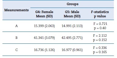
The final sample consisted of 60 dry skulls, whose death occurred at ages ranging between 16 and 64 years and a mean age of 28.4 } 12.3 years. Mean and standard deviation (SD) values for all samples, as well as 95% confidence intervals (CIs), are presented in Table 1. A coefficient variation analysis revealed that all three measurements (i.e., A, B, and C) and age were homogeneous (< 25%).
Descriptive results were obtained (Tables 2 and 3) by separating the skulls into the following groups: G1, younger than 22 years (n = 21); G2, 22 to 30 years old (n = 20); G3, older than 30 years (n = 19); G4, all females (n = 25); and G5, all males (n = 35). However, age and sex did not interfere with the results of the measurements A (age, p=0.833; sex, p=0.400), B (age, p=0.065; sex, p=0.152), and C (age, p=0.165; sex, p=0.564).
Level of significance: p<0.05
Level of significance: p<0.05
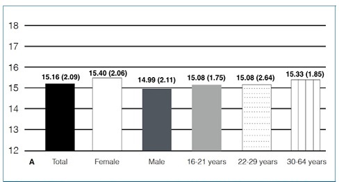
There were no correlations between the distances A and B or between B and C, and only a weak correlation was noted between measurements A and C (Table 4). Measurement A exhibited a relatively constant value of 15 mm for all groups (Figure 4).
Discussion
Since the artificial maxillary anterior teeth set aesthetic, phonetic, and functional patterns, determining and maintaining their proper position is essential for adequately constructing both tooth‑supported and implant‑supported prostheses.6
However, the facial contours of edentulous patients offer only indirect indications for artificial anterosuperior teeth arrangement.6 Furthermore, the use of references such as the smile line and the distance between mouth corners reflects significant variation and does not facilitate precise or reliable positioning of artificial teeth.6
A study23 attempted to establish the ideal oral cavity anatomical structures with which to locate the palatal foramen and reported linear distances between the front teeth and the landmark in question but did not provide data about the IF.
Those authors, using computed tomography (CT) images, questioned the use of dry skulls in studies due to the frequent lack of information available about sex and the difficulty with assessing angular measurements, leading to a low level of accuracy.
In our study, the whole sample had been classified and identified by both sex and age at the time of death. Also, our study considered the previous data and involved only direct linear measurements.
Another study18 assessed 34 CT skull scans and found that sex, age, and the presence or absence of teeth did not affect the anatomy of the nasopalatine canal, including the IF. However, these authors reported the need for studies with larger samples to assess the influence of these factors better. Similarly, we herein did not observe any differences in bone and dental measurements when considering sex and age. It should be noted that the sample size of our study (n = 60 skulls) was almost twice as large as that study’s.
Many authors12,14,19,20,24,25reported no statistical differences in the anatomy of the IF when comparing toothed and edentulous samples. Conversely, in one study,13 the appearance of an enlarged incisive canal was observed after the teeth were lost; however, the posterior wall of the incisive canal remained relatively stable. It is noteworthy, though, that these results were achieved using a heterogeneous sample in which no pairing for comparison of anatomical changes was conducted.
Therefore, the findings could be attributed to anatomical variations of the evaluated structure.
In the current study, both age and sex were not statistically significantly correlated with the measurements obtained.
In another study,19 the IF exhibited no statistical differences in terms of sex, age, or the presence or absence of the central incisors. A small increase in the IF in males was noted. Elsewhere, two studies8,20observed differences between males and females in terms of the size of the IF and the dimensions of the remaining buccal bone in edentulous patients. However, these authors did not identify significant differences regarding age.
No correlations were observed between the measurements A, B, and C (Table 4), so it was not possible to establish an approximate mathematical correlation between them. Nevertheless, the high consistency level obtained from measurement A, exhibiting widely uniform and constant results for all groups (Figure 4), suggests that the 15‑mm distance from the central incisors to the IF posterior wall could be an additional parameter to help guide anteroposterior positioning of superior anterior artificial teeth in cases of prosthetic rehabilitation.
Moreover, these data exhibited constant means independently of sex and age. The clinical relevance of this study was the presentation of an additional measurement to be used in planning prosthetic replacements of edentulous anterior teeth in the maxilla with extensive alveolar bone resorption, which should be incorporated not as a fixed measurement but rather as a reference value to assist clinicians.
One particular limitation of this study was that the anteroposterior measurements were not purely horizontal because the dry skull points used were not positioned in the same axial plane. Therefore, other studies should test the correlation between measurements A and B considering perpendicular projections of the points to only one axial plane. This assessment is possible using CT images and could allow a direct application in the clinic. Finally, because the study sample was nonprobabilistic, covering only one specific origin, caution should be taken when generalizing the findings to larger populations.
Conclusions
The following conclusions can be drawn from this study:
1. It was not possible to establish a mathematical ratio between the distance from the labial surface of the maxillary central incisors to the incisive foramen and the distance from the incisive foramen posterior wall to the posterior nasal spine because there is no correlation between these measurements when obtained directly from dry skulls.
2. The 15‑mm distance from the central incisors to the incisive foramen posterior wall could be an additional parameter to guide the anteroposterior positioning of the maxillary anterior artificial teeth in cases of prosthetic rehabilitation.
3. Age and sex did not interfere with the anatomical distances from the labial surface of the maxillary central incisors to the IF posterior wall, from the IF posterior wall to the distal portion of the posterior nasal spine, nor with the width of the maxillary central incisors.
4. Other studies are necessary to test the correlation between these measurements in only one axial plane using maxillary CT images.


