SciELO - Scientific Electronic Library Online

 
vol.61 número3Prevalência de erosão, cárie dentária e traumatologia orofacial em atletas de hóquei em patins: Estudo preliminar no distrito de Lisboa índice de autoresíndice de assuntosPesquisa de artigos
Home Pagelista alfabética de periódicos  

Serviços Personalizados

Journal

Artigo

Indicadores

Links relacionados

  • Não possue artigos similaresSimilares em SciELO

Compartilhar


Revista Portuguesa de Estomatologia, Medicina Dentária e Cirurgia Maxilofacial

versão impressa ISSN 1646-2890versão On-line ISSN 1647-6700

Rev Port Estomatol Med Dent Cir Maxilofac vol.61 no.3 Lisboa set. 2020  Epub 30-Set-2020

https://doi.org/10.24873/j.rpemd.2020.11.713 

Original Research

6-month clinical performance of a universal adhesive on non-carious cervical lesions: self-etch and etch-and-rinse techniques

Desempenho clínico a 6 meses de adesivo universal em lesões cervicais não cariosas: técnicas self-etch e etch-and-rinse

Joana Cruz1 

Ana Luísa Silva1 

Raquel Eira1 

Catarina Coito1 

Manuela Lopes1 

Alexandre Cavalheiro1 

1Faculdade de Medicina Dentária, Universidade de Lisboa, Lisbon, Portugal


Abstract

Objectives:

The purpose of this study was to evaluate the 6-month clinical performance of Adhese Universal applied with two different application strategies (self-etch vs. etch-andrinse technique) when restoring non-carious cervical lesions.

Methods:

Twenty-six patients participated in this study. Restorations of 117 non-carious cervical lesions were assigned to 2 groups: 1) Adhese Universal in the etch-and-rinse mode (n=59) and 2) Adhese Universal in the self-etch mode (n=58). The same resin composite (Tetric EvoCeram) was used for all restorations. The restorations were evaluated at baseline and at 6 months, using the World Dental Federation criteria. The results were analyzed statistically by the McNemar test (α=0.05 and power of 80%) to compare the differences between baseline and 6 months and a generalized estimating equation to compare the differences between the 2 techniques.

Results:

No differences were found in restoration performance between the baseline and the end of the 6-month period in the self-etch mode (marginal coloring: p=0.1366; fractures/retention: p=1.000; marginal adaptation: p=1.000; hypersensitivity: p=0.4795; recurrence of caries: p=1.000). On the other hand, in the etch-and-rinse mode, for both fractures/retention (p=0.0028) and marginal adaptation (p=0.0016), significant differences were found. Significant differences were also detected between groups at 6 months for fractures/retention and marginal adaptation (p<0.01). Nine restorations were lost at 6 months in the etch-and-rinse group.

Conclusions:

The tested universal adhesive obtained better results in the self-etch technique than in the etch-and-rinse technique, both on fractures/retention and marginal adaptation.

Keywords: Clinical trial; Etch-and-rinse; Self-etch; Universal adhesive

Resumo

Objetivos:

O objetivo deste estudo foi avaliar o desempenho clínico do Adhese Universal a 6 meses aplicado no modo self-etch e no modo etch-and-rinse em lesões cervicais não cariogénicas.

Métodos:

Vinte e seis participantes participaram neste estudo. As restaurações das 117 lesões cervicais não cariogénicas foram distribuídas por dois grupos: 1) Adhese Universal no modo etch-and-rinse (n=59) e 2) Adhese Universal no modo self-etch (n=58). Foi utilizada a mesma resina composta em todas as restaurações. As restaurações foram avaliadas no baseline e passado seis meses, com recurso aos critérios da Federação Dentária Internacional. Os resultados foram analisados estatisticamente pelo teste de McNemar (α=0,05 poder de 80%) para comparar a diferença entre o baseline e os 6 meses e uma equação de estimativa generalizada

para comparar a diferença entre as duas técnicas.

Resultados:

Não foram encontradas diferenças no desempenho das restaurações entre o baseline e os 6 meses no modo self-etch (Coloração marginal: p=0,1366; Fraturas/retenção: p=1,000; Adaptação marginal: p= 1,000; Hipersensibilidade: p=0,4795; Recorrência de lesões de cárie: p=1,000). No entanto, no modo etch-and-rinse, tanto para fraturas/retenção (p=0,0028) como para adaptação marginal (p=0,0016), foram encontradas diferenças significativas entre o baseline e os seis meses. Diferenças significativas também foram encontradas entre os dois grupos para fraturas/retenção e adaptação marginal (p<0,01). No grupo etch-and-rinse

foram perdidas 9 restaurações nos 6 meses de avaliação.

Conclusão:

Este adesivo universal obteve melhores resultados na técnica self-etch do que na técnica etch-and-rinse para os critérios fraturas/retenção e adaptação marginal.

Palavras-chave: Estudo clinico; Etch-and-rinse; Self-etch; Adesivo universal

Introduction

New adhesives called universal or multimode adhesives have been recently developed. These adhesives are more versatile and, theoretically, can be used with or without acid etching in both dentin and enamel.1,2

Universal adhesives applied on dentin have been shown to have high bond strength values using both the self‑etch (SE) and etch‑and‑rinse (ER) techniques. This feature may be due to the presence of special amphiphilic monomers, such as methacryloyloxydecyl dihydrogen phosphate (MDP) or glycerol phosphate dimethacrylate (GPDM), that promote chemical bonding to the tooth.3,4,5Some studies have shown that both the SE and ER techniques achieve comparable bond strength values on dentin.3,5On enamel, the bond strengths of non‑universal adhesives are always higher with the ER mode compared to the SE mode.6,7

Laboratory and clinical results do not always show a direct correlation. Whether in vitro results also occur in vivo is yet to be confirmed. Moreover, clinical trials investigating the effectiveness of universal adhesives are limited, despite the importance of evaluating their clinical performance.8,9In general, non‑carious cervical lesions (NCCLs) are considered ideal for determining adhesives’ clinical effectiveness because they provide minimal, if any, macro‑retention; thus, all retention relies solely on the adhesion effectiveness of the adhesives tested and an ineffective bonding results in restoration loss.10

Furthermore, in these lesions, the restoration is bonded to both enamel and dentin, access is simple and does not require complicate restorative techniques, and restoration is also possible with a low C‑factor.10,11

Some clinical studies8,9,12conducted with universal adhesives on NCCLs reported no differences in the universal adhesive behavior when applied using the SE or the ER technique.

Other studies have demonstrated the superiority of the ER technique compared to the SE technique.1,13This randomized, double‑blind clinical study aimed to evaluate the clinical effectiveness of a universal adhesive applied with two different application strategies SE and ER on NCCLs.

The null hypothesis was that there was no difference in clinical performance between the ER and SE application modes.

Material and methods

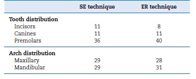
This study followed the Consolidated Standards of Reporting Trials (CONSORT) statement.14 The study was a double‑blind, randomized clinical trial that took place in the clinic of the Faculty of Dental Medicine of the University of Lisbon. All participants were informed about the study’s nature and objectives, but were not aware of what lesion received the treatments under evaluation. The Local Ethics Committee reviewed and approved the protocol and the consent form for this study. Based on pre‑established criteria, 26 participants, 15 females and 11 males, with NCCLs in incisors, canines, and premolars (Table 1) were selected. Written informed consent was obtained from all participants before treatment.

Table 1 Distribution of restorations per tooth and arch. 

As inclusion criteria, participants had to be at least 18 years old and in good general health. They needed to have at least 20 teeth in occlusion and an acceptable oral hygiene level.

Their lesions had to be nonretentive, non‑carious, and deeper than 1 mm. The lesions had to involve both the enamel and dentin of vital teeth without mobility. The cavosurface margin could not involve more than 50% of the enamel.15

Every tooth included in the study was in occlusion and proximal contact with the adjacent tooth. All patients were given oral hygiene instructions before operative treatment.

Patients with heavy bruxism habits, xerostomia, poor oral hygiene, severe or chronic periodontitis, or smoking habits were excluded from the study.8,9

The same operator restored all lesions. The operator was not blinded to group assignment when administering interventions, but the participants were. Each patient received at least two cervical restorations: one with the ER technique and the other with the SE technique.

Before isolation with the rubber dam, the operator anesthetized the teeth with lidocaine 2% with epinephrine 1:80,000 (XilonibsaR 2%; Inibsa, Barcelona, Spain). All teeth were then cleaned with pumice and water using a rubber prophylactic cup to remove the salivary pellicle and dental plaque. They were then rinsed with water and dried. The operator did not prepare any additional retention or bevel, following the American Dental Association (ADA) guidelines.16

The teeth were randomly assigned, using randomization tables, for restoration with either of two application procedures: Adhese Universal (ADH, Ivoclar Vivadent, Schaan, Liechtenstein) in the ER mode (ADH‑ ER) or Adhese Universal in the SE mode (ADH‑SE). A total of 117 cervical lesions were restored: 59 with ADH‑ER and 58 with ADH‑SE. Only a maximum of three restorations per group was placed in one patient so that, per patient, restorations prepared following the two different protocols were mutually compared.

The adhesive Adhese Universal (ADH) was used according to the manufacturer’s instructions (Table 2). The resin composite (Tetric EvoCeram, Ivoclar Vivadent, Schaan, Liechtenstein) was applied in increments of up to 2 mm, each one light‑cured for 40 seconds under an LED light‑curing unit (Elipar S10; 3M ESPE, Seefeld, Germany) with a light intensity of 600 mW/cm2 (6 J/cm2). The curing light’s output was periodically verified at >600 mW/cm2 with a radiometer (Curing Radiometer P/N 10503, Kerr, Orange, CA, USA) throughout the study. The restorations were finished immediately with fine‑grain diamond burs (Diatech Dental AG, Heerbrugg, Switzerland). Polishing was performed with rubber points (Astropol, Ivoclar Vivadent, Schaan, Liechtenstein).

Table 2 Components, composition (information supplied by the manufacturer), and application mode of the tested adhesive. 

Two calibrated independent experienced dentists evaluated the restorations with the aid of a 2.5x‑magnification dental loupe at baseline and after 6 months. They were unaware of which material had been used; thus, the study was double‑blind.

Each restoration was documented by photographs. The examiners were calibrated before the baseline evaluation, evaluating 15 restorations representing each score for each criterion, from 15 different patients with cervical restorations that did not participate in this study. Each examiner evaluated each restoration on two different time points, on two consecutive days. Cohen’s kappa statistic was used to analyze the interexaminer agreement. An intraexaminer and interexaminer agreement of at least 85% was required for the evaluation to begin.17

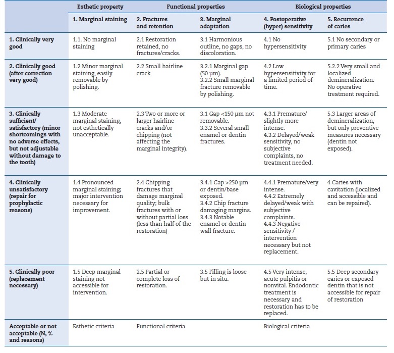
The restorations were evaluated under the World Dental Federation (FDI) criteria (Table 3).18,19Both examiners evaluated all the restorations once and independently; any discrepancy between evaluators was resolved chairside.

Table 3 World Dental Federation (FDI) Criteria Used for Clinical Evaluation. 

Sample size calculations were performed using the G*Power Program Statistical Analysis (G*Power Program, Dusseldorf, Germany) with an α=0.05, a power of 80%, and a two‑sided test.20,21The minimal sample size was 50 restorations per group in order to detect a difference of 20% among the tested groups.

The results were analyzed statistically by a paired chi‑square test - McNemar test (SAS Institute Inc., SAS/STAT 9.3 User’s Guide, Cary, NC: SAS Institute Inc., 2002‑2010) - with an α=0.05, to compare differences between baseline and 6 months. A generalized estimating equation modeling analysis was also used to compare the two techniques while controlling potential clustering problems due to multiple teeth from the same patient.

Results

Results from the restorations’ evaluation are presented in Table 4, summarized as frequencies and proportions. Strong agreement between the examiners was found, with a kappa value of 0.87. Recall rates were at 100% for all follow‑ups.
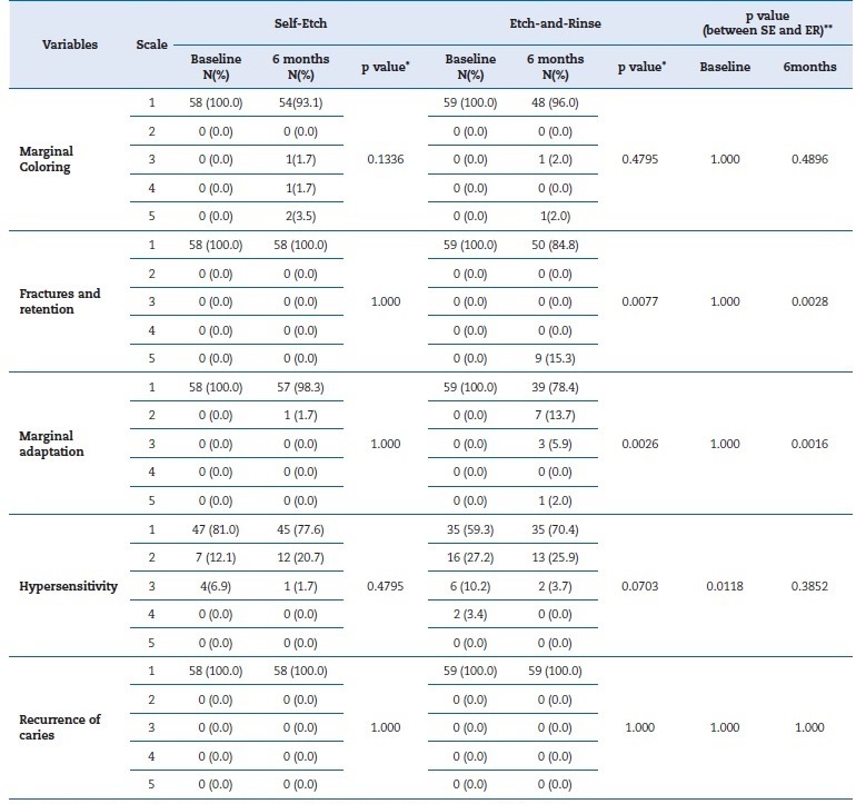
Table 4 Number of evaluated restorations for each experimental group classified according to the World Dental Federation (FDI) criteria 

* From McNemar test, and categories 2‑5 were combined for the test; ** From GEE model analysis.

In the ADH‑SE group, no differences were found in the performance of restorations between baseline and the 6‑month follow‑up (marginal coloring: p=0.1366; fractures/retention: p=1.000; marginal adaptation: p=1.000; hypersensitivity: p=0.4795; recurrence of caries: p=1.000). However, in the ADH‑ER group, significant differences (p<0.01) were found regarding both fractures/retention (p=0.0028) and marginal adaptation (p=0.0016).

At baseline, significant differences were found between the two techniques on hypersensitivity (p=0.0118) (proportion of no hypersensitivity: 81% in the ADH‑SE vs. 59% in the ADH‑ER group). However, at 6 months, no differences were observed in postoperative sensitivity between these techniques (p=0.3852).

At 6 months, significant differences were detected between groups regarding fractures/retention and marginal adaptation (p<0.01). The ER technique had a lower proportion of FDI criteria’ level 1 than the SE technique (84.8% vs. 100% for fractures/retention and 78.4% vs. 98.3% for marginal adaptation). Nine restorations were lost at 6 months in the ADH‑ER group according to the FDI criteria. No restorations were lost due to caries.

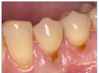
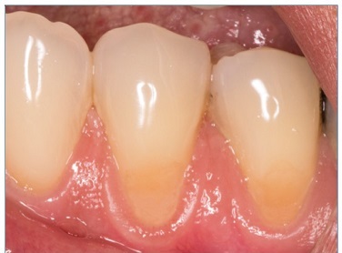
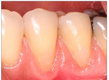
There were no dropouts in this study, so all patients were evaluated at baseline and at 6 months. Representative images of restorations are presented in Figures 1, 2, 3, 4, 5, and 6.

Figure 1 Preoperative photograph of the non‑carious cervical lesions of teeth 34 and 35 

Figure 2 Photographs after 6 months of tooth 34’s restoration by the etch‑and‑rinse technique and tooth 45’s restoration by the self‑etch technique 

Figure 3 Preoperative photograph of the non‑carious cervical lesions of teeth 44 and 45 

Figure 4 Photographs after 6 months of tooth 44’s restoration by the self‑etch technique and tooth 45’s restoration by the etch‑and‑rinse technique 

Figure 5 Preoperative photograph of the NCCLs of teeth 34 and 35 

Figure 6 Photographs after 6 months of tooth 34’s restoration by the etch‑and‑rinse technique and tooth 35’s restoration by the self‑etch technique 

Discussion

The clinical success of resin composite restorations depends on effective adhesion to enamel and dentin. Clinical studies are the first level of scientific evidence to evaluate the effectiveness of dental adhesives.22

Since universal adhesives have only recently been introduced, most studies found in the literature are laboratory tests, mainly microtensile bond strength tests. The results from these in vitro studies on dentin are very similar between the ER and SE techniques.3,8,23For enamel, it seems that etching the enamel prior to universal adhesive application improves bond strength because etching creates microporosities that are readily penetrated by the adhesive.24,25,26,27,28,29

Although in vitro studies can help us understand the behavior of adhesives,30,31clinical trials with controlled and standardized study designs are the ultimate test to evaluate the clinical effectiveness of universal adhesives, preferably in NCCLs.32

This study was designed following the recommendations of the ADA. These indicate that each group should have at least 30 restorations, with a minimum of 25 patients in the initial phase of the study and 20 patients after six months, as well as a gender and age balance between study groups. In this study, a universal adhesive’s clinical performance was evaluated at baseline and after 6 months. One hundred seventeen NCCLs were restored in 26 patients, with the adhesive applied in SE and ER modes, combined with a resin composite. Each patient received at least two cervical restorations to ensure that they had a restoration from each technique, to control various environmental factors (such as oral hygiene, saliva composition, and diet).33

Due to the expulsive configuration of the NCCLs, the retention of restorations depends on a strong and stable bond of restorative material to dentin. The occurrence of structural changes in enamel and dentin resulting from age, such as dentin sclerosis, may negatively impact the quality of that bond and, consequently, the retention and longevity of cervical restorations.34This is of special concern with NCCLs where dentin is often sclerotic and, thus, more mineralized than normal dentin.35,36In fact, Mjor27 attributed the rather poor success scored with adhesives in clinical trials (in contrast to laboratory results) to the extreme variety of dentin composition and structure found clinically.37,38

Reactive sclerosis occurs in response to slowly progressive or mild irritations like mechanical or chemical erosion and abrasion in response to severe insults, like aggressive operative procedures, attrition, and caries.37,39 Several studies show that dentin sclerosis increases with age,37,39,40which may explain why greater restoration losses have been found in older patients: patients aged 21‑40, 41‑60, and 61‑ 80 years had restoration losses of 31%, 62%, and 75%, respectively.41However, other studies have shown that retention failures cannot be associated with substrate type only,34confirming that the process of adhesion involves multifactorial aspects. Indeed, a clinical study (2000)42had an equal number of restoration failures in sclerotic and non‑sclerotic lesions, indicating that the negative interaction between dentin sclerosis and the clinical retention of adhesive systems is yet to be confirmed. In this study, there was no relationship between age and restoration loss.

A period of 6 months to 1 year seems to be sufficient to predict an adhesive’s clinical behavior accurately.43 In fact, in this study, the 6‑month evaluation period was sufficient to detect significant differences in the performance of the tested adhesive system, which belongs to a novel family of universal adhesives for which there are insufficient clinical studies.

In this study, after 6 months, nine restorations failed as a result of debonding, which highlights the poor bonding efficacy of ADH when used with the ER strategy. Furthermore, at 6 months, the ER technique had poorer results than the SE technique for marginal adaptation (78.4% vs. 98.3%). The good performance of the SE restorations is likely due to the presence of an acidic functional monomer, 10‑MDP, because calcium ions (released upon the partial dissolution of hydroxyapatite) diffuse within the hybrid layer and assemble the MDP molecules into nanolayers.44This chemical interaction between hydroxyapatite and MDP creates a stable nanolayer, which can form a stronger area at the adhesive interface to both enamel and dentin, as both contain hydroxyapatite.45,46,47Results obtained with the ER technique can be explained by the incomplete infiltration of the deeply demineralized collagen network by the bonding resin, which occurs because the phosphoric acid can decalcify dentin more deeply than the adhesive can infiltrate.48,49 Due to this incomplete impregnation of the demineralized substrate, the adhesive interface is not impermeable, and, as a result, water and dentinal fluid can easily move through the adhesive interface with consequent nanoinfiltration.50,51,52

Marginal discoloration was observed with both techniques, but no statistically significant differences were found. In the ADH‑ER group, one restoration exhibited deep marginal staining and another presented moderate marginal staining; these were not esthetically unacceptable. Discolorations were observed in the gingival margins, where cementum or dentin are more likely found than enamel margins.53In the SE technique, two restorations showed deep marginal staining, one restoration exhibited pronounced marginal staining, and one restoration presented moderate marginal staining; these were not esthetically unacceptable. The discoloration was located at the enamel margin, which may suggest the importance of including enamel’s selective conditioning with phosphoric acid to obtain the best marginal seal of restorations.54ADH is considered a mild SE adhesive, as other available universal adhesives, because it presents a pH of 2.5. Due to their moderately high pH, these adhesives have limited interaction with enamel as they cannot condition enamel as effectively as in the ER technique, resulting in increased marginal changes.45In fact, some studies concluded that additional etching of the enamel cavity margins resulted in an improved marginal adaptation on the enamel side. However, this was not critical and did not affect the overall clinical success of restorations.55,56

Marginal discoloration may be a clinical sign of future restoration failure, but it does not imply the imminent need for replacement because these discolorations, if superficial, can be removed by polishing and routine finishing.10,57,58

In this study, no restoration had secondary caries, maybe because the participants selected for this study had good oral hygiene habits.57

In this study, there was a significant difference in postoperative sensitivity between the SE and ER techniques at baseline.

Postoperative sensitivity was higher with the ER technique, possibly because phosphoric acid removes the peritubular dentin and fully opens the dentin tubules,59 which the adhesive may not be able to seal completely afterward. In contrast, with the SE technique, the dentin surface is smear‑layer sealed, and there is a lesser tubule opening.60Nevertheless, there was no difference in postoperative sensitivity between the ER and SE modes, which may be explained by thepulp’s capacity to recover in cases of reversible pulpitis.61 Results from the literature indicate that a decrease or absence of hypersensitivity may occur over time in those with NCCL restorations.57,62,63

Regarding the effect of clinical co‑variables (degree of sclerosis, patient age, tooth type, and gender), no correlation was found between these co‑variables and the results presented in the two groups at the 6‑month evaluation.

For this study, FDI criteria were used as opposed to the United States Public Health Service (USPHS) modified criteria because authors of recent publications comparing the 6‑month clinical performance of adhesion strategies using FDI and USPHS‑modified criteria concluded that the FDI criteria are more sensitive than the USPHS‑

modified criteria to small variations in clinical outcomes.1,8,9,64

Although significant differences were found in this study with the 6‑month evaluation, it may be interesting to consider a longer follow‑up in future investigations. It would also be important to evaluate this adhesive system’s behavior with the selective‑etch technique, comparing it with the SE and ER techniques, to better evaluate its clinical performance.

Conclusions

The SE technique performed better than the ER technique for the tested universal adhesive; thus, the null hypothesis is rejected. The 6‑month clinical performance of Adhese Universal depends on the bonding strategy used.

References

1. Lopes LS, Calazans FS, Hidalgo R, Buitrago LL, Gutierrez F, Reis A, et al. Six‑month Follow‑up of Cervical Composite Restorations Placed With a New Universal Adhesive System: A Randomized Clinical Trial. Oper Dent. 2016;41:465‑80. [ Links ]

2. Carrilho E, Cardoso M, Marques Ferreira M, Marto CM, Paula A, Coelho AS. 10‑MDP Based Dental Adhesives: Adhesive Interface Characterization and Adhesive Stability‑A Systematic Review. Materials (Basel). 2019;12:790. [ Links ]

3. Munoz MA, Luque I, Hass V, Reis A, Loguercio AD, Bombarda NH. Immediate bonding properties of universal adhesives to dentine. J Dent. 2013;41:404‑11. [ Links ]

4. Loguercio AD, Munoz MA, Luque‑Martinez I, Hass V, Reis A, Perdigao J. Does active application of universal adhesives to enamel in self‑etch mode improve their performance? J Dent . 2015;43:1060‑70. [ Links ]

5. Cruz J, Sousa B, Coito C, Lopes M, Vargas M, Cavalheiro A. Microtensile bond strength to dentin and enamel of self‑etch vs. etch‑and‑rinse modes of universal adhesives. Am J Dent . 2019;32:174‑82. [ Links ]

6. Marchesi G, Frassetto A, Mazzoni A, Apolonio F, Diolosa M, Cadenaro M, et al. Adhesive performance of a multi‑mode adhesive system: 1‑year in vitro study. J Dent . 2014;42:603‑12. [ Links ]

7. Perdigao J, Sezinando A, Monteiro PC. Laboratory bonding ability of a multi‑purpose dentin adhesive. Am J Dent . 2012;25:153‑8. [ Links ]

8. Loguercio AD, de Paula EA, Hass V, Luque‑Martinez I, Reis A, Perdigao J. A new universal simplified adhesive: 36‑Month randomized double‑blind clinical trial. J Dent . 2015;43:1083‑92. [ Links ]

9. Mena‑Serrano A, Kose C, De Paula EA, Tay LY, Reis A, Loguercio AD, et al. A new universal simplified adhesive: 6‑month clinical evaluation. J Esthet Restor Dent. 2013;25:55‑69. [ Links ]

10. Peumans M, De Munck J, Mine A, Van Meerbeek B. Clinical effectiveness of contemporary adhesives for the restoration of non‑carious cervical lesions. A systematic review. Dent Mater. 2014;30:1089‑103. [ Links ]

11. Faye B, Sarr M, Bane K, Aidara AW, Niang SO, Kane AW. One‑year clinical evaluation of the bonding effectiveness of a one‑step, self‑etch adhesive in noncarious cervical lesion therapy. Int J Dent . 2015;2015:984065. [ Links ]

12. Perdigao J, Kose C, Mena‑Serrano AP, De Paula EA, Tay LY, Reis A, et al. A new universal simplified adhesive: 18‑month clinical evaluation. Oper Dent . 2014;39:113‑27. [ Links ]

13. Oz FD, Ergin E, Canatan S. Twenty‑four‑month clinical performance of different universal adhesives in etch‑and‑rinse, selective etching and self‑etch application modes in NCCL - a randomized controlled clinical trial. J Appl Oral Sci. 2019;27:e20180358. [ Links ]

14. Schulz KF, Altman DG, Moher D, CONSORT Group. CONSORT 2010 statement: updated guidelines for reporting parallel group randomised trials. Int J Surg. 2011;9:672‑7. [ Links ]

15. Loguercio AD, Reis A, Barbosa AN, Roulet JF. Five‑year double‑blind randomized clinical evaluation of a resin‑modified glass ionomer and a polyacid‑modified resin in noncarious cervical lesions. J Adhes Dent. 2003;5:323‑32. [ Links ]

16. American Dental Association Council on Scientific Affairs. Chicago I (2001) Acceptance program guidelines: dentin and enamel adhesive materials. American Dental Association. [ Links ]

17. Cvar JF, Ryge G. Reprint of criteria for the clinical evaluation of dental restorative materials 1971. Clin Oral Investig.2005;9:215‑32. [ Links ]

18. Hickel R, Peschke A, Tyas M, Mjor I, Bayne S, Peters M, et al. FDI World Dental Federation - clinical criteria for the evaluation of direct and indirect restorations. Update and clinical examples. J Adhes Dent . 2010;12:259‑72. [ Links ]

19. Hickel R, Roulet JF, Bayne S, Heintze SD, Mjor IA, Peters M, et al. Recommendations for conducting controlled clinical studies of dental restorative materials. Science Committee Project 2/98-FDI World Dental Federation study design (Part I) and criteria for evaluation (Part II) of direct and indirect restorations including onlays and partial crowns. J Adhes Dent . 2007;9(Suppl 1):121‑47. [ Links ]

20. Faul F, Erdfelder E, Buchner A, Lang AG. Statistical power analyses using G*Power 3.1: tests for correlation and regression analyses. Behav Res Methods. 2009;41:1149‑60. [ Links ]

21. Faul F, Erdfelder E, Lang AG, Buchner A. G*Power 3: a flexible statistical power analysis program for the social, behavioral, and biomedical sciences. Behav Res Methods . 2007;39:175‑91. [ Links ]

22. Hafer M, Jentsch H, Haak R, Schneider H. A three‑year clinical evaluation of a one‑step self‑etch and a two‑step etch‑and‑rinse adhesive in non‑carious cervical lesions. J Dent . 2015;43:350‑61. [ Links ]

23. Wagner A, Wendler M, Petschelt A, Belli R, Lohbauer U. Bonding performance of universal adhesives in different etching modes. J Dent . 2014;42:800‑7. [ Links ]

24. Rosa WL, Piva E, Silva AF. Bond strength of universal adhesives: A systematic review and meta‑analysis. J Dent . 2015;43:765‑76. [ Links ]

25. McLean DE, Meyers EJ, Guillory VL, Vandewalle KS. Enamel Bond Strength of New Universal Adhesive Bonding Agents. Oper Dent . 2015;40:410‑7. [ Links ]

26. Taschner M, Nato F, Mazzoni A, Frankenberger R, Kramer N, Di Lenarda R, et al. Role of preliminary etching for one‑step self‑etch adhesives. Eur J Oral Sci. 2010;118:517‑24. [ Links ]

27. Van Landuyt KL, Peumans M, De Munck J, Lambrechts P, Van Meerbeek B. Extension of a one‑step self‑etch adhesive into a multi‑step adhesive. Dent Mater . 2006;22:533‑ 44. [ Links ]

28. Torii Y, Itou K, Nishitani Y, Ishikawa K, Suzuki K. Effect of phosphoric acid etching prior to self‑etching primer application on adhesion of resin composite to enamel and dentin. Am J Dent . 2002;15:305‑8. [ Links ]

29. Li N, Nikaido T, Alireza S, Takagaki T, Chen JH, Tagami J. Phosphoric acid‑etching promotes bond strength and formation of acid‑base resistant zone on enamel. Oper Dent . 2013;38:82‑90. [ Links ]

30. Van Meerbeek B, De Munck J, Yoshida Y, Inoue S, Vargas M, Vijay P, et al. Buonocore memorial lecture. Adhesion to enamel and dentin: current status and future challenges. Oper Dent . 2003;28:215‑35. [ Links ]

31. Peumans M, Munck J, Van Landuyt K, Lambrechts P, Van Meerbeek B. Three‑year clinical effectiveness of a two‑step self‑etch adhesive in cervical lesions. Eur J Oral Sci . 2005;113:512‑8. [ Links ]

32. Van Meerbeek B, Perdigao J, Lambrechts P, Vanherle G. The clinical performance of adhesives. J Dent . 1998;26:1‑20. [ Links ]

33. Tuncer D, Yazici AR, Ozgunaltay G, Dayangac B. Clinical evaluation of different adhesives used in the restoration of non‑carious cervical lesions: 24‑month results. Aust Dent J. 2013;58:94‑100. [ Links ]

34. Santiago SL, Passos VF, Vieira AH, Navarro MF, Lauris JR, Franco EB. Two‑year clinical evaluation of resinous restorative systems in non‑carious cervical lesions. Braz Dent J. 2010;21:229‑34. [ Links ]

35. Powell LV, Johnson GH, Gordon GE. Factors associated with clinical success of cervical abrasion/erosion restorations. Oper Dent . 1995;20:7‑13. [ Links ]

36. Yoshiyama M, Carvalho RM, Sano H, Horner JA, Brewer PD, Pashley DH. Regional bond strengths of resins to human root dentine. J Dent . 1996;24:435‑42. [ Links ]

37. Van Meerbeek B, Braem M, Lambrechts P, Vanherle G. Morphological characterization of the interface between resin and sclerotic dentine. J Dent . 1994;22:141‑6. [ Links ]

38. Mjor I (1987) Reaction patterns of dentin. In: Thylstrup A. Lesch SA, Qvist V (ed) Dentine and Dentine Reactions in the Oral Cavity. Oxford University Press 27‑31. [ Links ]

39. Stanley HR, Pereira JC, Spiegel E, Broom C, Schultz M. The detection and prevalence of reactive and physiologic sclerotic dentin, reparative dentin and dead tracts beneath various types of dental lesions according to tooth surface and age. J Oral Pathol. 1983;12:257‑89. [ Links ]

40. Heymann HO, Sturdevant JR, Bayne S, Wilder AD, Sluder TB, Brunson WD. Examining tooth flexure effects on cervical restorations: a two‑year clinical study. J Am Dent Assoc. 1991;122:41‑7. [ Links ]

41. Bayne SC, Heymann HO, Sturdevant JR, Wilder AD, Sluder TB. Contributing co‑variables in clinical trials. Am J Dent . 1991;4:247‑50. [ Links ]

42. van Dijken JW. Clinical evaluation of three adhesive systems in class V non‑carious lesions. Dent Mater . 2000;16:285‑91. [ Links ]

43. Blunck U, Knitter K, Jahn KR. Six‑month clinical evaluation of XP BOND in noncarious cervical lesions. J Adhes Dent . 2007;9(Suppl 2):265‑8. [ Links ]

44. Yoshida Y, Yoshihara K, Nagaoka N, Hayakawa S, Torii Y, Ogawa T, et al. Self‑assembled Nano‑layering at the Adhesive interface. J Dent Res. 2012;91:376‑81. [ Links ]

45. Van Meerbeek B, Yoshihara K, Yoshida Y, Mine A, De Munck J, Van Landuyt KL. State of the art of self‑etch adhesives. Dent Mater . 2011;27:17‑28. [ Links ]

46. Yoshida Y, Van Meerbeek B, Nakayama Y, Yoshioka M, Snauwaert J, Abe Y, et al. Adhesion to and decalcification of hydroxyapatite by carboxylic acids. J Dent Res. 2001;80:1565‑9. [ Links ]

47. Hashimoto M, Ohno H, Yoshida E, Hori M, Sano H, Kaga M, et al. Resin‑enamel bonds made with self‑etching primers on ground enamel. Eur J Oral Sci . 2003;111:447‑53. [ Links ]

48. Perdigao J, Carmo AR, Anauate‑Netto C, Amore R, Lewgoy HR, Cordeiro HJ, et al. Clinical performance of a self‑etching adhesive at 18 months. Am J Dent . 2005;18:135‑40. [ Links ]

49. Pashley DH, Tay FR. Aggressiveness of contemporary self‑etching adhesives. Part II: etching effects on unground enamel. Dent Mater . 2001;17:430‑44. [ Links ]

50. Perdigao J, Frankenberger R. Effect of solvent and rewetting time on dentin adhesion. Quintessence Int. 2001;32:385‑90. [ Links ]

51. Tay FR, Pashley DH, Suh B, Carvalho R, Miller M. Single‑step, self‑etch adhesives behave as permeable membranes after polymerization. Part I. Bond strength and morphologic evidence. Am J Dent . 2004;17:271‑8. [ Links ]

52. Tay FR, Pashley DH, Suh BI, Carvalho RM, Itthagarun A. Single‑step adhesives are permeable membranes. J Dent . 2002;30:371‑82. [ Links ]

53. Mirmohammadi H, Khosravi K, Kashani K, Kleverlaan C, Feilzer A. Influence of filler existence on microleakage of a self‑etch adhesive system. J Conserv Dent. 2014;17:175‑8. [ Links ]

54. Miguez PA, Castro PS, Nunes MF, Walter R, Pereira PN. Effect of acid‑etching on the enamel bond of two self‑etching systems. J Adhes Dent . 2003;5:107‑12. [ Links ]

55. Peumans M, De Munck J, Van Landuyt KL, Poitevin A, Lambrechts P, Van Meerbeek B. Eight‑year clinical evaluation of a 2‑step self‑etch adhesive with and without selective enamel etching. Dent Mater . 2010;26:1176‑84. [ Links ]

56. Peumans M, De Munck J, Van Landuyt K, Lambrechts P, Van Meerbeek B. Five‑year clinical effectiveness of a two‑step self‑etching adhesive. J Adhes Dent . 2007;9:7‑10. [ Links ]

57. Yaman BC, Dogruer I, Gumustas B, Efes BG. Three‑year randomized clinical evaluation of a low‑shrinkage silorane‑based resin composite in non‑carious cervical lesions. Clin Oral Investig . 2014;18:1071‑9. [ Links ]

58. Gladys S, Van Meerbeek B, Lambrechts P, Vanherle G. Marginal adaptation and retention of a glass‑ionomer, resin‑modified glass‑ionomers and a polyacid‑modified resin composite in cervical Class‑V lesions. Dent Mater . 1998;14:294‑306. [ Links ]

59. Rosales‑Leal JI, de la Torre‑Moreno FJ, Bravo M. Effect of pulp pressure on the micropermeability and sealing ability of etch & rinse and self‑etching adhesives. Oper Dent . 2007;32:242‑50. [ Links ]

60. Oliveira SS, Marshall SJ, Hilton JF, Marshall GW. Etching kinetics of a self‑etching primer. Biomaterials. 2002;23:4105‑12. [ Links ]

61. Goldberg M, Farges JC, Lacerda‑Pinheiro S, Six N, Jegat N, Decup F, et al. Inflammatory and immunological aspects of dental pulp repair. Pharmacol Res. 2008;58:137‑47. [ Links ]

62. Can Say E, Ozel E, Yurdaguven H, Soyman M. Three‑year clinical evaluation of a two‑step self‑etch adhesive with or without selective enamel etching in non‑carious cervical sclerotic lesions. Clin Oral Investig . 2014;18:1427‑33. [ Links ]

63. Tuncer D, Celik C, Yamanel K, Arhun N. 1 year clinical evaluation of microhybrid composites used in the restoration of non‑carious cervical lesions. Oral Health Dent Manag. 2014;13:366‑71. [ Links ]

64. Perdigao J, Dutra‑Correa M, Saraceni CH, Ciaramicoli MT, Kiyan VH, Queiroz CS. Randomized clinical trial of four adhesion strategies: 18‑month results. Oper Dent . 2012;37:3‑11. [ Links ]

Received: January 08, 2020; Accepted: September 25, 2020

* Corresponding author. Joana Cruz E-mail address: joanacruz2@gmail.com

Conflict of interest The authors have no conflicts of interest to declare.

Creative Commons License This is an open-access article distributed under the terms of the Creative Commons Attribution License