Introduction
The restoration of endodontically treated teeth requires a proper clinical and radiographic examination of the remaining structure, the bone implantation, and the periapical status.1,2Besides adequate endodontic treatment, it comprises the complete removal of decayed tissue, previous restorations, and enamel without dentin support.1,2 Whenever the amount of remaining coronal structure is insufficient to support the prosthetic restoration, the use of an intracanal retainer is necessary for retention purposes.2-4
However, selecting the optimal treatment option is difficult because it must consider different clinical factors, such as the amount of remaining tooth structure and the complexity of the case. Therefore, different intracanal retainers have been suggested to restore endodontically treated teeth.2-4 For instance, metallic retainers are still the most frequent type of retainer used in the dental clinic.5-7These retainers demand a series of meticulous clinical steps, essential to maintain tooth resistance, asepsis, and root canal sealing.1,5If the professional does not properly follow this clinical protocol, they will compromise the restoration’s longevity.6,7Moreover, thorough knowledge of the root anatomy, the type and status of the endodontic filling material, the method of filling material removal during the root canal preparation, and the ability of the operator are crucial for the clinical success of the therapy using intracanal retainers.8,9
Before starting the restoration of an endodontically treated tooth with insufficient dentinal support, it is primordial to observe the quality of the endodontic treatment to ensure the success and longevity of the intracanal retainer.10-14The endodontic treatment must not present radiolucent spaces inside the root canal, and a minimal quantity of remaining endodontic filling material (around 3-4 mm) should be kept to preserve the apical sealing.15-17 Furthermore, the operator must carefully analyze important aspects, such as the root canal walls’ inclination after preparation and the retainer’s length, diameter, and surface characteristics.1,11
One of the most important requirements for the fabrication of an intracanal retainer is its length, which must achieve two thirds (2/3) of the root canal length, maintaining a 4- to 5-mm apical sealing.1,3,7,8 In teeth with bone loss, the intracanal retainer length must reach half of the root bone anchorage.7,8 On the other hand, some authors claim that the retainer’s length must always be as long as possible, maintaining a 4- to 5-mm apical sealing.3,7 However, in short or bent roots, the post will be shorter, and thus, retention will be compromised.9,10Short intracanal retainers associated with large clinical crowns may lead to root fracture or post displacement due to an unfavorable crown-to-root ratio.10
The literature seems to agree that a correct length of the retainer within the root is essential to prevent root fractures.10-14In fact, the greater the post’s length, the greater its retention will be.10,12-14A proper extension of the intracanal retainer is associated with adequate dissipation of forces and longevity of the restoration.10,12-14However, the post extension cannot compromise the apical sealing or the remaining root’s strength and integrity.15-18 Authors suggest the conservation of at least 3 mm of filling material for short roots,9 whereas others claim that 4 to 5 mm are needed to maintain apical sealing integrity.7,19
To the best of our knowledge, there is little evidence correlating the length of the intracanal retainer with the quality of the remaining endodontic sealing.15,16,19,20Furthermore, few surveys have assessed the quality of restorations performed in endodontically treated teeth at Dental Schools.15,16Therefore, this retrospective study aimed to radiographically evaluate the length of metallic cast retainers performed in a Dental School from Southern Brazil and its correlation with the quality of the remaining apical sealing.
Material and methods
This study was previously approved by the Ethics Committee for Research with Human Beings of the Federal University of Santa Catarina (Protocol N. 2269/12), in full accordance with the Declaration of Helsinki.
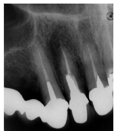
Clinical records of patients from the Dental School of the Federal University of Santa Catarina were randomly selected, with no specific population, for this study. This study’s radiographic images came from initial radiographic examinations performed in patients submitted to dental treatment. The inclusion criteria were based on previous studies.16,20 Periapical radiographs containing cast retainers placed on single-rooted teeth, as well as adequate processing and storage quality, were considered for the final sample selection (Figure 1).

Figure 1 Periapical radiography used for prosthetic/endodontic aspects assessment. Cast metal posts and cores placed on superior teeth
The radiographic images were selected randomly, with no influence of the investigator on the results. When two or more single-rooted teeth containing intraradicular retainers were found in the same radiographic image, they were individually assessed. Patients’ identity was not disclosed during data collection,
assuring its confidentiality. The radiographs were placed on a negatoscope and photographed with a 12.1-megapixel digital camera (Cyber-Shot, Sony, New York, NY, USA). The images were taken in a black-and-white mode within a 15-cm distance (Figure 1). Adobe Photoshop 7.0 software (Abobe System, SJ, USA) was used to calibrate the magnification and contrast of the images.
Two independent, pre-calibrated examiners blindly evaluated the images. Kappa index was used to check the agreement between examiners (0.91). Both prosthetic and endodontic aspects were recorded using standardized scores in a database for later analysis.
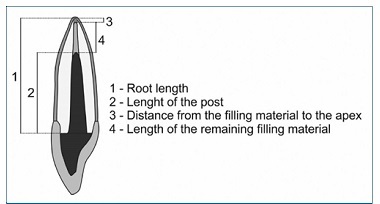
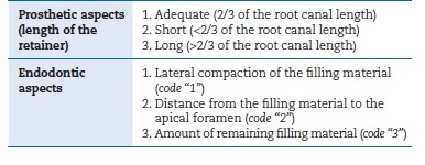
The intracanal retainer’s length should follow the proportion of 2/3 of the root canal length (Figure 2).1,3,8 Intracanal retainers were classified as “adequate,” “short,” and “long” when presenting lengths equivalent to 2/3, <2/3, and >2/3 of the root canal length, respectively.20 The data were codified with “a,” “s,” and “l,” for adequate, short, and long retainers, respectively (Table 1).
The first endodontic aspect assessed was the quality of the filling material’s lateral compaction (1), where compaction was considered adequate in the absence of radiolucent spaces in the remaining endodontic filling material. Secondly, a 1-2 mm distance from the filling material to the apical foramen (2) was accepted as correct. The third aspect assessed was the amount of remaining filling material (3), where a minimum of 3 mm was considered adequate (Table 1).
After the analysis by each examiner and registration on separate spreadsheets, the indicators whose answers differed between examiners were verified. The examiners reached a consensus for a final decision. When consensus was not reached, the opinion of a third examiner was sought. With the data tabulated, a descriptive analysis of the results was performed.
The data were submitted to statistical analysis through a chi-square test at a 5% significance level.
Results
A total of 320 periapical radiographs comprising 443 teeth with cast intracanal retainers met the eligibility criteria and were selected. The overall results of the study are summarized in Table 2.
Regarding the intracanal retainers’ length, only 66 (14.9%) were considered adequate, whereas the other 377 (85.1%) were considered inadequate, either due to being short (n=365, 82.4%) or long (n=12, 2.7%) (Table 2). Overall, 321 (72.5%) endodontic treatments were considered inadequate. Namely, the following results were obtained regarding the endodontic aspects observed: an insufficient amount of remaining filling material in 92.5% of the cases (n=297), inadequate lateral compaction in 92.2% (n=296), and incorrect distance from the remaining filling material to the apical foramen in 53.3% (n=171). Two or three aspects were observed in the same tooth.
In 148 cases (46.1%), endodontic treatment was considered incorrect in all aspects assessed, regardless of the retainer length. The absence of endodontic treatment was observed in 21 cases (4.7%), and four of them (0.9%) presented adequate retainers (Table 2).
The chi-square test was used to assess the association between short intracanal retainers in the cases where endodontic treatment was missing. The hypothesis that short retainers are more often in teeth with missing endodontic treatment was tested, and no statistical significance was found (p=0.537).
On the other hand, the same statistical test could not be applied to long retainers due to the limited number of cases (12 retainers). The chi-square test was also applied to test the hypothesis that inadequate intracanal retainers are more often in cases where endodontic treatment is inadequate, and statistical significance was found (p=0.0003), correlating inadequate retainers with inadequate root canal filling.
Discussion
The purpose of this retrospective study was to radiographically evaluate the quality of intracanal retainers, considering their length and the condition of the endodontic treatment performed before retainers’ fixation.
Our findings demonstrated that the intracanal retainer’s length was adequate in only 66 cases (14.9%). The great majority of the assessed cases (82.4%) presented short intracanal retainers. Conversely, Klautau et al.15 reported inadequate length in less than half (44.7%) of the retainers in their study.
The conflicting results might result from these authors having considered retainers that presented half of the root length as adequate. On the other hand, Jamani et al.,16 who assessed a total of 320 retainers, found that only 3.21% (18 retainers) had adequate length. They also reported that 57.15% of the retainers were short, due to showing a length shorter than that of the crowns, and that only 32.14% (180 retainers) were longer than expected.
Regarding endodontic treatment, this clinical procedure had not been done in 4.7% of the cases in this study, which corroborates the results by Klautau et al. (4.16%).15On the other hand, Jamani et al.,16who examined 560 radiographic images, found no evidence of filling material in 16.79% of the cases. The authors justified the high percentage of endodontic treatment absence with pulp mummification, a common procedure in Jordania, where the investigation was conducted.
In the present study, 72.5% of the endodontic treatments detected were considered inadequate. The most common failures in the 321 inadequate endodontic treatment observed were the amount of remaining endodontic filling material (92.5%) and the lateral compaction quality (92.2%). On the other hand, in the study by Al-Hamad et al.,19only 4.7% of the 129 retainers assessed showed deficient lateral compaction, and only 4.7% presented less than 3 mm of remaining filling material.
In our study, the retainers selected for evaluation were fabricated and fixed by dental students, under clinical lecturers’ supervision, which might explain the conflicting results. In addition, the distance from the endodontic filling material to the apical foramen had a high (53.3%) prevalence of failure.
In the study by Klautau et al.,15 40.21% of the cases did not show homogeneity of the filling material, 27.71% showed less than 3 mm of remaining apical sealing, and 53.64% presented inadequate endodontic treatment. Jamani et al.16found more than 5 mm of remaining filling material in 70.71% of the assessed cases, whereas 10.36% had between 4 to 5 mm and 2.14% between 1 to 3 mm.
In this study, adequate retainers were 2.5 times more often in teeth with adequate endodontic treatment. When inadequate endodontic treatments were considered, the rate was maintained; i.e., the partial or total inadequacy of the endodontic treatment did not change the proportion of inadequate retainers. This finding contributes to the hypothesis that professionals who perform inadequate intracanal retainers, in most of the cases, do not properly evaluate the quality of the endodontic treatment. Jamani et al.16 also observed this, stating that the data found represented the poor quality of the endodontic treatment in the studied population. However, in the present study, the association between the presence or absence of endodontic treatment and the fabrication of adequate or short intracanal retainers was not statistically significant.
Therefore, the presence of endodontic treatment did not interfere with the adequacy of the retainer.
Some factors may explain the inadequate fabrication of these retainers. One factor is intracanal preparation failure, especially if it is performed using the indirect technique.11,16
During the impression, the entire radicular portion of the preparation must be copied for a faithful reproduction of the intracanal preparation.11,16Otherwise, the impression will be shorter than the desired length and, consequently, will result in a shorter intracanal retainer.11,16When a radiographic examination is performed before the prosthetic crown cementation, as recommended by the clinical protocol,1,9the mistake might be easily detected and corrected with a new impression.1,9 However, if the professional skips this clinical step and only checks the retainer’s clinical settlement, it may be cemented with an inadequate length.1,9
The failure in intracanal retainers’ fabrication is also likely to result from the fear of perforating the root canal during its preparation.11,16Therefore, if the professional is
not safe enough to perform this procedure, they should avoid using burs within the root canal and/or refer the patient to a specialist.1,9On the other hand, excessive caution should also be considered in the fabrication of short intracanal retainers, as it results in inadequate root canal emptying.1,9 In any case, the lack of knowledge and mastery of the technique may be considered the main reason for fabricating inadequate retainers.11,16
Only 5.9% of the cases in this study were considered totally adequate (both endodontic and prosthetic results). However, when considering the cases where endodontic treatment was considered adequate and short or long retainers were present (inadequate prosthetic results), the number increases to 16.9%.
In these cases where retainers removal may represent risks, such as radicular perforation and crack or fracture of the root, the retainers could be kept and followed up, making the case clinically acceptable.21-23However, 77.2% of the cases are still unacceptable, i.e., inadequate retainers with inadequate endodontic treatment, which justifies a clinical re-intervention.
In this study, the patient’s dental history was not evaluated. Therefore, the presence of periapical lesions was not considered.
The presence of radiolucency at the root apex does not provide sufficient information about the case since it may mean either a lesion undergoing remission or a well-performed endodontic retreatment where the next step would be parendodontic surgery to remove the periapical lesion.24 Furthermore, this study included only cast retainers due to the need for sample standardization. Nonetheless, the study could have been conducted with other types of retainers, as in previous studies.16,19
Some other limitations have been found in this study. Too dark or too light radiographic examinations, where adjustment by digital resources was not sufficient, were eliminated from the sample. Elongated or shortened radiographs were also eliminated due to the need to control the image acquisition - preferably by a single operator; however, this is practically impossible in a Dental School, where the referred patients bring their exams or dental students take the initial radiographs. Even though cast retainers are traditionally the most often used retainers,1,4clinicians have shown a lack of mastery of the technique, either by lack of knowledge or skill or excessive prudence or negligence. A great number of cases considered as inadequate may compromise the longevity of the treatments performed, either by endodontic failure, making the tooth susceptible to infections, or by prosthetic failure, favoring the occurrence of a tooth fracture, which usually leads to extraction.
Conclusions
Considering the findings of this retrospective study, it can be concluded that clinically acceptable intracanal retainers are generally associated with adequate endodontic treatments. When endodontic treatments are inappropriate, intracanal retainers are also inappropriate, usually shorter than recommended.


